
NATIONAL INDUSTRY ALLIANCE FOR MARINE DRUGS AND BIOPRODUCTS
Chitosan (CS) has emerged as a promising alternative against antimicrobial resistance due to its broad-spectrum antimicrobial properties. CS's antimicrobial activity is supposed to be influenced by its molecular weight, degree of deacetylation, chemical functionalization, and biological origin. Despite extensive research, the mechanisms underlying its bioactivity remain poorly understood, with hypotheses including membrane disruption, ion chelation, and intracellular interference. In this study, we systematically evaluated the antimicrobial efficacy and mechanisms of action of various CS samples having different molecular weight (from 2 to 300 kDa), different functionalization (carboxyl and lactate moieties) or different origin (from crustacean or fungal source). Through various chemical and microbiological assays, we identify the most effective structural features leading to the highest performance and its mechanism of action, corresponding to lactate-functionalised CS. Similarly, 7 kDa CS displays the best activity among pristine CS with different MWs. This systematic analysis offers valuable insights for the rational design of more efficient, sustainable CS-based antimicrobial therapies, contributing to developing alternative strategies against microbial infections.
Chitosan
;
Antimicrobial
;
C. elegans
;
Chitosan oligosaccharide lactate
Antimicrobial resistance represents a pressing global health challenge, accounting for approximately 5 million deaths annually worldwide [1]. The reduced development of new antimicrobials in the pipeline and the increase in the number of resistant species have only aggravated this scenario [1], [2], [3]. The discovery and production of effective antimicrobials is a process that demands substantial time and resources and often entails significant environmental implications and reduce long-term effectivity due to the rapid acquisition of antimicrobial resistant of these novel compounds [4].
Chitosan (CS), a chitin derivative composed of β-(1 → 4)-linked D-glucosamine units with varying degrees of N-acetylation [5], has demonstrated promising antimicrobial properties [6]. CS is a significant by-product recovered mainly from the seafood industry. Thereby, its repurpose contributes to a sustainable and circular economy [7], [8], [9]. Structurally, CS is a cationic polymer (in physiological pH range), whose physical properties heavily depend on its degree of deacetylation (DD), molecular weight (MW), and chemical functionalization [10], [11]. In this regard, CS with relatively large and broad MW distributions (MW > 100 kDa), typically characterized by high dispersity indices, together with a wide range of degrees of deacetylation (DD, ca. 75 to 90%). Moreover, very low-MW CS molecules (MW < 5 kDa) have been studied in the literature, most often produced by controlled depolymerization of high-MW precursors using chemical (acid, basic, or enzyme-catalysed hydrolysis) or physical (microwave assisted or gamma-radiation) methods. [12], [13], [14] However, less attention has been devoted to the synthesis and commercialization of intermediate-MW CS (MW = 5–10 kDa) due to the need of high controlled processed.
Moreover, beyond average MW values, the control and reporting of MW dispersity (polydispersity index, Đ) has received limited consideration across both high- and low-MW CS systems, despite its critical influence on physicochemical properties, solubility, bioactivity, and reproducibility. Consequently, there remains a clear need for CS materials with well-defined MW, narrow dispersity, and precisely controlled DD, particularly in the intermediate-MW regime.
The ability of CS to inhibit bacterial, fungal, and yeast growth has been previously reported revealing promising result for its implementation in biomedical applications [6], [15], [16]. Nevertheless, the mechanism of action of these biopolymers remains poorly elucidated. On the one hand, the main hypothesis involves the electrostatic interaction between the positively charged amino groups of the biopolymer with the negatively charged microbial membrane, as it happened with other positively charged macromolecules, as AMPs [15], [17]. This interaction would lead to membrane fluidization, intracellular content leaking, and hence, cell death. Pore formation by positively charged CS in microbial membranes has also been proposed as a potential mechanism of action [18], [19]. On the other hand, the ability of CS to scavenge divalent cations present in the bacterial membrane has been reported, as well as the effect on membrane destabilization in the osmotic balance of microbial cells [20]. Noteworthy, other mechanisms of action, such as enzyme deactivation, induction of metabolic changes, DNA transcription hindering, or efflux pump inhibition, have also been postulated [21], [22], [23]. Taken together, this mechanistic diversity, combined with the strong dependence of CS activity on structural parameters, provides a plausible explanation for the variability and apparent inconsistencies reported across different studies [24], [25].
Several factors influence the antimicrobial efficacy of CS, with MW and surface charge at physiological pH conditions being among the most significant, as they govern both CS solubility and its interactions with microbial cell membranes [26]. Low MW-CS have been hypothesized to penetrate microbial membranes more efficiently, enabling interactions with intracellular targets and potentially disrupting nutrient uptake as well as RNA and DNA replication and translation processes [27]. However, while some studies report enhanced activity of low MW-CS against Gram-positive bacteria, others observe comparable antimicrobial effects for higher-MW CS [26]. These apparently contradictory findings highlight the need for a more systematic evaluation of how MW modulate the antimicrobial activity of CS.
The amine and hydroxyl groups of the glucosamine repeating unit in CS can be chemically modified or exploited to anchor functional moieties that provide additional properties while simultaneously affecting CS solubility [28]. Such chemical functionalization represents a powerful strategy to enhance antimicrobial activity by increasing interactions with microbial cells and improving affinity toward target structures [29]. For instance, functionalization with quaternary ammonium groups has been shown to improve the antimicrobial performance compared with the pristine CS [30], [31]. However, the introduction of cationic-groups is often associated with reduced biocompatibility, which can limit their practical applicability [32]. Other functionalization, such as N-lactate, have excelled in wound healing and haemostasis-related applications [33], [34], [35], and promising antimicrobial properties have been reported [36]. Yet, their antimicrobial activity has not been systematically investigated. In addition to deliberate chemical modification, structural variability in CS can also arise from its biological origin. CS can be obtained from various sources, including crustacean shells (shrimp, crab), fungal cell walls, and insect exoskeletons [5], [8]. While animal-derived CS typically exhibits a linear polymeric structure, fungal-derived CS often displays a branched architecture, in which β(1 → 4)-linked D-glucosamine backbones are substituted with β(1 → 6) linked glucan side chains at the C6 position [37].
According to the above discussion, the antimicrobial activity of CS is likely governed by the interplay of multiple structural and physicochemical factors rather than by a single parameter such as MW. Notably, the lack of a systematic and comparative studies in this field largely stems from the difficulty of evaluating CS samples that differ in MW, chemical functionalization, or biological origin sharing a common structural background. On this basis, this work investigates the antimicrobial activity and mechanism of action of a well-defined library of CS materials differing in MW, chemical functionalization, and origin. In particular, low-MW CS samples (CS7 and CS1) were produced by controlled depolymerization of the same high-MW precursor (CS300), thereby maintaining a constant DD. In addition, commercially available low-MW CS (CS1-c) and chemically modified derivatives, including lactate-functionalized (CSlac) and carboxymethylated CS (CScm), were examined. The inclusion of CSlac was motivated by the reported antimicrobial potential of lactate-containing compounds, [36] whereas the negatively charged groups in CScm were expected to attenuate antimicrobial activity and thus provide insight into the role of chemical functionalization. The antimicrobial performance of all CS samples was systematically evaluated, and the biocompatibility of the most active material was further assessed using Caenorhabditis elegans as a novel alternative method model following the EU 3R directives (Directive 2010/63/EU). Overall, this study advances the understanding of the structure-activity relationships governing CS antimicrobial behaviour and provides a rational framework for the design of more effective CS-based antimicrobial materials, contributing to the development of sustainable alternatives to combat antimicrobial resistance.
Commercial CSs were acquired form different sources; high MW CSs (CS300 and CS50) and, lactate functionalized CS (CSlac) were purchased form Sigma-Aldrich (St Louis, MO, USA), carboxymethyl functionalized CS (CScm) from Heppe Medical Chitosan (HMC+, Halle, Germany), low MW (CS1-c) from Qingdao Hehai Biotech Co. Ltd. (Qingdao, China). Information about their MW distribution and DD can be found in the main text (Table 1). Sodium chloride (NaCl), sodium hydroxide (NaOH), hydrogen peroxide (H2O2) (30%, v/v), Phosphate Buffered Saline (PBS) solution (0.1 M, pH 7.4), ethylendiaminetetraacetic acid disodium salt dihydrate (Na2EDTA·H2O), sodium acetate trihydrate and Propidium Iodide (PI) were purchased from Merck Sigma-Aldrich (St Louis, MO, USA). LIVE/DEAD BlackLight Bacterial viability kit and agarose were purchased from Invitrogen (Thermo Fisher, Waltham, Massachusetts, USA Bacterial DNA extraction was performed with a GeneJEt DNA purification kit (Thermo Fisher, Waltham, Massachusetts, USA). The GeneRuler™ 1 kb DNA ladder was purchased from Thermo Fisher Scientific (Waltham, Massachusetts, USA). European bacteriological agar, triptone, pentose, glucose and yeast extract were purchased from Condalab (Torrejón de Ardoz, Spain). Ethanol (EtOH, 96% v/v for clinical diagnosis), acetone (technical grade), and dimethyl formamide (DMF) were purchased from PanReac ApplicChem (ITW Reagents, Castellar del Vallès, Spain). Acetic Acid (Glacial 99.8% AGR ACS ISO Reag. Ph. Eur.) was supplied by Labkem (Premia de Dalt, Spain). All aqueous solutions were prepared with Milli-Q water (Milli-Q IQ 7000 Ultrapure Water Purification System, Millipore Burlington, Massachusetts, USA). All chemicals were used as received without further purification.
Table 1. Summary of the different structural parameters (MW, dispersity Đ, deacetylation degree DD and functionalization) obtained from NMR and electrophoretic mobility for all the CS materials.
| Empty Cell | MW (kDa) | Đ | DD (%) | ζ-potential (pH 7) (mV) | Functionalization |
|---|---|---|---|---|---|
| CS300a, c | 100-300b | – | >80b | +3 ± 2 | |
| CS50c | 30-50b | – | 80b | +18 ± 2 | |
| CS7 | 7.5 ± 0.6 | 2.2 ± 0.3 | 75.8 | +45 ± 5 | |
| CS1 | 1.7 ± 0.3 | 2.0 ± 0.2 | 76.6 | +35 ± 1 | |
| CS1-cc | 2.0 ± 0.2 | 1.7 ± 0.3 | 88.2 | +7 ± 2 | |
| CSlacc | 1075 ± 25 | 2.7 ± 0.2 | 62.1 | +23 ± 3 | N-lactate |
| CScmc | 790 ± 20 | 1.4 ± 0.1 | 23.5 | −20 ± 4 | N,O-carboxymethyl |
Not measurable due to lack of solubility in aqueous media.
Information provided by the supplier.
Commercially acquired samples.
Low MW CSs (CS7 and CS1) were synthesized from CS300 via a microwave-assisted oxidative hydrolysis process with some modifications (See Supplementary Data).
NMR spectra were measured in a Bruker Avance III 500 spectrometer equipped with an inverse-detection TBI (1H/13P/BB) probe head. NMR samples of CS7, CS1, CS1-c, CSlac, and CScm were prepared by weighting 1 mg in 0.5 mL of D2O in oven-dried 5 mm NMR tubes. CS300 could not be analyzed by NMR due to its insolubility in deuterated water. CS50 was commercially acquired and used as received without further purification. Diffusion coefficients were determined via pulse-field gradient spin-echo (PGSE) diffusion NMR experiments that were conducted using the stimulated echo sequence within bipolar pair pulses [38]. Gradient pulses were applied using a smoothed rectangular shape (SMSQ) and were automatically adjusted in strength (G) throughout the experiments. The diffusion coefficients (D values) were extracted from the slope of the regression line plotting ln(I/I0) against G2. This regression approach linearly rises the inverse Laplace transformation based on the Stejskal-Tanner equation for SMSQ gradient pulses [39]. To check reproducibility and lack of convection, three different measurements with different diffusion times (Δ) were always carried out. The contribution of convection to the calculated D values seems to be negligible since it remains always constant under the three different Δ assayed. The conversion into MW values was achieved using previously established universal calibration approaches, which are independent of concentration and solvent viscosity [40], [41]. Molecular weight distributions and dispersity (Đ) were calculated as previously reported [42]. The degree of acetylation (DA) of all the CS samples was determined from their respective 1H NMR spectra recorded using a water-presaturation pulse sequence (noesygppr1d), and was based on the relative integrals of the N-acetyl methyl protons and the H2 proton (see Fig. 1) of glucosamine units, following established procedures reported in the literature [43]. The degree of deacetylation (DD) was calculated as DD (%) = 100 − DA (%).
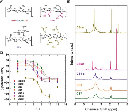
Fig. 1. (A) Chemical structures of the investigated CS derivatives. Hydrogens atoms have been omitted for clarity. (B) Stacked 1H NMR spectra (D2O, 500 MHz) of the studied polymers: CS7 (magenta), CS1cm (teal), CS1 (blue), CS1lac (red), and CS1-c (black). (C) ζ-potential measurements of the studied polymers at different pH values (n = 3). Experimental data were fitted using a Boltzmann sigmoidal function (R2 > 0.95). (For interpretation of the references to colour in this figure legend, the reader is referred to the web version of this article.)
The pka of the different CSs was determined by measuring the ζ-potential of aqueous solutions (1 mg/mL) at 25 °C using a Zetasizer ZS instrument (Malvern Panalytical, Malvern, United Kingdom) equipped with a H Ne 633 nm laser and with a backscattering of 173 0 controlled by with a Malvern DTS 7.03 software at a constant 2.5 mS/cm conductivity. ζ-potential was calculated based on Henry's equation with a Smoluchowski approximation for Henry function. Obtained data were fitted following a Boltzmann function with a Pearson correlation coefficient (R2) > 0.95. The pka value was estimated as the minimum of the derivative of the fitting. Data were analyzed using OriginPro v.2024b (OriginLab Corporation, Northampton, MA, USA) software.
ATR-FTIR spectroscopic analysis was performed in a FT/IR 4700 (JASCO Co., Tokio, Japan) instrument. All CS samples were dried overnight at 60 °C before the measurement and pure samples were used for the measurements.
TGA-DSC analysis was performed in a NETZSCH -STA 449 F1 Jupiter instrument (Selb, Germany) from 30 °C to 800 °C at 5 °C/min under N2/O2 atmosphere. Samples were dried overnight at 60 °C prior analysis.
MIC values for Staphylococcus (S.) aureus (ATCC 12600), Escherichia (E.) coli (K12), Pseudomonas (P.) aeruginosa (PA01), Salmonella (S.) tiphymurium (ATCC 14028) and Candida (C.) albicans (ATCC 10231) were obtained via the standard broth-dilution method in 96-well microlitre plates. Lysogeny Broth (LB) and Sabouraud media were used for bacterial cells and fungal cells, respectively. The MIC of each compound was evaluated using a 2-fold decreasing concentration of each sample between 60 and 0.5 mg/mL against a bacterial/fungal inoculum with an optical density (OD600) of 0.005. Note that depending on the microbial species, this OD600 corresponded to experimentally determined concentrations ranging from 107 to 109 colony forming units per mL (CFU/mL). Plates were incubated at 37 °C for 18 h. MIC values were determined as the lowest concentration where no turbidity was observed [44]. Simultaneously, the optical density at 600 nm (OD600) was measured with a SpectraMax iD3 (Molecular Devices, San José, CA, USA) plate reader. MIC reported values are based on three biological replicates.
Surface charge of S. aureus (ATCC 12600), E. coli (K12), P. aeruginosa (PA01), S. tiphymurium (ATCC 14028) and C. albicans (ATCC 10231) in absence or presence of biopolymers was measured with a Zetasizer (ZS, Malvern Panalytical Ltd., Malvern, UKm) equipped with a HNe 633 nm laser and with a back-scattering of 173 0 using PBS (0.1 M, pH 7.4) as dispersant at 25 °C. Overnight cell inoculum of 107–109 CFU/mL in PBS were incubated with 1 mg/mL of each CS sample in PBS (0.1 M, pH 7.4) for 4 h at 37 °C under mild shaking. Then, samples were centrifuged (5000 rpm, 5 min), washed with fresh PBS and the ζ-potential measured. Depicted values (average ± SD) are based on three biological replicates.
The integrity of the cell membrane in the presence of the CS's was evaluated following a procedure previously described by H. E. Smith & J. M. A. Blair [45]. Briefly, overnight inoculum of S. aureus (ATCC 12600), E. coli (K12), P. aeruginosa (PA01), S. tiphymurium (ATCC 14028) and C. albicans (ATCC 10231) were incubated for 4 h under mild shaking at 37 °C in PBS (0.1, pH 7.4, OD600 = 0.2). The buffer solution contained Propidium Iodide (PI, 5 mg/L for bacterial and fungal cultures) and the CS of choice at a final concentration 1 mg/mL. Then, the samples were centrifuged (5000 rpm, 5 min), washed with fresh PBS and finally suspended in PBS containing 5% glucose (w/v). 200 μL of each culture was transferred to a 96 well black microlitre tray (Corning, Amsterdam, the Netherlands) and fluorescence was measured (λexc = 575 nm, λem = 635 nm) with a SpectraMax iD3 plate reader (Molecular Devices, San José, CA, USA). Results (average ± SD) are based on three biological replicates. A PBS solution of Na2DTPA·2H2O (10%, w/v) was employed as positive control.
Bacterial viability experiments were performed using E. coli (K12) bacterial reference species and C. albicans (ATCC 10231). Overnight inoculate of microorganisms were washed and diluted in PBS buffer (0.1 M, pH 7.4) to a final concentration of 107–109 CFU/mL, and incubated with the different CS molecules at a concentration of 1 mg/mL for 4 h under magnetic stirring (150 rpm) at 37 °C. Then, cultures were centrifuged (6500 rpm, 3 min), washed with fresh PBS, and stained using the LIVE/DEAD BackLight bacterial viability test kit. Fluorescence imaging was performed in an Olympus IX73-DP75 (Olympus, Tokyo, Japan) fluorescence microscope. Image analysis was performed with OriginPro v.2024b (OriginLab Corporation, Northampton, MA, USA) software. Depicted results (average ± SD) are based on three technical replicas from three different biological replicates (N = 3, n = 3).
E. coli (K12) DNA was extracted and purified using a GeneJET DNA purification kit (Thermo Fisher Scientific Inc., Waltham, USA) following the protocols established by the kit manufacturer. Gel electrophoresis was performed on a 1% agarose-TBE gel using 100 V for 90 min in 1xTBE buffer. [DNA] = 4.5 ng/mL.
CD measurements were performed in a J-815 CD Spectrometer (JASCO Co., Tokio, Japan) using Milli-Q water as a dispersant. Extracted E. coli DNA (40 ng/μL) was incubated for 10 min at room temperature with the desired CS in a 1:1 mass ratio. Depicted data were obtained after five accumulations.
These experiments were performed with the N2 wild-type strain of Caenorhabditis (C.) elegans provided by the Caenorhabditis Genetics Centre (CGC, University of Minnesota). C. elegans survival, growth in and the reprotoxicity in the presence of CS7 or CSlac were studied following a previously reported methodology [46]. Early developmental-stage nematodes (L1) were exposed to CS7 or CSlac solutions for 72 h at 20 °C. The concentration ranges of CS7 or CSlac were determined from MIC values (0–15 mg/mL and 0–5 mg/mL, respectively). Experiments were performed in the presence or the absence (for survival studies) of heat-inactivated E. coli OP50 (OD600 = 0.1) in S-medium. Depicted results (average ± SD) are based on three biological replicates (15 ± 5 worms/replica) and the statistical significance was determined by 1-way ANOVA.
Data are presented as mean ± standard deviation (SD) of at least three independent biological replicates. Statistical significance was determined by one-way or two-way ANOVA followed by a Tukey's HSD post-hoc test. ns p > 0.05; *: 0.01 < p ≤ 0.05; **: 0.001 < p ≤ 0.01; ***: p ≤ 0.001. Detailed p-values for comparisons across conditions are summarized in Tables S2 and S4.
Parameters such as MW, DD, and chemical functionalization are supposed to play a crucial role in the antimicrobial activity of CS. To cover all these features, different CS molecules having different MW, dispersities, chemical functionalization and origin were studied (Fig. 1.A). Nuclear Magnetic Resonance (NMR), and diffusion NMR spectroscopy were employed to determine the chemical composition, molecular weight (MW) distribution and dispersity (Đ) of the different CS samples. Fig.S2 show the monomodal MW distribution of each type of CS assayed herein. First, a set of CS molecules with different MWs (CS1, CS7, CS50 and CS300) but showing the same DD were explored. Note that CS1 and CS7 were produced from CS300 by a microwave-assisted hydrolysis method with some modifications. [13] CS50 was commercially acquired and used as received without further purification. Then, and in order to further investigate the effect of the biopolymer origin on its antimicrobial activity, these samples were directly compared to a commercially available CS obtained from fungal origin (CS1-c) and CS molecules bearing different functional groups such as lactate (CSlac) and carboxymethyl (CScm). All CS samples present a narrow Đ besides the CScm (Table 1).
CS7, and CS1 exhibited highly similar 1H NMR spectra, characterized by diagnostic resonances at δH 1.93, 2.75, and 4.42 ppm, which were assigned to the CH3 group of the N-acetyl-D-glucosamine unit, the H-2 proton of the D-glucosamine unit, and the anomeric H-1 protons of both units, respectively (Fig. 1B). As expected, all these CS samples displayed identical degrees of deacetylation (DD), as summarized in Table 1. These results confirm that the production of CS7 and CS1 from CS300 via microwave-assisted oxidative hydrolysis does not alter the chemical structure of the polymer backbone nor affect the DD of the resulting materials. MW of 7.5 ± 0.6 kDa, and 2.0 ± 0.2 kDa were determined for CS7 and CS1, respectively by NMR analysis. Furthermore, successful chemical functionalization of commercial CSlac and CScm was confirmed by 1H NMR spectroscopy (Fig. 1B). In the case of CSlac, new resonances appearing at δH 1.30 and 4.05 ppm were observed and assigned to the terminal CH3 and CH protons of the lactate moiety, respectively. The 1H NMR spectrum of CScm displayed an additional signal at ca. δH 3.8 ppm, which was attributed to the CH2 protons of the carboxymethyl group. Interestingly, an averaged MW of 1075 ± 25 kDa was determined by NMR analysis for CSlac, with only small fraction of the sample having the reported MW by the supplier. This discrepancy can be due to the different analytical techniques employed in the CSlac production and characterization, inherit calibration error, or batch-to-batch discrepancies. Complementary SEC-MALS analysis only showed a population with average MW of 40 kDa, also detected by DOSY (Fig. S1, Fig. S2 and Table S1). The guard columns and on-line filters prior the SEC column might trap hight MW CSlac, and, in that way, not send it in the chromatograph. Based on these results, we decided to used 1075 kDa as the average MW for CSlac. Determined MW for CScm (790 ± 20 kDa) is in good agreement with the commercially determined one.
The presence of lactate and carboxymethyl functionalities in CSlac and CScm was further corroborated by 13C NMR spectroscopy (Fig. S3). The 13C NMR spectrum of CSlac showed additional resonances at δC 20–22 and 68.1 ppm, which were assigned to the methyl and methane carbons of the lactate moiety, respectively. A characteristic signal in the carbonyl region at δC 181.7 ppm corresponding to the lactate carboxylic carbon was also observed. In the case of CScm, the 13C NMR spectrum displayed an additional resonance at δC 69.5 ppm, attributed to the methylene carbon of the carboxymethyl substituent, along with a signal at δC 179.9 ppm associated with the carboxylic carbon. This confirms the successful carboxymethylation of the CS backbone.
Finally, structural differences between the synthesized CS1 and the commercial CS1-c sample (fungal origin) were investigated by combined 1H and 13C NMR analysis in solution (Fig. S3). In the 1H NMR spectrum of CS1-c, an additional resonance was detected at δH 5.36 ppm, assigned to the H-1 of the glucan residue, which was not present in CS1. Another characteristic resonance at δH 3.28 was assigned to the H-2 proton of the same residue; typical of fungal-derived CS. [37] The difference in the integral intensity between H-2 from the glucan residue (δH 3.28) and H-2 from the glucosamine (δH 2.90 ppm) in the 1H NMR spectrum is 1:15. Establishing a degree of branching of approximately 6.7% (i.e., 1 glucan branch per 15 backbone units). Consistently, 13C NMR spectroscopy revealed extra signals in CS1-c beyond those corresponding to the CS backbone, notably resonances at δC 98.1 ppm (anomeric C1), 69–77 ppm (ring carbons C2–C5), and 60–62 ppm (C6), which appear in the same region as those of CS1. These features are in agreement with the reported branched architecture of fungal CS, consisting of a β-(1 → 4)-linked D-glucosamine backbone decorated with β-(1 → 6)-linked glucan chains/oligomers. [37].
The functionalization of CSlac and CScm was further confirmed by FT-IR spectroscopy (Fig. S4). Peaks corresponding to C O (1638 cm−1), CO (1363 cm−1), and CN (1105 cm−1) stretching bonds of the lactate moieties were identified in CSlac, [47] while vibrations of the terminal acid group (1591 cm−1 and 1410 cm−1) were found for CScm. The chemical differences between CS1 and CS1-c were also investigated by FT-IR (Fig. S4). The observed broadening of the vibration band centred at 3310 cm−1 in CS1-c can be associated with the higher density of hydroxyl groups from the glucan branching [37]. Similarly, the FT-IR spectrum of CS1-c presents a stronger vibration at 1510 cm−1 compared with CS1, which is associated with OH deformation vibration from the hydroxyl groups of the glucan branches. Thermogravimetric analysis coupled with differential scanning calorimetry (TGA-DSC) revealed a multi-step degradation process for both samples but with remarkable differences. On the one hand, CS1 exhibits a sharp mass loss around 648 °C, preceded by a gradual monotonic degradation. On the other hand, CS1-c shows earlier degradation events at approximately 375 °C and 610 °C (Fig. S5). The higher thermal stability of CS1 could be attributed to its greater packing efficiency of lineal polymers, in contrast to the lower packing density of the branched CS1-c, in line with reported literature [48].
Finally, the ζ-potential of all the CS samples in aqueous solution was evaluated as a function of pH at a fixed conductivity of 2.5 mS/cm (Fig. 1C). The corresponding isoelectric points or apparent pKa values (Table 1) were estimated from the ζ-potential curves, acquired as described in the experimental section and Supplementary Data (Fig. S6). These parameters are particularly relevant, as the net charge of CS is expected to play a central role in its interaction with microbial membranes and, consequently, in its antimicrobial performance. A clear correlation between the MW and apparent pKa is observed for the unfunctionalized CS samples. Specifically, the pKa decreases with increasing MW, from 8.6 for CS1 to 6.4 for CS300. This behaviour can be rationalized by considering the increasing relative contribution of terminal sugar units in low-MW CS. Shorter polymer chains present a higher fraction of terminal units, which possess greater conformational freedom, and enhanced solvent accessibility compared to units embedded within long polymer chains. As a result, these terminal amino groups are more readily protonated as they experience reduced intramolecular electrostatic repulsion compared with amino groups embedded within long polymer chains. In contrast, in high-MW CSs, the higher local charge density and reduced accessibility of internal amino groups favour earlier deprotonation, leading to lower apparent pKa values.
A markedly lower apparent pKa was observed for the fungal-derived CS1-c compared with CS1 of similar MW and DD. This difference can be attributed to the distinct macromolecular architecture of fungal CS, which contains β-(1 → 6)-linked glucan side chains attached to the CS backbone (see discussion above). The presence of these branched glucan units increases structural complexity and alters the intramolecular hydrogen-bonding network and polymer–polymer interactions in solution. As a consequence, the amino groups of the CS backbone in CS1-c experience a modified local microenvironment and reduced effective contribution to the electrokinetic potential compared with the linear CS1, which disfavours protonation from an electrokinetic standpoint. In addition, the more heterogeneous and branched polyelectrolyte architecture of CS1-c enhances collective electrostatic and interfacial effects, stabilizing the deprotonated state and resulting in a lower apparent pKa as observed from ζ-potential measurements. Accordingly, distinct antimicrobial behaviour can be anticipated for these two samples, despite their comparable MW and DD.
ζ-potential measurements as a function of pH provide not only an estimation of the apparent pKa or isoelectric point, but also valuable information on the colloidal stability of the different CS samples in aqueous solution (Fig. 1C). In general, high absolute ζ-potential values are associated with enhanced electrostatic repulsion between particles and, therefore, greater dispersion stability, whereas values close to zero indicate a higher tendency toward aggregation. In contrast to the unfunctionalized CS samples, which exhibit a clear transition from positive to negative ζ-potential values and reach an isoelectric point within the investigated pH range, the chemically functionalized samples display markedly different electrokinetic behaviours. In particular, CSlac shows a ζ-potential profile similar in shape to that of unmodified CS, reaching near-neutral values at pH 7, consistent with the presence of both protonable amino groups and lactate functionalities that moderately modulate the net charge. By contrast, CScm exhibits a fundamentally different response, characterized by persistently negative ζ-potential values across the entire near-neutral pH range and the absence of an observable isoelectric point. This behaviour can be attributed to the high density of carboxymethyl groups, which are largely deprotonated above their intrinsic pKa (ca. 4.5) [49], thereby dominating the electrokinetic response and overpassing the contribution of protonated amino groups on the CS backbone. As a result, CScm remains negatively charged and electrostatically stabilized over a broad pH window, indicating enhanced colloidal stability under physiological and near-neutral conditions.
Previous studies have already demonstrated the antimicrobial properties of CS, CS derivatives biopolymers, and CS-based materials [27]. Efforts have also been made to correlate antibacterial performance with specific chemical and structural parameters of CS, such as MW or DD [6], [24]. However, these studies are generally limited to narrow sets of CS materials and often focus on individual parameters in isolation, rather than addressing the combined and interdependent effects of multiple variables, including MW, DD, dispersity, and chemical functionalization. Given the considerable potential of CS-based systems as antimicrobial agents, the present work investigates the antimicrobial properties and mechanisms of action of a comprehensive library of CS samples differing in MW, chemical functionality, and biological origin (animal or fungal).
First, the Minimum Inhibitory Concentration (MIC) of all samples against representative bacterial species (S. aureus, E. coli, P. aeruginosa, and S. tiphymurium) and fungus C. albicans was determined. MIC values were determined in Lysorgenic Broth (LB) for bacterial species and Sabouraud media for C. albicans (Table 2, Fig. S7–8). The highest-MW biopolymer, CS300, exhibited moderate to low antibacterial activity, with MIC values of 30 mg/mL against S. aureus and 60 mg/mL or higher against Gram-negative bacteria tested. Against C. albicans, CS300 showed moderate activity (MIC = 15 mg/mL). CS50 displayed a comparable antibacterial profile, although a slight improvement was observed against E. coli (MIC = 30 mg/mL). In contrast, a marked enhancement in activity was observed for lower-MW samples. Specifically, CS7 showed a 4-fold decrease in MIC values against S. aureus (7.5 mg/mL) and C. albicans (2.5 mg/mL), and an 8-fold decrease against E. coli (3.2 mg/mL). CS7 also showed improved activity against P. aeruginosa (MIC = 7.5 mg/mL), whereas its activity against S. typhimurium remained moderate (MIC = 30 mg/mL). Remarkably, CS1 showed even higher MIC values than CS7 across most strains. In contrast, the fungal-derived CS1-c displayed consistently lower activity than CS1 against all tested microorganisms, which can be attributed to its branched macromolecular architecture, in agreement with previous reports [50]. MIC values for C. albicans were also determined in LB media at neutral pH (7.4) (Table S2 and Fig. S8). As expected, most samples exhibited reduced antifungal activity under these conditions compared with assays performed in Sabouraud medium (pH 5.5), reflecting the lower degree of protonation of CS at neutral pH. Overall, these results highlight the strong influence of MW on the antimicrobial performance of non-functionalized CS, which is closely linked to its solubility and self-aggregation behaviour in aqueous solution [14]. In general, an increase in MW correlates with a progressive decrease in antimicrobial activity.
Table 2. Minimum inhibitory concentration (MIC) values (mg/mL) for explored CS against representative bacterial (S. aureus, E. coli, P. aeruginosa, S. typhimurium) and fungal (C. albicans) species. MICs were determined by the 2-fold dilution method in LB (bacteria) and Sabouraud medium (fungus) at 37 °C under mild agitation (150 rpm). Data represent the mean of three biological replicates.
| Empty Cell | Bacteria | Fungi | |||
|---|---|---|---|---|---|
| Gram+ | Gram- | ||||
| S. aureus | E. coli | P. aeruginosa | S. tiphymurium | C. albicans | |
| CS300 | 30 | 60 | > 60 | > 60 | 15 |
| CS50 | 30 | 30 | > 60 | > 60 | 15 |
| CS7 | 7.5 | 3.2 | 7.5 | 30 | 2.5 |
| CS1 | 15 | 15 | 30 | 30 | 7.5 |
| CS1-c | 30 | 30 | 30 | >60 | 15 |
| CSlac | 3.2 | 0.9 | 3.2 | 15 | 0.2 |
| CScm | > 60 | > 60 | > 60 | > 60 | 30 |
As discussed above, a peak in antimicrobial performance was observed for CS7, particularly against E. coli, P. aeruginosa, and C. albicans. At first glance, this observation may appear to contradict previous reports suggesting that antimicrobial activity generally decreases with increasing MW [24]. However, this apparent discrepancy highlights the existence of an optimal MW window in which multiple physicochemical factors are favourably balanced. The antimicrobial activity of CS mainly relies on electrostatic interactions between the positively charged amino groups and negatively charged components of the microbial cell envelope [6]. These interactions destabilize membrane integrity, ultimately leading to cell death. Noteworthy, the molecular conformation of CS in solution strongly influences the accessibility and effective exposure of these amino groups [14]. High-MW CS tends to form aggregates due to reduced solubility and increased chain entanglement, which limits amino-group exposure and diminishes antimicrobial efficacy, in line with the poor activity observed for CS300 and CS50 [24]. In contrast, CS7 appears to represent an optimal compromise between chain length and solubility. Its intermediate MW is sufficient to ensure multivalent interactions with microbial membranes, while still preventing extensive aggregation in solution. This combination maximizes the availability of protonated amino groups and enhances membrane interaction, resulting in superior antimicrobial and antifungal activity. Further reduction of MW, as in CS1, likely decreases the cooperative and multivalent character of these interactions, leading to a partial loss of activity.
In addition to polymer-related factors, the composition and architecture of the microbial cell envelope play a crucial role in determining susceptibility to CS [26], [51]. Gram-positive bacteria such as S. aureus possess a relatively simple and permeable membrane, while Gram-negative species (E. coli, P. aeruginosa, and S. tiphymurium) feature an additional outer membrane that restricts the penetration of antimicrobial agents and confers intrinsic resistance [3]. Moreover, variations in membrane composition and surface charge among bacterial species and strains directly influence their interaction with positively charged polymers and peptides [17], [52], [53]. These differences likely contribute to the particularly pronounced activity of CS7 against E. coli and P. aeruginosa, as well as against C. albicans, whose cell envelope presents distinct negatively charged components susceptible to electrostatic disruption.
As shown in Fig. 1.B, CS7 exhibit a higher positive ζ-potential than CS50 and CS300 at pH 7.4, which corresponds to the bacterial growth medium. This higher surface charge is expected to enhance both colloidal stability in aqueous solution and electrostatic interactions with negatively charged components of the microbial cell envelope, thereby contributing to its superior antimicrobial performance. Interestingly, CS1 displays a ζ-potential comparable to that of CS7 under the same conditions, despite showing higher MIC values. This apparent discrepancy suggests that surface charge alone is not sufficient to fully explain antimicrobial efficacy. This behaviour is consistent with previous reports and theoretical models indicating that low-molecular-weight polymers and oligomers interact with biological membranes through mechanisms distinct from those of longer chains [19]. In particular, smaller molecules may cross or insert into microbial membranes with limited membrane disruption, resulting in reduced membrane fluidization and weaker bactericidal effects. Instead, their activity may rely on interactions with intracellular targets, a mechanism that has been proposed for small cationic species but remains largely unexplored experimentally in the case of low-MW CS.
CS1-c displayed a lower ζ-potential at pH 7.4 (Fig. 1.C, Table 1), which is consistent with its higher MIC values compared with linear CS1. In addition, as discussed above, 1H NMR analysis revealed clear compositional differences between fungal-derived CS1-c and the synthesized CS1. In particular, the presence of bulky β-(1 → 6)-linked glucan side chains increases the structural complexity of CS1-c, which can hinder effective biopolymer–cell interactions and reduce antimicrobial activity [37]. In the case of C. albicans, an overall increase in antifungal activity was observed for all unfunctionalized CS samples. The fungal cell wall is mainly composed of chitin, β-glucans, and mannoproteins [54]. The mannoproteins in the outer cell wall layer carry a high negative charge, which facilitates electrostatic interactions with cationic CS macromolecules. Moreover, direct interactions between exogenous CS and chitin present in the fungal cell wall have also been reported [55] The combined contribution of these interactions likely accounts for the enhanced antifungal activity observed for the unfunctionalized CS samples. Differences among individual CSs can therefore be rationalized using the same structure–charge–conformation arguments discussed for their antibacterial activity.
As summarized in Table 2, CSlac displays the lowest MIC values against all the microbial species explored. Besides its high MW (Table 1), CSlac displays a markedly enhanced antimicrobial performance respect to the rest of CSs. Specifically, a twofold decrease in MIC was observed for P. aeruginosa (MIC = 7.5 mg/mL) and S. typhimurium (MIC = 15 mg/mL), a fourfold decrease for S. aureus (MIC = 3.2 mg/mL), and a sixteen-fold decrease for E. coli (MIC = 0.9 mg/mL) was observed respect to CS7 (the second explored CS with highest activity). Most notably, the MIC value against C. albicans decreased by 64-fold (MIC = 0.2 mg/mL) compared with CS7. Based on previous reports demonstrating the ability of lactate-containing compounds to sequester membrane-associated divalent cations, [18] we hypothesize that CSlac may exert its antimicrobial activity, at least in part, through the scavenging of Ca2+ and Mg2+ ions from the bacterial membrane. Furthermore, this mechanistic is consistent with the pronounced improvement observed for CSlac against S. aureus, E. coli, and C. albicans. In contrast, CSlac showed only a modest enhancement in activity against P. aeruginosa compared with CS7. This behaviour can be explained by the well-known adaptive features of P. aeruginosa. In particular, P. aeruginosa secretes large amounts of lipopolysaccharides into the extracellular medium, which can interact with antibacterial polymers and prevent their direct contact with the bacterial outer membrane [56]. Moreover, several P. aeruginosa strains, including the PAO1 strain used in this study, harbour mutations that promote aminoarabinosylation of the lipid A region of the outer membrane's lipopolysaccharides. [57] This modification, driven by the arn operon, results in the covalent modification with a 4-amino-4-deoxy-L-arabinose molecule to the lipid A region's phosphate groups, protecting membrane divalent cations. Such adaptations, also associated with colistin resistance, diminish cation chelation by exogenous agents and could be a plausible explanation for the limited improvement in CSlac activity against P. aeruginosa [58].
Finally, CScm did not exhibit antibacterial activity against any of the bacterial species evaluated up to 60 mg/mL and showed only moderate antifungal activity against C. albicans (MIC = 15 mg/mL). This lack of activity is consistent with its strongly negative ζ-potential at physiological pH values, which prevents effective electrostatic interactions with microbial cell envelopes and underscores the critical role of surface charge in CS-based antimicrobial systems.
After evaluating the antimicrobial performance of the CS samples, a series of complementary microbiological and imaging studies was conducted to validate the proposed mechanistic interpretations. Specifically, microbial cell-CS interactions were assessed by ζ-potential measurements, while membrane and cell wall integrity were examined by dye accumulation assay and fluorescence microscopy. In addition, circular dichroism (CD) spectroscopy was applied to bacterial DNA to determine whether the different CS samples are able to effectively interact with intracellular macromolecules.
As already mentioned, the interaction of cationic polymers with negatively charged microbial membranes leads to membrane fluidization, disruption, and intracellular content leak [53]. In order to evaluate whether the antimicrobial activity of CS results from membrane disruption, ζ-potential measurements of bacterial cells, dye accumulation assays, and fluorescence microscopy analyses were employed. First, the electrostatic interactions between the different CS biopolymers and the membrane of the different bacterial and fungal cells was monitored by ζ-potential measurements [59]. Microbial cells (106–107 CFU/mL) were incubated in PBS buffer containing 1 mg/mL of the biopolymer for 4 h at 37 °C. After isolation of microbial cells by centrifugation, the cell surface charge was measured in PBS and compared with untreated samples (Fig. 2A). Noteworthy, a clear correlation was observed between the antimicrobial activity of the CS biopolymers and the change in the surface charge of cells. In particular, the most active samples induced the strongest shift in the cells' surface charge toward neutral or even positive values (i.e., CSlac in E. coli and C. albicans). However, biopolymers with lower or no activity (i.e., CS300, CS50, and CScm) had little to no effect on the surface charge of the cells, suggesting that these compounds do not interact with the cells.
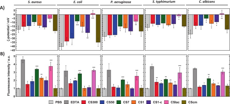
Fig. 2. (A) ζ-potential measurements of bacterial and fungal cells (106–107 CFU/mL) in PBS buffer (0.1 M, pH 7.4) before and after incubation with biopolymers (1 mg/mL) for 4 h at 37 °C. (B) Normalized propidium Iodide (PI) accumulation assays of bacterial and fungal cells (OD600 = 0.2) incubated in PBS buffer (0.1 M, pH 7.4) before and after incubation with each compound (1 mg/mL) for 4 h at 37 °C. EDTA PBS solution (10% w/v) was used as a positive control. Depicted values (average ± SD) are based on three biological replicas. Data has been normalized to the control experiment. For clarity, only the statistical significance between the control and the different CS conditions have been depicted. The rest of statistical comparisons can be found in Tables S3–5.
Beyond electrostatic surface interactions, cationic polymers can induce different degrees of membrane perturbation, including pore formation, membrane fluidization, and structural damage, ultimately leading to the leakage of intracellular components [18]. To directly assess membrane integrity, cell membrane disruption was explored using dye (propidium iodide, PI) accumulation assays as reported previously [45]. Note that PI is only internalized within the cells if their membrane has been compromised. The fluorescence intensity of internalized PI was measured in microbial inoculate (OD600 = 0.2) incubated for 4 h in PBS containing each CS (1 mg/mL) and it was compared with a control microbial inoculum in PBS (Fig. 2B). The dye's intensity increased in those scenarios where the change in membrane ζ-potential was more pronounce, while membrane/cell wall damage was not observed in the case of the CS that did not alter cell surface charge. Interestingly, membrane damage by CS7 in P. aeruginosa and S. typhimurium was less pronounced than that observed for CSlac, even though both CS displayed comparable antibacterial activity. This discrepancy suggests that, in addition to membrane disruption, alternative or complementary mechanisms may contribute to the antimicrobial activity of CS7.
Live/dead cell imaging of E. coli and C. albicans was performed to further elucidate their mechanisms of action. Microbial cells were incubated in PBS (0.1 M, pH 7.4) containing the studied CS (1 mg/mL) for 4 h at 37 °C. As a control for membrane permeability, cells were also treated with a 10% (w/v) EDTA solution in PBS (Fig. 3). The live/dead ratios (Fig. S9) obtained by fluorescence microscopy were in good agreement with the MIC results summarized in Table 2.
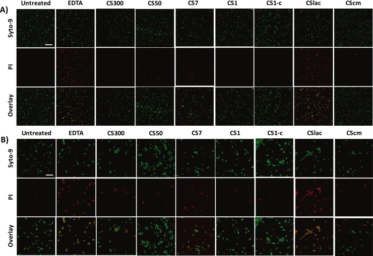
Fig. 3. Representative fluorescence microscopy of (A) E. coli and (B) C. albicans cells (106–107 CFU/mL) before and after incubation with biopolymers (1 mg/mL) in PBS buffer (0.1 M, pH 7.4) for 4 h at 37 °C. Green channel (SYTO-9) λexc = 460–495 nm, λem = 510–550 nm, red channel (PI) λexc 530–555 nm = nm, λem = 575–625 nm. Scale bar represents 20 μm. (For interpretation of the references to colour in this figure legend, the reader is referred to the web version of this article.)
CS has been reported to interact with charged biomolecules within the bacterial cytoplasm once internalized [21], [22], [23]. In particular, CS-DNA interactions have been reported as an additional mechanism contributing to antibacterial activity [22]. Nevertheless, the influence of MW and chemical functionalization on these interactions has not been systematically explored. Accordingly, circular dichroism (CD) analysis of bacterial DNA (extracted from E. coli, Fig. S10) in the presence of the different CS biomaterials was performed (Fig. 4). The interaction between CS and DNA was inferred from: i) a decrease in the intensity of circular dichroism signals, associated with partial loss of DNA chirality upon complex formation, and ii) changes in the spectral profile, indicative of DNA conformational rearrangements induced by interactions with CS molecules [60].
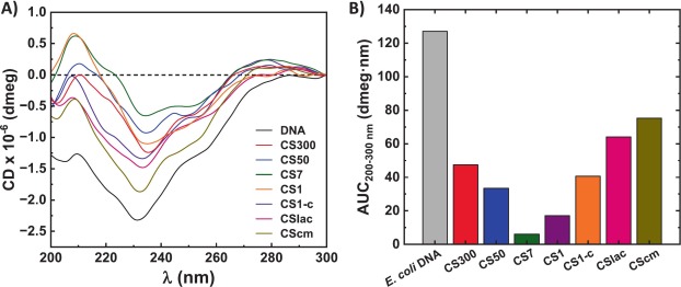
Fig. 4. (A) Circular dichroism (CD) spectra of E. coli DNA incubated with different CSs in PBS buffer (0.1 M, pH 7.4) at 37 °C for 1 h in a 1:1 mass ratio, and (B) calculated area under the curve (AUC) of each spectrum.
Among the CS with different MW, CS7 exhibited the strongest interaction with bacterial DNA, whereas higher-MW CS (CS300 and CS50) showed weaker interactions. The well-known tendency of high-MW CSs to self-aggregate in solution could hinder their accessibility to DNA binding sites [14]. Interestingly, both CS1 and CS1-c interacted less efficiently with DNA than CS7, despite their lower MW. This behaviour may be related to the reduced number of available binding sites in very short CS chains, which limits multivalent interactions with the polyanionic DNA backbone. Indeed, the relationship between the number of binding sites and CS-DNA interaction strength has been previously described [61]. However, that study focused exclusively on variations in DD for already water-soluble CS and did not consider the aggregation behaviour of higher-MW samples in solution. Additionally, the branched architecture of CS1-c may further sterically hinder access to DNA binding sites, contributing to its reduced interaction.
In contrast, functionalized CS (CSlac and CScm) exhibited weak interactions with bacterial DNA compared with unfunctionalized CS. The strong negative ζ-potential of CScm (−20 ± 4 mV at pH 7) would electrostatically disfavour interactions with the negatively charged DNA. Similarly, the presence of electron-rich lactate moieties in CSlac may reduce its affinity toward DNA, even though CSlac remains positively charged at pH 7.4.
Although these ex-vivo experiment may present some limitations as the CS must cross the microbial membrane to reach intracellular components; the observed results indicate that for low-MW CS (CS1 and CS7), antimicrobial activity may preferentially involve interactions with intracellular components rather than direct membrane disruption, in contrast to CSlac, whose activity could be predominantly associated with membrane damage.
The biosafety of CS has been extensively reported and constitutes one of its main advantages for biomedical applications. Both in vitro and in vivo toxicity assays on CS biopolymers with different MW and chemical functionalizations have generally shown negligible cytotoxicity at the in therapeutically relevant concentrations [62]. In the present work we evaluated the in vivo toxicity of the promising CS compounds, CSlac and CS7, in the model organism Caenorhabditis (C.) elegans. This nematode was chosen due to its experimental simplicity, ease of maintenance, and high degree of genetic homology with humans (> 60–80%) [63], [64]. Moreover, C. elegans has been widely employed to study the toxicity of biopolymers such as cellulose and CS [46], [65], as well as other antimicrobial polymers, nanoparticles and macromolecules [66], [67], [68].
CS7 and CSlac were assessed by first incubating young (L1) C. elegans with different concentration of each CS (within the range of their corresponding MIC values) in growth medium supplemented with inactivated E. coli OP50 as a food source. After 72 h of incubation, nematodes reached the egg-laying adult stage and their viability was assessed as previously described [46]. Survival rates were calculated from population counts after exposing C. elegans to different CS concentrations (Fig. 5). No significant decrease in survival was observed in the presence of CS7 or CSlac at concentrations up to 15 mg/mL and 5 mg/mL, respectively, when OP50 was present. These results indicate good biocompatibility of both samples under nutritionally supported conditions and support their potential for biomedical applications. Additional experiments were conducted in the absence of OP50 as acute treatment. Under these conditions, no significant toxicity was observed for CS7 across the tested concentration range. In contrast, a concentration-dependent decrease in viability was detected for CSlac at 5 mg/mL. This reduction in survival at high CSlac concentrations, exceeding the MIC values, may be attributed to an acute and toxic effect.
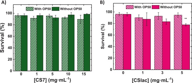
Fig. 5. Cytotoxicity of (A) CS7 and (B) CSlac on C. elegans in presence and the absence of inactivated OP50 E. coli. Depicted results (average ± SD) are based on three biological replicates. Statistical significance was determined by one-way ANOVA. ns p > 0.05, * p < 0.05, ** p < 0.01, *** p < 0.001.
In this work, the antimicrobial activity of a series of CS biomaterials with different MW, chemical functionalities, and biological origins was explored via a range of microbiological and molecular biology approaches. The results indicate demonstrate that the antimicrobial activity of CS is not governed by a single parameter, but rather arises from the interplay of multiple, interconnected factors.
On the one hand, MW plays a central role in determining antimicrobial performance. Unfunctionalized high-MW CS polymers tend to aggregate in aqueous media, which limits their accessibility and interaction with microbial membranes. In contrast, very low-MW polymers exhibited enhanced colloidal stability at physiological pH, facilitating their interaction with microbial cells. Notably, our data reveal that low-MW CS can strongly interact with bacterial DNA, suggesting that their antimicrobial mechanism extends beyond membrane disruption and may involve intracellular targets.
On the other hand, chemical functionalization an effective means to modulate antimicrobial activity. In particular, the introduction of lactate groups in CSlac enhances CS-microbe interactions and promotes efficient membrane disruption, leading to markedly improved antimicrobial performance, besides its high MW. Conversely, carboxymethyl functionalization introduces a negative charge that can hinder electrostatic interactions with microbial membranes, resulting in reduced activity, as observed for CScm.
Finally, the biological origin of CS was found to have significant impact on antimicrobial efficacy. Even at comparable MW and DD, fungal-derived CS (CS1-c) displays markedly lower antimicrobial activity compared to its animal-derived analogue (CS1). This behaviour is attributed to the presence of glucan side chains in fungal CS, which alter the macromolecular architecture and impair effective interactions with microbial cells. While the microbial strains evaluated in this work serve as established reference models, the results should not be directly generalized to all clinically relevant or multidrug-resistant pathogens. Furthermore, while the C. elegans assays provide robust preliminary in vivo biocompatibility data, further studies in mammalian models will be required to fully establish safety profiles for clinical translational applications.
This work provides a comprehensive and systematic assessment of the key variables governing the antimicrobial activity of CS. Our findings demonstrate that CS activity emerges from a complex balance among MW, chemical functionality, molecular charge, and biological origin. These findings provide valuable mechanistic insight into the structure–activity relationships of CS derivatives, and support forward-looking design recommendations for next-generation CS-based antimicrobial polymers aimed at combating bacterial resistance (Fig. 6). In particular, our results suggest that the development of highly active CS materials should consider the use of lineal low-to-intermediate MW to improve colloidal stability and microbial accessibility, and the introduction of functional groups that enhance interactions with microbial membranes.
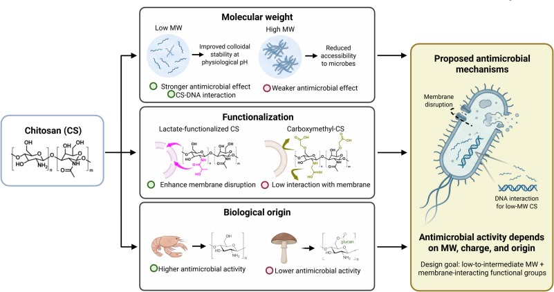
Fig. 6. Scheme summarizing the interplay between key macromolecular parameters of CS (i.e., Mw, degree of deacetylation, and linear vs. branched architecture), chemical functionalization, surface charge/colloidal behaviour, and their corresponding antimicrobial outcomes (membrane adhesion, cell disruption, and hypothesized intracellular effects).
Asier R. Muguruza: Writing – review & editing, Writing – original draft, Methodology, Investigation, Conceptualization. Liqiong Li: Writing – review & editing, Writing – original draft, Investigation. Carlos López-Orts: Writing – review & editing, Methodology, Investigation. Francisco M. Arrabal-Campos: Writing – review & editing, Writing – original draft, Investigation. Rafael Contreras-Caceres: Writing – review & editing, Writing – original draft, Investigation. Anna Laromaine: Writing – review & editing, Writing – original draft, Supervision, Resources. Ignacio Fernández: Writing – review & editing, Writing – original draft, Supervision, Resources, Funding acquisition. Pablo Guardia: Writing – review & editing, Writing – original draft, Supervision, Resources, Funding acquisition, Conceptualization.
The authors declare that they have no known competing financial interests or personal relationships that could have influenced the work reported in this paper.
This work was supported by the State Research Agency of the Spanish Ministry of Science and Innovation under Grant Nos. CNS2022-135583, PID2021-122645OB-I00 and CPP2022-009967 by the Spanish Government (MCIN/AEI/10.13039/501100011033) and the European Union “Next Generation EU”/PRTR. The authors also acknowledge financial support from the Generalitat de Catalunya through AGAUR Grants No. 2021SGR00446 and from the “Severo Ochoa” Programme for Centers of Excellence in R&D funded by MCIN and FEDER (“A way of making Europe”) under Grant No. CEX2023-001263-S. Individual funding is acknowledged from the MCIN/AEI “Ramón y Cajal” Programme and the European Social Fund “FSE invierte en tu futuro” under Grant Nos. RYC2019-028414 and RYC2021-03447-I. Financial support from the Plan Propio de Investigación y Transferencia 2023 of the University of Almería is also acknowledged (ref. ARyC2023_01). L.L. acknowledges the Chinese Scholarship Council for her predoctoral fellowship (Grant No. 202206650005). C.L.O acknowledges the financial support from the Generalitat de Catalunya through the FI-STEP 2025 AGAUR Grant, as well as funding by the European Union as part of the European project NextGEM, under Grant Agreement Number 101016567.
Data will be made available on request.
C.J. Murray, K.S. Ikuta, F. Sharara, L. Swetschinski, G. Robles Aguilar, A. Gray, C. Han, C. Bisignano, P. Rao, E. Wool, S.C. Johnson, A.J. Browne, M.G. Chipeta, F. Fell, S. Hackett, G. Haines-Woodhouse, B.H. Kashef Hamadani, E.A.P. Kumaran, B. McManigal, R. Agarwal, S. Akech, S. Albertson, J. Amuasi, J. Andrews, A. Aravkin, E. Ashley, F. Bailey, S. Baker, B. Basnyat, A. Bekker, R. Bender, A. Bethou, J. Bielicki, S. Boonkasidecha, J. Bukosia, C. Carvalheiro, C. Castañeda-Orjuela, V. Chansamouth, S. Chaurasia, S. Chiurchiù, F. Chowdhury, A.J. Cook, B. Cooper, T.R. Cressey, E. Criollo-Mora, M. Cunningham, S. Darboe, N.P.J. Day, M. De Luca, K. Dokova, A. Dramowski, S.J. Dunachie, T. Eckmanns, D. Eibach, A. Emami, N. Feasey, N. Fisher-Pearson, K. Forrest, D. Garrett, P. Gastmeier, A.Z. Giref, R.C. Greer, V. Gupta, S. Haller, A. Haselbeck, S.I. Hay, M. Holm, S. Hopkins, K.C. Iregbu, J. Jacobs, D. Jarovsky, F. Javanmardi, M. Khorana, N. Kissoon, E. Kobeissi, T. Kostyanev, F. Krapp, R. Krumkamp, A. Kumar, H.H. Kyu, C. Lim, D. Limmathurotsakul, M.J. Loftus, M. Lunn, J. Ma, N. Mturi, T. Munera-Huertas, P. Musicha, M.M.M. ussi-Pinhata, T. Nakamura, R. Nanavati, S. Nangia, P. Newton, C. Ngoun, A. Novotney, D. Nwakanma, C.W. Obiero, A. Olivas-Martinez, P. Olliaro, E. Ooko, E. Ortiz-Brizuela, A.Y. Peleg, C. Perrone, N. Plakkal, A. Ponce-de-Leon, M. Raad, T. Ramdin, A. Riddell, T. Roberts, J.V. Robotham, A. Roca, K.E. Rudd, N. Russell, J. Schnall, J.A.G. Scott, M. Shivamallappa, J. Sifuentes-Osornio, N. Steenkeste, A.J. Stewardson, T. Stoeva, N. Tasak, A. Thaiprakong, G. Thwaites, C. Turner, P. Turner, H.R. van Doorn, S. Velaphi, A. Vongpradith, H. Vu, T. Walsh, S. Waner, T. Wangrangsimakul, T. Wozniak, P. Zheng, B. Sartorius, A.D. Lopez, A. Stergachis, C. Moore, C. Dolecek, M. Naghavi
Global burden of bacterial antimicrobial resistance in 2019: a systematic analysis
R. Laxminarayan, A. Duse, C. Wattal, A.K.M. Zaidi, H.F.L. Wertheim, N. Smpradit, E. Vlieghe, G. Levy Hara, I.M. Gould, H. Goossens, C. Greko, A.D. So, M. Bigdeli, G. Tomson, W. Woodhouse, E. Ombaka, A. Quizhpe Peralta, F. Naz Qamar, F. Mir, S. Kariaki, Z.A. Bhutta, A. Coates, R. Bergstorm, G.D. Wright, E.D. Brown, O. Cars
Antibiotic resistance-the need for global solutions
E.M. Darby, E. Trampari, P. Siasat, M.S. Gaya, I. Alav, M.A. Webber, J.M.A. Blair
Molecular mechanisms of antibiotic resistance revisited
M.A.T. Blaskovich, M.A. Cooper
Antibiotics re-booted — time to kick back against drug resistance check for updates
M. Rinaudo
Chitin and chitosan: properties and applications
M. Kong, X.G. Chen, K. Xing, H.J. Park
Antimicrobial properties of chitosan and mode of action: a state of the art review
K. Kurita
Chitin and chitosan: functional biopolymers from marine crustaceans
V.P. Santos, N.S.S. Marques, P.C.S.V. Maia, M.A.B. de Lima, L. de O. Franco, G.M. de Campos-Takaki
Seafood waste as attractive source of chitin and chitosan production and their applications
P.S. Bakshi, D. Selvakumar, K. Kadirvelu, N.S. Kumar
Chitosan as an environment friendly biomaterial – a review on recent modifications and applications
C.K.S. Pillai, W. Paul, C.P. Sharma
Chitin and chitosan polymers: chemistry, solubility and fiber formation
V. Zargar, M. Asghari, A. Dashti
A review on chitin and chitosan polymers: structure, chemistry, solubility, derivatives, and applications
C. Gonçalves, N. Ferreira, L. Lourenço
Production of low molecular weight chitosan and chitooligosaccharides (COS): a review
H. Jafari, C. Delporte, K.V. Bernaerts, G. De Leener, M. Luhmer, L. Nie, A. Shavandi
Development of marine oligosaccharides for potential wound healing biomaterials engineering
O.E. Philippova, E.V. Volkov, N.L. Sitnikova, A.R. Khokhlov, J. Desbrieres, M. Rinaudo
Two types of hydrophobic aggregates in aqueous solutions of chitosan and its hydrophobic derivative
D. Raafat, K. Von Bargen, A. Haas, H.G. Sahl
Insights into the mode of action of chitosan as an antibacterial compound
R. Jayakumar, D. Menon, K. Manzoor, S.V. Nair, H. Tamura
Biomedical applications of chitin and chitosan based nanomaterials - a short review
W. Jiang, J. Liu, A. Chen, T. Zhao, Z. Xu, L. Xu, Y. Du, X. Guan, J. Ding, Y. Liu, D. Sun
Antimicrobial peptides as novel ecological approaches to caries prevention
B. Bertrand, R. Garduño-Juárez, C. Munoz-Garay
Estimation of pore dimensions in lipid membranes induced by peptides and other biomolecules: a review
S.L. Regen
Membrane-disrupting molecules as therapeutic agents: a cautionary note
D.H. Young, H. Kauss
Release of calcium from suspension-cultured Glycine max cells by chitosan, other polycations, and polyamines in relation to effects on membrane permeability
A. Bozkir, O.M. Saka
Chitosan nanoparticles for plasmid DNA delivery: effect of chitosan molecular structure on formulation and release characteristics
Drug Deliv., 11 (2004), pp. 107-112
A. Bozkir, O.M. Saka
Chitosan–DNA nanoparticles: effect on DNA integrity, bacterial transformation and transfection efficiency
Sonia, Komal, S. Kukreti, M. Kaushik
Exploring the DNA damaging potential of chitosan and citrate-reduced gold nanoparticles: physicochemical approach
M. Másson
The quantitative molecular weight-antimicrobial activity relationship for chitosan polymers, oligomers, and derivatives
S. Gabriel Kou, L. Peters, M. Mucalo
Chitosan: a review of molecular structure, bioactivities and interactions with the human body and micro-organisms
M. Nasaj, M. Chehelgerdi, B. Asghari, A. Ahmadieh-Yazdi, M. Asgari, S. Kabiri-Samani, E. Sharifi, M. Arabestani
Factors influencing the antimicrobial mechanism of chitosan action and its derivatives: a review
H. Atay Yilmaz
Antibacterial Activity of Chitosan-Based Systems
S.-L. Zou, X.-R. Liu, J.-H. Zhang, Y.-C. Xu, Q. Wang, L.-P. Xiao, R.-C. Sun
Advanced functionalization strategies of chitin and chitosan toward sustainable nanocomposites
A. Plota-Pietrzak, M. Pęśko, R. Fotiadou, I.V. Pavlidis, A. Masek
Strategies in chitin and chitosan functionalization – from the production of defined oligos to the synthesis of blends
Y.L. Qiu, Y. Li, G.L. Zhang, H. Hao, H.M. Hou, J. Bi
Quaternary-ammonium chitosan, a promising packaging material in the food industry
W. Tan, Q. Li, F. Dong, Q. Chen, Z. Guo
Preparation and characterization of novel cationic chitosan derivatives bearing quaternary ammonium and phosphonium salts and assessment of their antifungal properties
C.F. de Moura Junior, D. Ochi, E.D. Freitas, J. Kerwald, M.A. d'Ávila, M.M. Beppu
Synthesis and characterization of n-phosphonium chitosan and its virucidal activity evaluation against coronavirus
Y. Jiang, X. Tang, T. Li, J. Ling, Y. Ge, Y. Yang
Chitosan lactate particles for non-compression hemostasis on hepatic resection
Y. Lin, W. Lun, Y. Bai, Q. Che, H. Cao, J. Guo, Z. Su
Chitosan lactate mitigates metabolic-associated fatty liver disease via TGR5-mediated anti-inflammatory mechanisms and the gut-hepatic-bile acid axis
M.R. Bazaz, H.P. Padhy, M.P. Dandekar
Chitosan lactate improves repeated closed head injury-generated motor and neurological dysfunctions in mice by impacting microbiota gut-brain axis
L.A. Shelef
Antimicrobial effects of lactates: a review
Q. Cheng, M.C.D. Widanage, J.R. Yarava, J.-P. Latgé, P. Wang, T. Wang
Molecular architecture of chitin and chitosan-dominated cell walls in Zygomycetous fungal pathogens by solid-state NMR
D.H. Wu, A. Chen, C.S. Johnson
An improved diffusion-ordered spectroscopy experiment incorporating bipolar-gradient pulses
D. Sinnaeve
The Stejskal–Tanner equation generalized for any gradient shape—an overview of most pulse sequences measuring free diffusion
F.M. Arrabal-Campos, P. Oña-Burgos, I. Fernández
Molecular weight prediction with no dependence on solvent viscosity. A quantitative pulse field gradient diffusion NMR approach
F.M. Arrabal-Campos, M. González-Lázaro, J.M. Pérez, J.A. Martínez Lao, I. Fernández
Concentration-independent molecular weight determination of polymers via diffusion NMR: a universal approach across solvents
F.M. Arrabal-Campos, L.M. Aguilera-Sáez, I. Fernández
Algebraic reconstruction technique for diffusion NMR experiments. Application to the molecular weight prediction of polymers
A. Hirai, H. Odani, A. Nakajima
Determination of degree of deacetylation of chitosan by 1H NMR spectroscopy
I. Wiegand, K. Hilpert, R.E.W. Hancock
Agar and broth dilution methods to determine the minimal inhibitory concentration (MIC) of antimicrobial substances
E.E. Whittle, S.W. Legood, I. Alav, P. Dulyayangkul, T.W. Overton, J.M.A. Blair
Flow cytometric analysis of efflux by dye accumulation
A. Muñoz-Juan, A. Assié, A. Esteve-Codina, M. Gut, N. Benseny-Cases, B.S. Samuel, E. Dalfó, A. Laromaine
Caenorhabditis elegans endorse bacterial nanocellulose fibers as functional dietary Fiber reducing lipid markers
M.F. Cervera, J. Heinämäki, N. de la Paz, O. López, S.L. Maunu, T. Virtanen, T. Hatanpää, O. Antikainen, A. Nogueira, J. Fundora, J. Yliruusi
Effects of spray drying on physicochemical properties of chitosan acid salts
I. Farinha, P. Duarte, A. Pimentel, E. Plotnikova, B. Chagas, L. Mafra, C. Grandfils, F. Freitas, E. Fortunato, M.A.M. Reis
Chitin-glucan complex production by Komagataella pastoris: downstream optimization and product characterization
S. Kalliola, E. Repo, V. Srivastava, F. Zhao, J.P. Heiskanen, J.A. Sirviö, H. Liimatainen, M. Sillanpää
Carboxymethyl chitosan and its Hydrophobically modified derivative as pH-switchable emulsifiers
A. Khayrova, S. Lopatin, B. Shagdarova, O. Sinitsyna, A. Sinitsyn, V. Varlamov
Evaluation of antibacterial and antifungal properties of low molecular weight chitosan extracted from Hermetia illucens relative to crab chitosan
Y. Xing, X. Wang, X. Guo, P. Yang, J. Yu, Y. Shui, C. Chen, X. Li, Q. Xu, L. Xu, X. Bi, X. Liu
Comparison of antimicrobial activity of chitosan nanoparticles against bacteria and fungi
Y. Yang, Z. Cai, Z. Huang, X. Tang, X. Zhang
Antimicrobial cationic polymers: from structural design to functional control
Z. Si, W. Zheng, D. Prananty, J. Li, C.H. Koh, E.T. Kang, K. Pethe, M.B. Chan-Park
Polymers as advanced antibacterial and antibiofilm agents for direct and combination therapies
W.L. Chaffin, J.L. López-Ribot, M. Casanova, D. Gozalbo, J.P. Martínez
Cell wall and secreted proteins of Candida albicans: identification, function, and expression
T. Pusztahelyi
Chitin and chitin-related compounds in plant–fungal interactions
J.E. Cadieux, J. Kuzio, F.H. Milazzo, A.M. Kropinski
Spontaneous release of lipopolysaccharide by Pseudomonas aeruginosa
A. Lo Sciuto, M. Cervoni, R. Stefanelli, C. Mancone, F. Imperi
Effect of lipid a aminoarabinosylation on Pseudomonas aeruginosa colistin resistance and fitness
A. Lo Sciuto, F. Imperi
Aminoarabinosylation of lipid a is critical for the development of colistin resistance in Pseudomonas aeruginosa
Antimicrob. Agents Chemother., 62 (2018), Article e01820–17, 10.1128/AAC.01820-17
S. Halder, K.K. Yadav, R. Sarkar, S. Mukherjee, P. Saha, S. Haldar, S. Karmakar, T. Sen
Alteration of zeta potential and membrane permeability in bacteria: a study with cationic agents
N.C. Garbett, P.A. Ragazzon, J.O.B. Chaires
Circular dichroism to determine binding mode and affinity of ligand-DNA interactions
L.M. Bravo-Anaya, J.F.A. Soltero, M. Rinaudo
DNA/chitosan electrostatic complex
T. Kean, M. Thanou
Biodegradation, biodistribution and toxicity of chitosan
E. Moreno-Arriola, N. Cárdenas-Rodríguez, E. Coballase-Urrutia, J. Pedraza-Chaverri, L. Carmona-Aparicio, D. Ortega-Cuellar
Caenorhabditis elegans: a useful model for studying metabolic disorders in which oxidative stress is a contributing factor
L. Gonzalez-Moragas, A. Roig, A. Laromaine, C.
Elegans as a tool for in vivo nanoparticle assessment
Z. Ma, A. Garrido-Maestu, C. Lee, J. Chon, D. Jeong, Y. Yue, K. Sung, Y. Park, K.C. Jeong
Comprehensive in vitro and in vivo risk assessments of chitosan microparticles using human epithelial cells and Caenorhabditis elegans
S.H.O. Zarroug, J.S. Bajaman, F.N. Hamza, R.A. Saleem, H.K. Abdalla
Caenorhabditis elegans as an in vivo model for the discovery and development of natural plant-based antimicrobial compounds
A. Desalermos, M. Muhammed, J. Glavis-Bloom, E. Mylonakis
Using Caenorhabditis elegans for antimicrobial drug discovery
S.Y. Srinivasan, P.A. Illera, D. Kukhtar, N. Benseny-Cases, J. Cerón, J. Álvarez, R.I. Fonteriz, M. Montero, A. Laromaine
Arrhythmic effects evaluated on Caenorhabditis elegans: the case of polypyrrole nanoparticles
来源 查看原文
版权申明 本网站立足国家海洋药物和生物制品产业联盟,致力于打造海洋生物医药“产学研用”一站式、综合性资讯共享服务平台,促进我国海洋药物和功能制品“资源链、科技链、产业链、应用链”协同创新、深度融合发展。本文转载自原文来源。版权归原作者及原来出处所有,部分文章未能联系到原作者,在此深表歉意。如涉及版权问题,请留言联系我们协商处理(邮箱:czhang@tio.org.cn),侵权内容将即刻删除。
免责声明 本文仅代表作者个人观点,与本平台立场无关。本站不对内容真实性、完整性、时效性作保证,仅供参考,请读者自行核实。