
NATIONAL INDUSTRY ALLIANCE FOR MARINE DRUGS AND BIOPRODUCTS
Developed ALG, CMC, and CHS-based hybrid films containing TAB@CuBTC MOFs
Demonstrated pH-dependent TAB release from TAB@CuBTC
Revealed matrix-dependent antibacterial activity against gram-positive and gram-negative bacteria
Polymer matrix composition influenced cytocompatibility and antibacterial performance.
Here, we report alginate (ALG), carboxymethyl cellulose (CMC), and chitosan (CHS) hybrid films incorporating CuBTC metal-organic frameworks (MOFs) containing a bioactive 2-aminothiophene derivative (TAB). Crystallite sizes estimated by the Debye–Scherrer and Williamson–Hall methods were 49.47/49.84 nm for CuBTC and 45.96/58.71 nm for TAB@CuBTC, respectively, suggesting possible strain-related broadening in TAB@CuBTC. UV–Vis mass-balance analysis gave a TAB loading capacity of 19.98 ± 0.92 wt% for the 45 mg feed. Nitrogen physisorption showed decreases in BET surface area from ~581.3 to ~123.2 m2/g and total pore volume from ~0.257 to ~0.086 cm3/g after TAB incorporation, indicating reduced accessible porosity. TAB release was pH-dependent, with the apparent zero-order rate constant (k0) increasing from 0.0983%·h−1 at pH 8.5 to 0.1514%·h−1 at pH 4.5, corresponding to an approximately 54% higher apparent release rate under acidic conditions. The hybrid films showed formulation- and time-dependent cytocompatibility, with cell viability at 10 wt% ranging from 57.6 ± 6.5% to 98.7 ± 0.3%. Antibacterial activity depended on polymer matrix and bacterial strain. CHS/TAB@CuBTC produced inhibition zones of 12.7 ± 1.1 mm against Pseudomonas sp. and 14.8 ± 2.5 mm against Serratia marcescens, whereas CMC/TAB@CuBTC showed 18.2 ± 1.7 mm against Bacillus subtilis and 11.0 ± 0.5 mm against Klebsiella sp. Overall, the results demonstrate a multicomponent hybrid-film platform combining pH-dependent TAB release with matrix-dependent cytocompatibility and antibacterial performance.
Alginate
;
Antimicrobial activity
;
Cellulose
;
Chitosan
;
Cytocompatibility
;
Hybrid films
;
Metal-organic frameworks
;
Natural polymer
Many studies have highlighted an important role of natural polymers such as sodium alginate (ALG), chitosan (CHS), and carboxymethyl cellulose (CMC) in biomedical applications [1], [2]. Chitosan (CHS) is a unique polycationic biopolymer with excellent biocompatibility, mucoadhesive properties, and high density of reactive amine groups, which used for controlled drug delivery [3], [4], [5], [6]. The carboxymethyl cellulose (CMC) is highly valued for its superior water-absorption capacity, abundant carboxyl functional groups, and versatility in formulating high-capacity active platforms [7], [8], [9]. Recent advances have further highlighted the high potential to use biopolymer matrices for development of advanced multifunctional films with enhanced mechanical and antimicrobial performance [10], [11]. When utilized as carrier matrices, these polymers can serve as protective barriers to effectively modulate drug release and control the cytotoxic effects associated with prolonged release of therapeutic agents. What is more, these polymer matrices (ALG, CHS and CMC) can be integrated with various porous materials to modulate multifunctional hybrid materials. In fact, this modification may lead to the following improvements: (1) stimuli-responsive effect [1]; (2) humidity- or moisture-responsive behavior [12], [13]; (3) controlled diffusion rates [2], [13], [14]; (4) improved stability and dispersibility [1], [15]; and (5) enhanced drug retention [12], [16]. Thus, the modification of polymer matrices with functional porous materials can formulate novel hybrid biomaterials with unique physicochemical properties.
Metal-organic frameworks (MOFs) have gained significant attention as promising porous materials due to their unique structural and functional characteristics, such as high specific surface area, tunable pore size, and easily modified framework structures [17], [18]. The application of MOFs demonstrates their remarkable performance in various fields of science. For example, MOF-derived 3D hierarchical matrices were designed to stabilize lithium plating and stripping in high-performance flexible Li S batteries [19]. In environmental engineering and water purification, stable MOFs serve as premier adsorbents and photocatalysts for the highly selective removal of hazardous pollutants, pharmaceuticals, organic dyes, and industrial waste from wastewater [20] as well as in advanced thin-film nanocomposite (TFN) reverse osmosis membranes for seawater desalination and highly selective boron rejection [21], [22]. Furthermore, MOF platforms have shown incredible promise in clinical diagnostics and therapy, such as serving as dual-action nanoplatforms for real-time diagnostic monitoring and tissue regeneration in diabetic wound healing [23], as well as in cosmetic and personal care applications, such as encapsulating chemical sunscreens within biocompatible frameworks (e.g., ZIF-8) to reduce skin irritation while maintaining high UV protection [24]. Additionally, they serve as excellent heterogeneous catalysts in organic synthesis, such as magnetic amine-functionalized Fe3O4@MOF nanocatalysts [25]. In the domain of environmental and chemical catalysis, various MOF nanocomposites—including BaTi0.85Zr0.15O3/MOF-5 [26], double Z-scheme BaTi0.85Zr0.15O3/ZIF-67 [27], bimetallic Co/Zn and Ni/Zn MOF-5 [28], g-C3N4/Cu-MOF/Nd2O3@NiO heterojunctions [29], and α-Fe2O3 supported on Cd(II)-terephthalic frameworks [30]—have been engineered to achieve superior photocatalytic degradation of persistent antibiotics (such as tetracycline or cefazolin) and dye pollutants under UV or visible light. The 2-aminothiophene derivative TAB was selected as the bioactive guest because 2-aminothiophene-based compounds are well-established medicinal scaffolds with reported antimicrobial potential [31], [32]. Chemically, TAB possesses a semi-flexible aromatic structure containing a phenyl ring and a fused tetrahydrobenzo[b]thiophene system, together with polar functional groups that allow its incorporation during CuBTC formation. These structural features may also promote non-covalent guest–host interactions with the BTC linkers, including π–π interactions and hydrogen bonding [33], [34], [35]. In the present study, TAB was therefore used as a chemically compatible and biologically active guest for evaluating the TAB@CuBTC/polymer hybrid platform rather than as a demonstrated alternative superior to conventional antibiotics.
Within the field of coordination chemistry, the overall physicochemical features, structural stability, and biological performance of MOFs depend heavily on the specific nature of their central metal cations and organic linker ligands. Ti-, Fe-, Zn-, and Zr-based MOFs have been investigated for diverse biomedical functions, including photocatalysis, Fenton-related activity, imaging, hyperthermia, and drug-delivery applications [36], [37], [38], [39]. CuBTC was selected in the present study because its well-established porous framework is suitable for guest incorporation and Cu-based MOFs have demonstrated antimicrobial activity in previous studies [40], [41], [42]. Among them, CuBTC or Cu3(BTC)2 (known as HKUST-1) is very promising for biomedical applications. It is constructed from copper (Cu2+) dimer clusters bridged by trimesic acid or benzene-1,3,5-tricarboxylic acid (BTC), forming a robust three-dimensional network. This architecture confers a high surface area, which varies markedly with synthesis and activation conditions [17], [43]. Reported values range from 692 m2/g [44] to 1615 m2/g [45]. In addition, CuBTC possesses a nanochannel system with pore diameters of approximately 1 nm [44], enabling high drug loading capacity. Furthermore, the accessible pore environment of CuBTC is amenable to functionalization [44], permitting modulation of drug-release kinetics and protection of labile compounds, such as DNAzyme-based systems, against premature degradation [46]. Therefore, these properties offer a high loading capacity, controlled release, and tunable functionality, making CuBTC as a promising candidate for biomedical engineering systems.
MOFs modified with polymers—such as silver (Ag)-based platforms [47], biomedical Zr-based (UiO-series) carriers [48], and Cu-based MOFs [39], [40], [41], [42], [49]—have emerged as state-of-the-art antimicrobial agents with broad-spectrum efficacy in wound healing, localized infection therapy, and food packaging applications. For instance, CuBTC MOFs can exhibit antibacterial and antifungal activities due to the release of Cu2+ ions [40], [41], [42]. It can effectively work against Saccharomyces cerevisiae and Geotrichum candidum [49], Escherichia coli, Staphylococcus aureus due to the controlled release of active Cu2+ ions [2], [42]. Localized drug-delivery systems may also have environmental relevance by reducing unnecessary drug use and limiting the release of active pharmaceutical compounds beyond the intended treatment site. In this context, minimizing pharmaceutical discharge could reduce the burden on downstream water-treatment processes. Photocatalytic degradation using multicomponent catalysts such as SnO2–BiVO4–CuO [50], Cu2O–CdS–BiVO4–WO3 systems [51], and magnetic Zn(II)/Ni(II) ferrite heterostructures [52] has been investigated for the removal of pharmaceutical contaminants. However, despite their advantages, the MOFs (including CuBTC) suffer from several limitations, such as high hydrolytic sensitivity and rapid structural degradation in aqueous environments [17], [53], [54]. To overcome these drawbacks, embedding or synthesizing MOFs within natural polymer matrices (such as CHS, ALG, or CMC) offers a highly effective encapsulation strategy. These polymeric coatings can serve as barrier layers that stabilize the MOF framework, moderate drug release, and enhance overall biocompatibility, while also opening new avenues for smart food preservation, such as CS/MOF packaging composites for meat freshness monitoring [12], [13], [55], [56] or biomimetic, insect-trapping-inspired MOF adsorbents for pesticide removal [57].
In this study, we focus on the design and fabrication of alginate (ALG), cellulose (CMC), and chitosan (CHS)-based hybrid films containing CuBTC MOFs loaded with a bioactive 2-aminothiophene derivative (TAB) as a unique multifunctional platform for prolonged release and enhanced combined antibacterial performance. While several studies have reported polymer/MOF antibacterial composites, the novelty of this work lies in the systematic comparison of three chemically distinct biopolymer matrices—cationic chitosan (CHS), anionic alginate (ALG), and carboxymethyl cellulose (CMC)—using the same TAB@CuBTC active phase. This comparative design allows direct evaluation of how polymer chemistry influences MOF spatial distribution, film morphology, cytocompatibility, and strain-dependent antibacterial performance. In addition, the TAB@CuBTC system exhibits pH-dependent release behavior, providing an additional functional dimension to the hybrid platform.
Unlike conventional binary polymer/MOF or polymer/drug composites, the developed materials comprise three functional components: a natural biopolymer matrix, the CuBTC coordination framework, and the bioactive guest TAB. The resulting multicomponent films combine matrix-dependent structural characteristics with drug-loading capability and antibacterial activity. Rather than implying a fully demonstrated pathogen-triggered or autonomous “smart” response, the present results support describing the system as a multicomponent hybrid material with pH-dependent release behavior. A schematic representation of the study design is shown in Fig. 1.
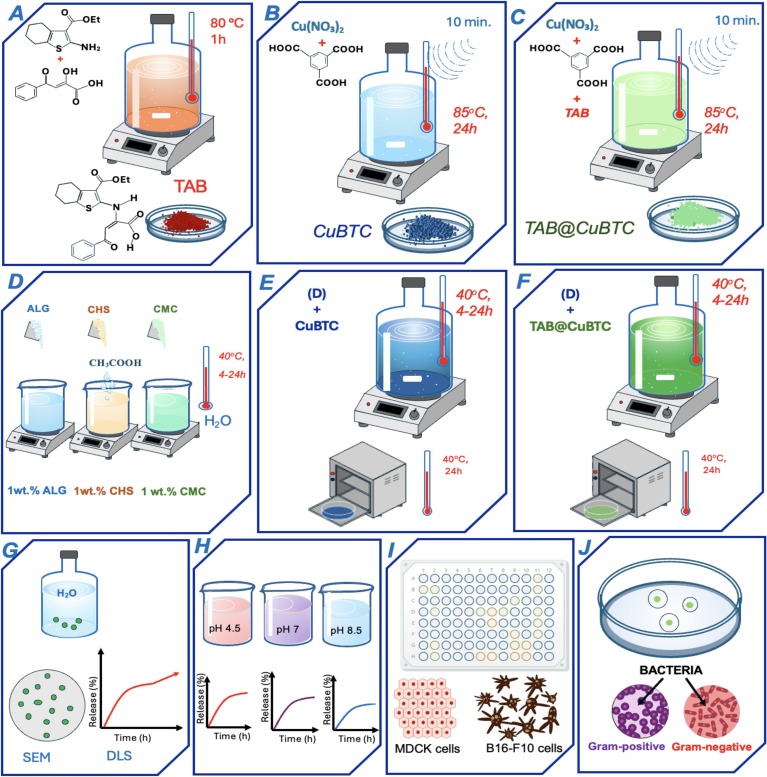
Fig. 1. Schematic illustration of the synthesis and characterization of natural polymer-based hybrid films incorporated with CuBTC and TAB@CuBTC MOFs: synthesis of (A) TAB, (B) CuBTC, and (C), and TAB@CuBTC; (D) preparation of ALG, CHS, and CMC polymer solutions; fabrication of (E) polymer/CuBTC and (F) polymer/TAB@CuBTC hybrid films; analysis of (G) stability, (H) drug loading and release efficiency, (I) cytotoxicity, and (J) antibacterial activity.
Comprehensive details on all chemical reagents and raw materials employed in the synthesis of TAB, CuBTC, and hybrid films were obtained from commercial suppliers and used without further purification unless otherwise specified. Detailed information on reagent suppliers, grades, and catalog numbers is provided in the Supplementary materials.
The scheme of synthesis of organic compounds is presented in Fig. S1. Ethyl-2-amino-4,5,6,7-tetrahydrobenzo[b]thiophene-3-carboxylate 2 (0.01 mol, 2.25 g) in ethanol (30 mL) was mixed with a solution of 2,4-dioxo-4-phenylbutanoic acid 1 (0.01 mol, 1.92 g) in ethanol (30 mL). The reaction mixture was heated at 80 °C under stirring for 1 h. The reaction was carried out under the air atmosphere. Upon completion, the resulting precipitate was filtered off and purified by recrystallization from acetonitrile. The yield of the product was 90%.
The structure of the obtained compound was characterized by 1H NMR (Fig. S1), 13C NMR (Fig. S2), and FTIR spectroscopy (Fig. 2G). The physicochemical characteristics of the obtained compound correspond to the data described previously [58]. Detailed information about the synthesis procedures of starting compounds is provided in the Supplementary materials.
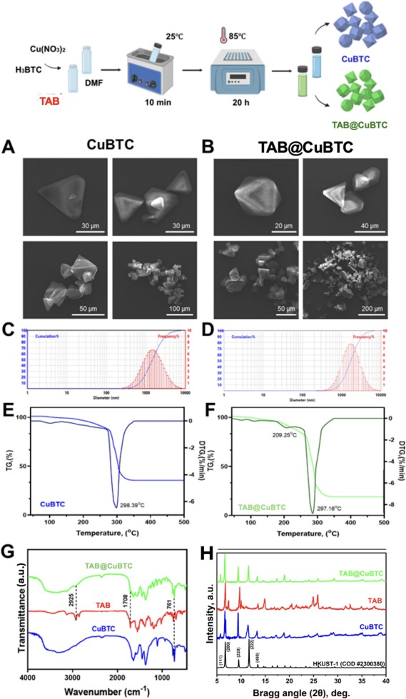
Fig. 2. Characterization of CuBTC and TAB@CuBTC MOFs: Scheme of synthesis (top row); SEM images of CuBTC (A) and TAB@CuBTC (B) MOFs; DLS analysis of CuBTC (C) and TAB@CuBTC (D) MOFs; (E, F) TGA curves of CuBTC (E) and TAB@CuBTC (F) MOFs; and (H) FTIR spectra of CuBTC, TAB and TAB@CuBTC; (H) PXRD analysis of CuBTC, TAB and TAB@CuBTC of TAB@CuBTC (top curve), free TAB drug (middle curve), and (bottom curve) composite pristine CuBTC.
The synthesis of CuBTC MOFs was carried out by the solvothermal procedure as described previously [59]. Copper (II) nitrate trihydrate Cu(NO3)2·3H2O (0.86 mmol, 207 mg) was dissolved in 1 mL of water H2O and benzene-1,3,5-tricarboxylic acid (0.48 mmol, 101 mg) was dissolved in 2 mL of a mixture of DMF:EtOH (1:1 v/v). After the mixing stage, the combined solutions were hermetically sealed and subjected to ultrasound treatment for 10 min. The sealed vessel was then heated to 85 °C for 24 h. After cooling to room temperature, the resulting blue solid product was isolated by filtration and thoroughly washed with a mixture of ethanol and water to remove unreacted species. The samples were dried in air, and the reaction yield was 95%.
The organic compound (TAB) (either 45 mg or 75 mg, depending on the batch) and benzene-1,3,5-tricarboxylic acid H3BTC (0.48 mmol, 101 mg) were dissolved in 1 mL of DMF using ultrasonic assistance, while copper(II) nitrate trihydrate Cu(NO3)2·3H2O (0.86 mmol, 207 mg) was dissolved in 1 mL of water H2O. The two prepared solutions were combined in a 4 mL glass vial, which was then sealed hermetically. After that, the reaction mixture was exposed to ultrasonic treatment for 10 min and subsequently heated at 85°С for 24 h. After cooling to room temperature, the resulting green precipitate was collected by filtration and washed three times with 2 mL portions of a DMF/H2O mixture. The samples were dried in air, and the reaction yield was 93%.
To determine the Drug Loading Efficiency (DLE) and Drug Loading Capacity (DLC), the reaction filtrate and subsequent washing solutions were collected and analyzed indirectly by UV–Vis spectrophotometry at the characteristic absorption wavelength of TAB (416 nm), using the corresponding calibration curve (Fig. S5). The mass of TAB incorporated into the TAB@CuBTC product was calculated by mass balance as:where Winitial is the initial mass of TAB added (45 or 75 mg), and Wfiltrate and Wwashings are the amounts of unbound TAB recovered from the reaction filtrate and washing solutions, respectively.
The Drug Loading Efficiency was calculated as:and the Drug Loading Capacity as:
where WCuBTC is the dry mass of the CuBTC host used for the loading-capacity calculation.
For in vitro release studies, 10 mg of the dried TAB@CuBTC was dispersed in 10 mL of PBS buffers (pH 4.5, 7.0, and 8.5) at 37 °C under orbital shaking (150 rpm). Cumulative released TAB concentrations were sampled at predetermined intervals and quantified by UV–Vis spectrophotometry at 416 nm using a NanoPhotometer C40 spectrophotometer and the corresponding calibration curves established in PBS at pH 4.5, 7.0, and 8.5.
Polymer-based films were obtained by pre-homogenization of all components in a porcelain mortar. Then, a 1 wt% aqueous ALG, CHS, or CMC solution was prepared, into which MOF-based materials were added in quantities of 0%, 0.5%, 1.0%, 5.0%, and 10.0% relative to the polymer mass. The mixtures were mixed using a magnetic stirrer at 400 rpm until a homogeneous mass was obtained, and then 5 mL of the solution was poured into Petri dishes with a diameter of 60 mm. The materials were formed by evaporating reagents at 40 °C under atmospheric pressure. The detailed procedures for preparation of hybrid films are given in the Supplementary materials.
The chemical composition and structural homogeneity of the obtained materials were examined using a scanning electron microscope (SEM, Quanta200, FEI, The Netherlands) equipped with energy-dispersive X-ray spectroscopy (EDAX nitrogen-free GENESIS XM 2 60). SEM analysis was performed at an accelerating voltage of 30 kV, and EDX analysis at 20 kV. Sample suspension in alcohol was applied to a clean silicon wafer and dried at room temperature. To quantitatively evaluate particle dispersion homogeneity, surface SEM micrographs (scale: 300 μm) were processed via ImageJ software using a 3 × 3 grid matrix (N = 9 regions of interest, ROIs). The mean particle count, standard deviation (SD), and coefficient of variation (CV% = (SD/Mean) x 100%) were systematically determined for each film.
The diffraction patterns were recorded using a Bruker D2 Phaser X-ray diffractometer. Scanning was performed over the angular range of 2θ = 5°–40°, with an angular resolution of 0.01° and a scanning speed of 1°/min. To investigate the crystalline domain parameters and understand the lattice state, the average crystallite size (D) of the synthesized MOFs was estimated using both the classical Debye–Scherrer equation and the Williamson–Hall (WH) model.
Debye–Scherrer equation [60], [61]:
Williamson–Hall (W–H) model [60], [61], [62]:where τ = average crystallite size, K = shape factor (0.90), λ = X–ray wavelength (1.54 Å), β = the line broadening at half the maximum intensity (FWHM) and θ = Bragg angle. The results regarding the crystallite size determination for pristine CuBTC and TAB@CuBTC are presented in Table S1.
Hydrodynamic diameters (Dh) were estimated using a DLS nanoparticle analyzer (BK Winner803, Jinan Winner Particle Instrument Stock Co., China).
Fourier transform infrared spectra (FTIR) were obtained on a spectrometer (WQF-530A) in the range of 400–4000 cm−1 in the transmission mode (KBr) with 32 scans.
The study of thermal stability was performed using TGA/DSC 1 equipment. Samples were heated from 35 to 500 °C at a ramping rate of 10 °C/min with air supplied at a rate of 30 cm3/min. During heating, mass (thermogravimetric) and thermal (differential-thermal) curves were recorded.
Nitrogen adsorption–desorption measurements were performed using a NOVA 1200e adsorption analyzer (Quantachrome Instruments, USA) at 77 K after degassing the samples at 120 °C for more than 10 h. The specific surface area was determined using the multipoint BET method, and the total pore volume was estimated from the N2 uptake at P/P0 close to 0.99. Pore-size distributions were evaluated using the DFT method.
To investigate the release behavior of TAB from TAB@CuBTC under different pH conditions (4.5, 7.0, and 8.5), the in vitro cumulative release data were fitted using four classical kinetic models:
Zero-order model: This model describes systems where the drug release rate is constant and independent of the active agent's concentration [63], [64]:
where Qt is the cumulative percentage of TAB released at time t; Q0 is the cumulative percentage of TAB released at the initial time (t = 0); and k0 is the zero-order release rate constant.
First-order model: This model represents a concentration-dependent release profile, where the release rate is proportional to the residual drug content remaining in the matrix [63], [64]:
where k1 is the first-order release rate constant.
Higuchi model: This model describes drug diffusion from porous structures or planar matrix systems based on Fick's law of diffusion [63], [64], [65]:
where KH is the Higuchi release rate constant; C is the intercept, representing the initial burst effect or lag time.
Korsmeyer–Peppas model: This semi-empirical model is widely utilized to analyze drug release from polymeric and porous networks, particularly when the exact release mechanism is complex or involves multiple coupled processes [63], [64], [66]:
where Qt/100 is the fraction of TAB released at time t (with the fitting restricted to the initial linear portion corresponding to cumulative release ≤60%); kKP is the Korsmeyer–Peppas release velocity constant; n is the diffusional exponent indicating the solute transport mechanism.
For spherical or near-spherical micro/nanoparticles, the physical significance of the exponent n is classified as follows:
n ≤ 0.43 corresponds to Fickian diffusion (concentration gradient-driven molecular transport).
0.43 < n < 0.85 indicates anomalous (non-Fickian) transport, representing a combined mechanism of diffusion and structural swelling/degradation of the carrier.
n ≥ 0.85 represents Super Case II transport, where the release rate is entirely controlled by the swelling or relaxation rate of the matrix.
All calculations and regression analyses were performed using linear and non-linear curve-fitting algorithms in OriginPro 2021 and Microsoft Excel softwares. The goodness-of-fit and reliability of each model were evaluated quantitatively using the coefficient of determination (R2).
MDCK cells were grown in Dulbecco's Modified Eagle Medium (DMEM) supplemented with 10% fetal bovine serum (FBS). The Murine melanoma cell line (B16-F10 obtained from the American Type Culture Collection) was cultured in Alpha-MEM containing 10 vol% FBS and 2 mM of UltraGlutamine IN. All cell line cultures were maintained in a sterile humidified atmosphere containing 95% air and 5% CO2 at 37 °C.
The cytotoxicity of the obtained materials was assessed using the Alamar Blue assay. To obtain a preliminary assessment of cellular responses to the developed materials, MDCK and B16-F10 cells were used as two distinct in vitro cell models. MDCK and B16-F10 cells were seeded into 96-well plates at a density of 1.0 × 10⁴ cells per well and incubated overnight at 37 °C under 5% CO2. The TAB and CuBTC were dissolved in the medium and added at concentrations of 2, 1, 0.5, 0.25, 0.125, 0.063, 0.031, 0.016, 0.008, and 0.004 mg/mL. The hybrid films were then added at a concentration of 10 mm × 10 mm per 1 mL of cell medium, cultured with the cells, and incubated for 24 or 48 h. Following incubation, the medium was removed and replaced with Alamar Blue (10% v/v). Absorbance was measured using a spectrophotometric microplate reader at 570 and 600 nm. Untreated cells incubated with culture medium only were used as negative cytotoxicity controls (representing 100% cell viability). All experiments were performed in triplicate (n = 3), and the results are presented as mean ± standard deviation. The details are presented in the Supplementary materials.
The antibacterial activity of polymer-based hybrid films containing CuBTC and TAB@CuBTC was evaluated using the agar diffusion method by measuring the diameter of the inhibition zones. Antibacterial activity was investigated against four gram-negative (Pseudomonas, Escherichia coli, Serratia marcescens, and Klebsiella) and one Gram-positive bacteria (Bacillus subtilis). All the bacterial cultures were obtained from the collection of the Institute of Biomedical Systems and Biotechnologies, Peter the Great St. Petersburg Polytechnic University (SPbPU). LB medium was used as a nutrient medium for bacterial cultivation, and all strains were incubated in a thermostat at 37 °C under standard growth conditions.
Bacterial cultures were grown in LB medium and incubated at 37 °C under standard growth conditions until reaching the logarithmic growth phase. The bacterial suspensions were then adjusted to a uniform density and evenly spread onto sterile LB agar plates. Polymer matrices (ALG, CHS, and CMC) containing CuBTC or TAB@CuBTC were cut into samples of defined size and gently placed onto the surface of the inoculated agar plates. Polymer platforms without MOF incorporation were used as negative controls. All procedures were carried out under sterile conditions.
After sample placement, the agar plates were incubated at 37 °C for 24 h. Antibacterial activity was assessed by measuring the diameter of the inhibition zones (mm) formed around the samples. The inhibitory properties of the obtained materials were studied by dilution in a liquid nutrient medium with spectrophotometric monitoring. Polymer-based hybrid films (ALG, CHS, and CMC) containing CuBTC or TAB@CuBTC were cut into 10 mm × 10 mm samples and carefully placed in a test tube containing the nutrient medium and bacteria. Based on the identical casting conditions used for all three polymer matrices, the nominal MOF areal loadings were approximately 0.0088, 0.0177, 0.0884, and 0.1768 mg/cm2 for the 0.5, 1.0, 5.0, and 10.0 wt% formulations, respectively. The optical density of the samples was measured after 1, 2, 5, 12, and 24 h. All experiments were performed in triplicate, and the results were expressed as mean ± standard deviation.
Cultivation was carried out in flasks with 20 mL of LB nutrient medium 500 μL of Pseudomonas, Escherichia coli, Serratia marcescens, Klebsiella and Bacillus subtilis overnight culture and composites were added to the nutrient medium. Optical density (OD590) was measured every 24 h.
All physicochemical experiments were performed in triplicate. For biological studies (cytotoxicity and antibacterial assays), evaluations were conducted in three independent biological replicates (n = 3), with each run containing three technical replicates (triplicate wells or plates) to ensure reproducibility. The statistical analyses were performed using the biological replicates (n = 3), and data are expressed as mean ± standard deviation (SD). Statistical analyses were carried out using a one-way analysis of variance (ANOVA) followed by Tukey's post hoc test. For comparisons between two groups, Student's t-test was applied. Differences were considered statistically significant at p < 0.05.
Detailed information on the synthesis and characterization of the biologically active compound 2-((3-(ethoxycarbonyl)-4,5,6,7-tetrahydrobenzo[b]thiophen-2-yl)amino)-4-oxo-4-phenylbut-2-enoic acid (TAB) is provided in the Supplementary material (Figs. S1–S3).
CuBTC and TAB@CuBTC were synthesized by the solvothermal method. SEM imaging revealed a heterogeneous structure of synthesized CuBTC and TAB@CuBTC. It shows crystalline octahedral and rectangular prismatic morphology of the developed MOFs with primary crystal sizes in the range of 10–40 μm (Fig. 2A, B). Quantitative image analysis using ImageJ yielded an average crystal size of 26.0 ± 10.2 μm for pristine CuBTC and 20.6 ± 7.3 μm for TAB@CuBTC (Fig. S4C, D). This morphological distribution is consistent with recent studies utilizing ultrasound-assisted crystallization to control HKUST-1 crystal structures [43]. Morphological and DLS analyses (Table S2) showed primary SEM crystal sizes of 26.0 ± 10.2 μm for pristine CuBTC and 20.6 ± 7.3 μm for TAB@CuBTC, with corresponding average hydrodynamic diameters (Dh) of 7.8 ± 4.1 μm (PDI = 0.5 ± 0.1) and 8.0 ± 3.2 μm (PDI = 0.3 ± 0.1), respectively. The smaller Dh values obtained by DLS relative to the SEM-derived particle sizes likely reflect preferential detection of the smaller suspended particle fraction, because larger microcrystals (>10 μm) can undergo rapid gravitational settling during DLS measurements.
The elemental analysis of developed MOFs was validated by EDX (Fig. S4). For CuBTC, the EDX spectrum shows the presence of Carbon (C), Oxygen (O), and Copper (Cu) peaks, consistent with the СuBTC lattice composition, while a Silicon (Si) signal is identified as an artifact from the silicon wafer used in sample mounting (Fig. S4A). The EDX spectrum of TAB@CuBTC (Fig. S4B) reveals a sulfur (S) signal at approximately 0.17 at.%, providing qualitative evidence for the presence of the sulfur-containing TAB compound.
The thermal behavior of pristine CuBTC and TAB@CuBTC was investigated by TGA in air (30–500 °C, 10 °C/min), and the main weight-loss stages are summarized in Table S3. Pristine CuBTC showed an initial mass loss of 11.65 wt% below 270 °C, assigned mainly to desorption of adsorbed water and residual solvents, followed by a 53.67 wt% loss between 270 and 350 °C associated with decomposition of the CuBTC framework, leaving a residual mass of 34.68 wt%. TAB@CuBTC showed a lower initial mass loss of 6.22 wt% below 200 °C, followed by an additional 9.12 wt% loss between 200 and 260 °C and a major 64.69 wt% loss between 260 and 350 °C, leaving a residual mass of 19.97 wt%. The additional 200–260 °C event is consistent with a TAB-related contribution to the thermal profile; however, overlap between TAB and framework decomposition prevents this step from being used as a direct quantitative measure of the total TAB content. Therefore, the TGA data are used as complementary evidence of compositional differences between CuBTC and TAB@CuBTC, whereas the drug loading capacity was quantified independently by UV–Vis mass-balance analysis.
The FTIR spectra of TAB, pristine CuBTC, and TAB@CuBTC are shown in Fig. 2G. Pristine CuBTC exhibited a characteristic Cu–O-related band at 761 cm−1 and carboxylate stretching bands in the 1373–1644 cm−1 region [1], [5], [13], [67], [68]. In TAB@CuBTC, the characteristic TAB-associated carbonyl band at approximately 1708 cm−1 remained detectable, together with the principal CuBTC bands [69], [70]. These observations support the incorporation of TAB into the CuBTC-containing material while indicating that the main Cu–BTC coordination features remain detectable after synthesis. Minor shifts and broadening in the carbonyl and carboxylate regions may be consistent with non-covalent guest–host interactions, including π–π interactions and hydrogen bonding [33], [34], [35]. However, FTIR alone cannot determine the precise location of TAB within the framework or establish its complete chemical integrity.
The crystalline structure and phase purity of the synthesized MOFs were investigated by PXRD (Fig. 2H). The PXRD pattern of the pristine CuBTC displays high-intensity diffraction peaks at low 2θ angles (notably around 6.6°, 9.5°, 11.5°, 13.3° and 19.0°, corresponding to the planes (200), (220), (222), (400) and (440)) [5], [13], [71], [72]. Upon TAB incorporation, TAB@CuBTC retained the characteristic diffraction pattern of pristine CuBTC, with no additional reflections attributable to a separate crystalline TAB phase. This indicates that the parent CuBTC crystalline structure was largely preserved during the one-pot synthesis. Differences in relative peak intensities were observed, as also reported for other HKUST-1-based composite systems [73]. However, since PXRD peak intensities can be influenced by several structural and experimental factors [74], these changes alone were not considered direct evidence of the location of TAB within the framework. Together with the N2 physisorption results showing pronounced decreases in accessible surface area and total pore volume, the PXRD data support preservation of the CuBTC framework while TAB incorporation substantially reduces the accessible microporosity. Because TAB is present during one-pot framework formation, a guest-assisted influence on CuBTC crystallization cannot be excluded from the present data, and the observed changes should therefore not be attributed exclusively to pore encapsulation.
Crystallite sizes estimated using the Debye–Scherrer and Williamson–Hall (WH) methods are summarized in Table S1. For pristine CuBTC, the two methods yielded similar values of 49.47 and 49.84 nm, respectively. However, the very low linear-regression coefficient of the WH fit (R2 = 0.0001) indicates that the corresponding microstrain value should not be interpreted quantitatively. For TAB@CuBTC, the Scherrer and WH estimates were 45.96 and 58.71 nm, respectively. The positive WH slope corresponds to an apparent tensile microstrain of 1.64 × 10⁻³, although the moderate fit quality (R2 = 0.5872) requires cautious interpretation. Therefore, the difference between the Scherrer and WH estimates suggests that strain-related broadening may contribute to the diffraction profile of TAB@CuBTC, but the present data do not allow this contribution to be assigned exclusively to TAB incorporation.
Nitrogen physisorption analysis provided further evidence for changes in the porous structure of CuBTC after TAB loading (Fig. S4E–G and Table S4). Pristine CuBTC showed typical microporous adsorption behavior, with a BET specific surface area of ~581.3 m2/g and a total pore volume of ~0.257 cm3/g. After TAB incorporation, the BET surface area decreased markedly to ~123.2 m2/g, corresponding to a reduction of approximately 78.8%, while the total pore volume decreased to ~0.086 cm3/g, representing a reduction of approximately 66.5%. The pore-size distribution was also markedly altered after TAB loading (Fig. S4F). Taken together, the pronounced decreases in accessible surface area and total pore volume, together with the altered pore-size distribution, demonstrate a substantial reduction in accessible porosity after TAB incorporation. These changes are consistent with occupation and/or blockage of the CuBTC pore system, although nitrogen physisorption alone does not establish the precise spatial location of TAB within the framework.
The loading efficiency of TAB is presented in Fig. 3A. The functional properties of CuBTC were assessed by measuring the free, unbound drug in washing solutions through UV–Vis spectrophotometry. The evaluation of drug encapsulation performance for the developed TAB@CuBTC composites was conducted by systematically analyzing key loading parameters under varying initial drug feeds of 45.0 mg and 75.0 mg, as summarized in Table 1.
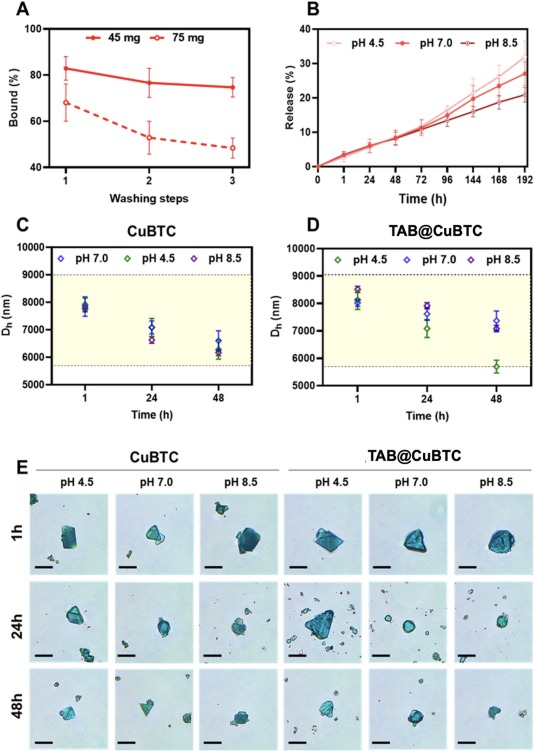
Fig. 3. TAB loading, its release and colloidal stability of TAB@CuBTC: (A) Drug loading efficiency of TAB into MOF (45 and 75 mg) after several wash steps; (B) time-dependent release profiles of TAB from TAB@CuBTC incubated at pH 4.5 (top curve), pH 7.0 (middle curve) and pH 8.5 (bottom curve); (С–D) colloidal stability of CuBTC and TAB@CuBTC at pH 4.5, 7.0 and 8.5 for 1, 24, 48 h; (E) optical images of CuBTC and TAB@CuBTC incubated in aqueous solutions at pH 4.5, 7.0 and 8.5 (scale bar: 50 μm).
Table 1. Quantitative drug loading parameters of TAB within the CuBTC MOFs.
| TAB initial feed (mg) | Dry weight of CuBTC host (mg)* | Drug loading efficiency (DLE, %)a | Absolute loaded TAB mass (mg) | Drug loading capacity (DLC, wt%)b | Drug loading capacity (DLC, mg/g) |
|---|---|---|---|---|---|
| 45.0 | 135.01 | 74.9 ± 4.3 | 33.71 ± 1.94 | 19.98 ± 0.92 | 199.8 ± 9.2 |
| 75.0 | 135.01 | 48.2 ± 4.8 | 36.15 ± 3.60 | 21.12 ± 1.66 | 211.2 ± 16.6 |
Note: Calculations are based on the anhydrous form of CuBTC (Cu3(BTC)2, MW = 604.87 g/mol) at a 93% reaction yield from Section 2.2.3.
DLE values are presented as Mean ± Standard Deviation (n = 3).
DLC (wt%) is defined as the weight percentage of loaded TAB relative to the total mass of the TAB@CuBTC composite.
The analysis revealed the DLE depends on the initial drug amount. Specifically, as the TAB feed increased from 45.0 mg to 75.0 mg, the DLE notably declined from 74.9 ± 4.3% to 48.2 ± 4.8%. Interestingly, this decline in DLE was coupled with a minimal change in the absolute mass of the loaded drug, which increased only slightly from 33.71 ± 1.94 mg to 36.15 ± 3.60 mg. Consequently, the drug loading capacity (DLC) stabilized within a range of 19.98 ± 0.92 wt% (equivalent to 199.8 ± 9.2 mg/g) to 21.12 ± 1.66 wt% (equivalent to 211.2 ± 16.6 mg/g), indicating a plateau in loading efficiency. The increase in initial TAB feed from 45.0 to 75.0 mg resulted in only a small increase in the absolute loaded TAB mass, from 33.71 ± 1.94 to 36.15 ± 3.60 mg, while the DLC remained within a relatively narrow range of 19.98 ± 0.92 to 21.12 ± 1.66 wt%. This behavior indicates that increasing the TAB feed beyond 45 mg did not substantially increase the amount incorporated under the present synthesis conditions. Together with the pronounced decreases in BET surface area and total pore volume after TAB incorporation, these results are consistent with substantial occupation and/or blockage of the accessible CuBTC pore system. However, the loading data do not by themselves establish a maximum pore-saturation capacity or distinguish intra-pore incorporation from external-surface adsorption. This evaluation of drug encapsulation performance aligned with the elemental sulfur detected by EDX, with further details in Section 2.2 of the Supplementary materials.
The release behavior of TAB from TAB@CuBTC was investigated in buffer solutions at pH 4.5, 7.0, and 8.5. The release profiles of TAB demonstrated pH-dependent behavior (Fig. 3B). The colloidal stability of CuBTC and TAB@CuBTC was investigated at pH 7, pH 4.5 and pH 8.5 (Fig. 3C, D). These pH values were selected to represent acidic (pH 4.5), near-neutral (pH 7.0), and alkaline (pH 8.5) conditions and thereby evaluate the influence of environmental pH on TAB release and MOF stability across a broad application-relevant range. We can observe no significant changes in colloidal stability between CuBTC and TAB@CuBTC. With increasing time, we can detect some degradation of MOFs which correspond to reduced Dh. To investigate drug release mechanisms, the release data of TAB from TAB@CuBTC under various pH conditions were analyzed using mathematical kinetic models, including Zero-order, First-order, Higuchi, and Korsmeyer–Peppas equations (the parameters are summarized in Table S5). Both the zero-order and first-order models showed high coefficients of determination (R2 > 0.970), although the best-fitting model varied slightly with pH (Table S5). The Korsmeyer–Peppas exponent increased from 0.3656 at pH 8.5 to 0.4676 at pH 4.5, indicating a pH-dependent change in the apparent release behavior. Because the Korsmeyer–Peppas model is semi-empirical and the present MOF system does not strictly conform to an idealized single geometry, these n values are not used to assign a unique molecular transport mechanism. The zero-order rate constant increased from 0.0983%·h−1 at pH 8.5 to 0.1514%·h−1 at pH 4.5, corresponding to an approximately 54% higher apparent release rate under acidic conditions.
Optical microscopy indicated time-dependent morphological changes in both CuBTC and TAB@CuBTC during aqueous incubation (Fig. 3E), while DLS revealed corresponding changes in the hydrodynamic diameter of the suspended particle fraction (Fig. 3C, D). Because the synthesized crystals are in the micrometer range and larger particles may sediment during DLS measurements, these trends alone cannot distinguish framework dissolution, fragmentation, sedimentation, or agglomeration. Therefore, the DLS and optical microscopy results are interpreted only as complementary indicators of time-dependent changes in the dispersed microcrystal population rather than direct evidence of a specific degradation mechanism.
SEM analysis showed clear pH- and time-dependent morphological changes in both pristine CuBTC and TAB@CuBTC after incubation at pH 4.5 and pH 7.0 for 24 and 48 h (Fig. S10 and Table S9). At pH 4.5, pristine CuBTC exhibited pronounced loss of its initial rod-like morphology after 48 h, whereas TAB@CuBTC retained more recognizable rod-like features. At pH 7.0, both materials showed comparatively less severe morphological alteration. These observations indicate different degrees of morphological stability under the tested conditions; however, SEM alone does not establish the underlying chemical degradation mechanism or long-range crystalline integrity.
Post-incubation FTIR spectra were additionally collected to evaluate changes in the CuBTC coordination environment after exposure to pH 4.5 and pH 7.0 for 24 and 48 h (Fig. S11). For both CuBTC and TAB@CuBTC, the characteristic carboxylate-related bands in the 1370–1650 cm−1 region and the Cu–O-related band in the low-wavenumber region remained detectable after incubation. Only moderate changes in band shape and relative intensity were observed. These results indicate that the principal copper–carboxylate coordination motifs remain detectable even after prolonged exposure. Importantly, the FTIR results should be considered together with the SEM observations: the pronounced morphological deterioration of pristine CuBTC at pH 4.5 after 48 h does not necessarily imply complete loss of the local Cu–BTC coordination environment. Thus, SEM and FTIR provide complementary information on morphological and chemical structural changes during incubation.
Hybrid films containing CuBTC and TAB@CuBTC MOFs were successfully synthesized using ALG, CHS, and CMC as polymer matrices. Optical microscopy revealed that the developed membranes formed cohesive, self-standing films that could be peeled and cut into defined shapes without visible cracking, allowing them to be easily peeled and cut into precise shapes without fracturing, regardless of the percentage of MOF incorporation (0.5–10 wt% relative to polymer mass). Based on the casting conditions, the nominal dry-film thickness was estimated to be approximately 12 μm. Fig. 4 (top row) illustrates the synthetic process for hybrid membranes impregnated with CuBTC and TAB@CuBTC.
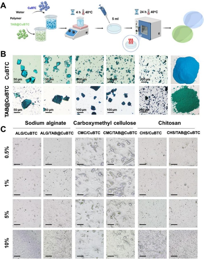
Fig. 4. Synthesis and characterization of hybrid films containing CuBTC and TAB@CuBTC: (A) Scheme of synthetic procedure; (B) optical images of CuBTC and TAB@CuBTC; and (C) optical images of hybrid films (CMC, ALG, and CHS) containing CuBTC and TAB@CuBTC MOFs (scale bar: 200 μm).
As shown in Fig. 4C, the distribution of MOFs crystals within the polymer matrices is significantly influenced by the nature of the polymers. In case of ALG and CHS, the TAB@CuBTC MOFs are mainly characterized by uniform distribution that indicates strong interaction between MOFs and the polymer matrix. In contrast, the CMC-based films show a distinct morphological feature characterized by the formation of MOF clusters. Quantitatively, grid-based ImageJ analysis on SEM micrographs (Fig. S8 and Table S8) confirmed highly uniform spatial dispersion in ALG/TAB@CuBTC (38.00 ± 5.69 particles/ROI, CV = 14.97%) and CHS/TAB@CuBTC (50.11 ± 9.18 particles/ROI, CV = 18.32%). Conversely, CMC/TAB@CuBTC exhibited higher spatial variability (11.22 ± 4.63 particles/ROI, CV = 41.26%), while pristine CuBTC films displayed CV values of 43.97%–59.95%, quantitatively corroborating the micro-clustered nature in CMC and the enhanced dispersibility upon TAB incorporation. The higher spatial variability observed for the CMC-based films indicates a more heterogeneous, micro-clustered distribution of MOF particles. Interactions between carboxymethyl cellulose and Cu2+ species have been reported previously and may contribute to the observed morphology [7], [8]; however, the present data do not directly establish the interfacial mechanism responsible for this clustering or demonstrate that such aggregation provides a functional advantage. The CMC-based films nevertheless retained antibacterial activity, including activity against Klebsiella strains (Table 2), indicating that the clustered morphology did not eliminate biological performance under the tested conditions. Further studies correlating cluster size, accessible MOF surface area, mechanical properties, and release kinetics are required to determine the functional consequences of this morphology.
Table 2. Antibacterial activity of hybrid films containing CuBTC and TAB@CuBTC MOFs against Gram +/− bacteria.
| Polymer matrix | Encapsulated compound | Pseudomonas | Escherichia coli | Bacillus subtilis | Serratia marcescens | Klebsiella |
|---|---|---|---|---|---|---|
| ALG | CuBTC | 0 | 0 | 9.5 ± 2.1 | 11.5 ± 0.5 | 5.3 ± 1.5 |
| TAB@CuBTC | 8.5 ± 2.0b | 11.8 ± 3.1a | 10.0 ± 0.1b | 11.0 ± 0.1b | 7.2 ± 1.1b | |
| CMC | CuBTC | 10.7 ± 1.3 | 11.0 ± 0.5 | 12.8 ± 0.5 | 0 | 10.3 ± 1.6 |
| TAB@CuBTC | 7.3 ± 5.0b | 13.2 ± 2.2a | 18.2 ± 1.7a | 6.7 ± 4.3c | 11.0 ± 0.5a | |
| CHS | CuBTC | 0 | 12.2 ± 2.3 | 11.8 ± 3.1 | 12.8 ± 2.1 | 9.5 ± 0.7 |
| TAB@CuBTC | 12.7 ± 1.1a | 13.0 ± 0.1a | 16.7 ± 0.5a | 14.8 ± 2.5a | 10.0 ± 1.4a |
Note: Values are expressed as mean ± standard deviation of the inhibition zone diameter (mm).
“0” indicates no detectable inhibition zone.
Gram-negative bacteria: Pseudomonas, E. coli, S. marcescens, Klebsiella.
Gram-positive bacterium: Bacillus subtilis.
Bold values indicate the highest antibacterial activity observed for each bacterial strain.
Different superscript letters (a, b, c) within the same column for the TAB@CuBTC-loaded films indicate statistically significant differences (p < 0.05) among the three polymer matrices (ALG, CMC, and CHS) determined by one-way ANOVA followed by Tukey's post-hoc test.
The cytotoxicity of free TAB and CuBTC toward MDCK and B16-F10 cells is shown in Fig. S7, and the exact cell viability values along with statistical significance analysis are compiled in Table S6. Both samples showed dose-dependent decrease in cell viability. Given the cytotoxic response of TAB and MOFs, further studies were conducted to assess whether incorporation into polymer matrices (ALG, CMC, and CHS) could modulate the biological response. The primary objective was to ensure that the encapsulation process did not induce significant cytotoxicity in healthy renal cells (MDCK).
The cytotoxicity of the hybrid films was evaluated using MDCK and B16-F10 cells via the Alamar Blue assay (Fig. 5).
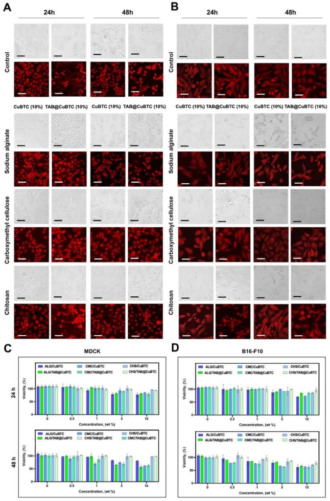
Fig. 5. Cytocompatibility of hybrid films (ALG/CuBTC, CMC/CuBTC, CHS/CuBTC, ALG/TAB@CuBTC, CMC/TAB@CuBTC, and CHS/TAB@CuBTC): (A, B) Optical and CLSM images of MDCK (A) and B16-F10 (B) cells after 24 and 48 h incubation with developed films (scale bar: 50 μm). (C, D) The viability of MDCK (C) and B16-F10 (D) cells determined by the Alamar Blue assay after 24 h and 48 h of incubation with film samples (cut to 10 mm × 10 mm size in 1 mL DMEM) containing different loading doses: Control, 0.5%, 1.0%, 5.0%, and 10.0% of pristine CuBTC or TAB@CuBTC. Data are presented as mean ± SD (n = 3).
The results of the Alamar Blue assay are depicted in Fig. 5C, D, with comprehensive numerical viability values and statistical significance relative to the untreated control provided in Table S7. The hybrid films exhibited loading-, formulation-, and time-dependent effects on cell viability. At lower loadings (0.5–1.0 wt%), most formulations maintained relatively high cell viability, although lower values were observed for some formulations after 48 h. At the highest loading level of 10 wt%, cell viability ranged from 57.6 ± 6.5% to 98.7 ± 0.3% across the tested formulations, cell lines, and incubation times. According to the quantitative criterion commonly applied under ISO 10993-5, a reduction in cell viability of more than 30% relative to the untreated control indicates cytotoxic potential. Accordingly, some formulations remained above the 70% viability threshold, whereas others fell below this threshold, particularly after 48 h, demonstrating formulation- and time-dependent cellular responses.
The comparatively higher viability observed for several hybrid-film formulations than for the corresponding free active components may reflect reduced cellular exposure when the active species are incorporated into the polymer/MOF matrix. However, because TAB and Cu2+ release from the individual polymer films was not directly quantified, a specific protective-reservoir or diffusion-barrier mechanism cannot be established from the present data.
These findings were generally consistent with the optical microscopy observations. MDCK cells retained a dense epithelial monolayer under several tested conditions compared with the untreated control (Fig. 5A), while B16-F10 cells generally preserved their characteristic spindle-like morphology and cell density (Fig. 5B). Nevertheless, the observed morphological features should be interpreted together with the quantitative Alamar Blue results, particularly for formulations showing viability below the 70% threshold.
The antibacterial activity of developed hybrid films containing CuBTC and TAB@CuBTC was systematically evaluated against gram-negative (Pseudomonas, Escherichia coli, Serratia marcescens, and Klebsiella) and gram-positive (Bacillus subtilis) bacteria. The results were summarized in Fig. 6 and Table 2. The antibacterial performance strongly depends on both the nature of the encapsulated MOF and type of polymer. The discussion related to antibacterial activity is presented in Section 3.6.1. Cooperative antibacterial action.
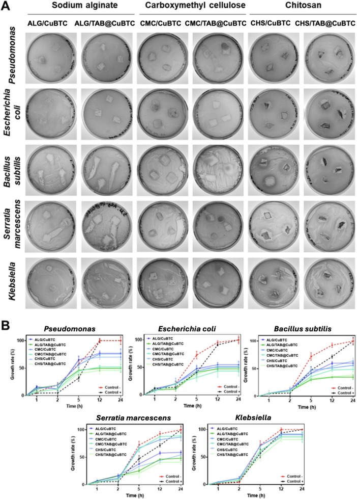
Fig. 6. Antibacterial activity of developed hybrid films (ALG/CuBTC, CMC/CuBTC, CHS/CuBTC, ALG/TAB@CuBTC, CMC/TAB@CuBTC, and CHS/TAB@CuBTC): Representative agar diffusion plates showing inhibition zones (A) and bacterial growth curves monitored over 24 h incubation at 37 °C against gram-negative (E. coli, Pseudomonas, S. marcescens, Klebsiella) and Gram-positive (B. subtilis) bacteria. (B) Quantitative bacterial growth kinetic curves monitored spectrophotometrically over 24 h in liquid LB medium at 37 °C. Sodium alginate – ALG, carboxymethyl cellulose – CMC, chitosan – CHS. Data are presented as mean ± SD (n = 3).
The pristine polymer films (pure ALG, CMC, and CHS) used as negative controls did not exhibit any detectable zones of inhibition (0 mm) against all tested gram-positive and gram-negative bacteria, confirming that the biopolymer matrices alone lack independent, diffusible antibacterial capability. Fig. 6 shows petri dishes with inhibition zones. We can observe that all hybrid films demonstrated inhibition of bacteria growth indicating the successful diffusion of antibacterial agents from polymer matrix. In several formulations, incorporation of TAB@CuBTC resulted in larger inhibition zones than the corresponding CuBTC-containing films (Table 2). In some cases, CuBTC-containing films showed no detectable inhibition zones against Gram-negative bacteria, including Pseudomonas sp. and S. marcescens.
The relatively large standard deviation observed for the CMC/TAB@CuBTC formulation against Pseudomonas sp. (7.3 ± 5.0 mm, Table 2) may partly reflect the greater spatial heterogeneity of MOF distribution in the CMC matrix observed by SEM and ImageJ analysis. However, the present data do not establish a direct causal relationship between MOF clustering and the variability of the inhibition zones. For comparison, the corresponding liquid-phase bacterial growth profile is shown in Fig. 6B.
TAB@CuBTC-containing films generally showed enhanced antibacterial activity relative to the corresponding CuBTC-containing formulations, although the magnitude of this effect depended on the polymer matrix and bacterial strain.
The antibacterial performance varied with the polymer matrix and bacterial strain (Table 2). Pairwise comparisons using one-way ANOVA followed by Tukey's post-hoc test showed that CHS/TAB@CuBTC produced significantly larger inhibition zones than ALG/TAB@CuBTC and CMC/TAB@CuBTC against Pseudomonas sp. and S. marcescens (p < 0.05). For B. subtilis and Klebsiella sp., CHS/TAB@CuBTC and CMC/TAB@CuBTC showed statistically comparable activity, whereas no significant differences among the three TAB@CuBTC-containing polymer matrices were observed for E. coli. These results demonstrate matrix-dependent differences in antibacterial performance; however, the present data do not establish the specific release or interfacial mechanisms responsible for these differences.
The hybrid films exhibited strain-dependent antibacterial responses. Chitosan contains protonatable amino groups and has been reported to interact electrostatically with negatively charged bacterial surfaces [3], [4]. Such interactions may contribute to the antibacterial activity observed for CHS-based films. However, zeta potential, bacterial membrane damage or permeability, ROS generation, and intracellular Cu2+ accumulation were not measured in the present study. Therefore, the observed strain- and matrix-dependent differences are not interpreted as direct evidence of membrane disruption, enhanced intracellular uptake, or any specific CHS-mediated antibacterial mechanism.
Fig. 6B shows the bacterial growth profiles over 24 h in liquid medium. Whereas the agar diffusion assay (Fig. 6A and Table 2) reflects both antibacterial activity and diffusion/release through the solid agar matrix, the liquid-phase assay avoids diffusion through agar because the films are fully immersed in the culture medium. The growth suppression observed in Fig. 6B therefore provides complementary evidence of antibacterial activity under liquid-phase conditions. The overall trends observed in the two assays are considered together; however, release from the polymer films still influences bacterial exposure, and neither assay fully separates intrinsic antibacterial potency from release kinetics. Accordingly, differences among formulations are not attributed exclusively to either antibacterial potency or diffusion/release behavior. The divergence between the growth curves of the composite-treated and control groups indicates sustained antibacterial activity over time. Local pH variations associated with bacterial biofilms have been reported in the literature [75] and could potentially influence the release behavior of TAB@CuBTC. Because TAB release from TAB@CuBTC was shown to be pH-dependent under controlled buffer conditions, such local pH changes may affect release during bacterial growth. However, the pH at the material–bacteria interface was not measured in the present study, and Cu2+ release was not directly quantified. Therefore, the bacterial growth data are not interpreted as direct evidence of a pH-triggered or stimulus-responsive release mechanism.
Overall, the data indicate that the combination of TAB@CuBTC with polymer matrix, especially CHS, leads to improved broad-spectrum antibacterial activity. Furthermore, the observed sensitivity of the typically resistant Klebsiella sp. to the CMC-based films containing CuBTC underscores the versatility of our hybrid platform, suggesting that the antibacterial profile can be strategically tailored by selecting appropriate polymer architectures. These findings demonstrate that combining the CuBTC framework, TAB, and different polymer matrices can produce distinct antibacterial responses. The pH-dependent release of TAB was directly measured (Table S5), while the release of copper species was not quantitatively determined in the present study. Therefore, the observed antibacterial activity is interpreted as the combined performance of the multicomponent formulations rather than as evidence for a quantitatively established dual-release mechanism. The results support further investigation of these hybrid films for biomedical and active-packaging applications, including direct measurement of copper release and material–bacteria interactions.
The developed hybrid films comprise three functional components: a biopolymer matrix (ALG, CMC, or CHS), the CuBTC framework, and the bioactive guest TAB. This ternary architecture provides a common platform for comparing how different polymer matrices influence film morphology, cytocompatibility, antibacterial performance, and the pH-dependent release behavior of TAB.
The TAB release profiles showed pH-dependent behavior, with the fitted zero-order rate constant (k0) increasing from 0.0983%·h−1 at pH 8.5 to 0.1514%·h−1 at pH 4.5, corresponding to an approximately 54% higher apparent release rate under acidic conditions (Table S5). The Korsmeyer–Peppas exponent also varied with pH, from 0.3656 at pH 8.5 to 0.4676 at pH 4.5, indicating a change in the apparent release mechanism. These results support a pH-dependent release behavior of TAB from the TAB@CuBTC system. Because Cu2+ release was not directly quantified, no quantitative conclusion is drawn regarding simultaneous Cu2+ release kinetics.
To examine the contributions of the different components, pure polymers, polymer/CuBTC, and polymer/TAB@CuBTC formulations were evaluated under the same antibacterial testing conditions. Pure biopolymer films showed no measurable inhibition zones, whereas CuBTC-containing films exhibited measurable antibacterial activity. Because Cu2+ release was not directly quantified, this activity cannot be assigned specifically to released copper species. Incorporation of TAB generally increased the inhibition zones; for example, the zone against B. subtilis increased from 12.8 ± 0.5 mm for CMC/CuBTC to 18.2 ± 1.7 mm for CMC/TAB@CuBTC. Activity was also observed against S. marcescens for the TAB@CuBTC-containing formulation where the corresponding CuBTC film showed no inhibition zone. These results indicate that TAB incorporation enhances the antibacterial performance of the hybrid films, while the magnitude of the effect depends on the polymer matrix and bacterial strain.
The antibacterial performance of the hybrid films varied with both polymer matrix and bacterial strain, indicating that the matrix composition influences the overall biological response. CHS-containing films showed strong antibacterial activity across several tested strains, while CMC-based formulations displayed a distinct activity profile, including measurable inhibition against Klebsiella strains. However, the present data do not directly establish the molecular mechanisms responsible for these matrix-dependent differences. They are therefore interpreted as experimentally observed formulation effects rather than as evidence for specific polymer–bacteria interaction pathways.
Overall, the results show that the choice of polymer matrix influences film morphology, cytocompatibility, and antibacterial performance within the same TAB@CuBTC platform. Together with the observed pH-dependent TAB release, these findings support the development of tunable multicomponent hybrid materials for further biomedical and packaging applications. However, site-specific or autonomous stimulus-responsive behavior was not directly demonstrated in the present study.
Although polymer/TAB@CuBTC hybrid films showed strong functional performance, several limitations should be addressed in future work.
First, direct ATR-FTIR, PXRD, and UV–Vis characterization of the hybrid films was limited by the dominant biopolymer matrices, polymer-related amorphous background, hydration bands, shallow ATR penetration depth, and light scattering at crystalline–amorphous interfaces. Therefore, future studies should employ advanced thin-film methods, such as GIXRD and micro-FTIR mapping, to better resolve the structural and interfacial states of the films.
Second, Cu2+ release was not directly quantified from CuBTC, TAB@CuBTC, or the corresponding hybrid films, and comparative TAB release from the individual ALG-, CMC-, and CHS-based films was not measured. Post-exposure SEM and FTIR analyses were performed for CuBTC and TAB@CuBTC after incubation at pH 4.5 and pH 7.0 for 24 and 48 h; however, post-incubation PXRD was not performed. Future work should combine direct elemental release profiling by ICP-OES or ICP-MS with HPLC or LC-MS analysis of TAB and post-exposure PXRD to further clarify framework degradation, drug stability, and release kinetics.
Third, the mechanical robustness and chemical homogeneity of the films were evaluated mainly through qualitative handling, incubation, and microscopy observations. Standard tensile testing, grid-based elemental profiling, cross-sectional EDX mapping, and surface profilometry are needed to quantitatively assess film durability and through-thickness uniformity.
Fourth, biological evaluation was limited to short-term cytocompatibility testing using MDCK and B16-F10 cells. The concentrations of released Cu2+ and TAB in the culture medium were not monitored during cytotoxicity assays, and normal human skin cell models, such as HaCaT keratinocytes and human dermal fibroblasts, were not included. Longer-term in vitro toxicity studies and in vivo biocompatibility assessments are therefore required before clinical or industrial translation.
Finally, although antibacterial activity was demonstrated in solid and liquid media, MIC/MBC values for the released active components were not determined, and no commercial antibiotic was used as a positive control. In addition, the proposed chitosan-mediated membrane disruption and acid-triggered release cascade were not directly verified by biophysical or bio-interface measurements. Future studies should include standardized MIC/MBC assays with free TAB, CuBTC, and antibiotic benchmark controls, together with zeta potential and ROS analyses, intracellular copper quantification, cellular TEM imaging, and in situ pH microelectrode mapping during pathogen co-culture.
In Conclusion, Hybrid Multicomponent films made of natural polymers (ALG, CMC, and CHS) containing TAB@CuBTC MOFs were successfully designed and developed via a controlled solution-casting method, producing uniform, self-standing membranes. The in situ solvothermal synthesis enabled incorporation of TAB into CuBTC, yielding drug loading capacities of 19.98 ± 0.92 to 21.12 ± 1.66 wt% based on the UV–Vis mass-balance analysis, while the parent octahedral morphology was largely retained. Crystallite sizes estimated using the Debye–Scherrer and Williamson–Hall (WH) methods were 49.47 and 49.84 nm for pristine CuBTC and 45.96 and 58.71 nm for TAB@CuBTC, respectively. The difference between the two estimates for TAB@CuBTC suggests that strain-related broadening may contribute to its diffraction profile; however, the moderate WH fit does not allow this contribution to be assigned exclusively to TAB incorporation. TAB@CuBTC exhibited pH-dependent TAB release, with the zero-order release rate constant (k0) increasing from 0.0983%·h−1 at pH 8.5 to 0.1514%·h−1 at pH 4.5, corresponding to an approximately 54% higher apparent release rate under acidic conditions. The hybrid films exhibited formulation- and time-dependent cytocompatibility, with cell viability at the highest loading level of 10 wt% ranging from 57.6 ± 6.5% to 98.7 ± 0.3% across the tested formulations, cell lines, and incubation times. According to the quantitative ISO 10993-5 criterion, some conditions remained above the 70% viability threshold, whereas others fell below this threshold, particularly after 48 h. Biological evaluations demonstrated matrix-dependent antibacterial activity against both gram-positive and gram-negative bacteria. Pairwise statistical comparisons (one-way ANOVA followed by Tukey's post-hoc test) showed that CHS/TAB@CuBTC produced significantly larger inhibition zones than the corresponding ALG- and CMC-based formulations against Pseudomonas sp. and S. marcescens (p < 0.05), while statistically comparable activity was observed among the top-performing formulations against E. coli, B. subtilis, and Klebsiella sp. Conversely, CMC/TAB@CuBTC exhibited relatively high antibacterial activity against B. subtilis (18.2 ± 1.7 mm) and Klebsiella sp. (11.0 ± 0.5 mm), illustrating the matrix-dependent variation in antibacterial performance. Collectively, these results demonstrate that the developed hybrid films provide a tunable multicomponent platform with potential for further investigation in biomedical and active-packaging applications.
Sergei A. Shipilovskikh: Writing – review & editing, Writing – original draft, Validation, Supervision, Resources, Methodology, Conceptualization. Thi H.N. Vu: Writing – original draft, Visualization, Formal analysis. Thanh S. Cam: Writing – review & editing. Aleksandra Brodskaia: Investigation, Data curation. Natalia A. Nosova: Investigation, Data curation. Anna Kuzminova: Investigation. Mariia Dmitrenko: Investigation. Anastasia Penkova: Writing – review & editing, Investigation. Alexander S. Timin: Writing – review & editing, Validation.
The authors declare that they have no known competing financial interests or personal relationships that could have appeared to influence the work reported in this paper.
The work was supported by the Russian Science Foundation (Project No. 25-73-10091 “The chemical part”). The work was supported by the Russian Science Foundation (Project No. 26-15-00376 “In vitro study: antibacterial activity and cell viability”).
Data will be made available on request.
G. Huang, Y. Li, Z. Qin, Q. Liang, C. Xu, B. Lin
Hybridization of carboxymethyl chitosan with MOFs to construct recyclable, long-acting and intelligent antibacterial agent carrier
S. Wang, F. Yan, P. Ren, Y. Li, Q. Wu, X. Fang, F. Chen, C. Wang
Incorporation of metal-organic frameworks into electrospun chitosan/poly (vinyl alcohol) nanofibrous membrane with enhanced antibacterial activity for wound dressing application
D. Yan, Y. Li, Y. Liu, N. Li, X. Zhang, C. Yan
Antimicrobial properties of chitosan and chitosan derivatives in the treatment of enteric infections
M.T. El-Saadony, A.M. Saad, M. Sitohy, S.S. Alkafaas, M. Dladla, S. Ghosh, D.M. Mohammed, T.N. Soliman, E.H. Ibrahim, M.A. Fahmy, J.S. AbuQamar, K.A. El-Tarabily
Chitosan nanoparticles: green synthesis, biological activities, and sustainable frontiers in targeted drug delivery and cancer nanomedicine – a comprehensive review
Z.H. Athab, A.F. Halbus, S.B. Mohammed, H.F. Alesary, A.S. Hasan, W.H. Hassan
Hybrid chitosan/HKUST-1 hydrogel with freezing and thawing modification as sustainable porous material for removal and selective separation of dye mixtures
Q. Yousefi, A. Nezamzadeh-Ejhieh
A chitosan-based magnetic system for response surface methodology (RSM) optimization of the influencing variables in ciprofloxacin loading/releasing
Z. Liu, J. Zhao, A. Wang, H. Yuan, Y. Chi
Adsorption behavior and mechanism of Cu(II) by sodium alginate/carboxymethylcellulose/magnesium hydroxide (SC-MH) hydrogel
L.A.S. Parolis, R. van der Merwe, G.V. Groenmeyer, P.J. Harris
The influence of metal cations on the behaviour of carboxymethyl celluloses as talc depressants
P. Ghahremani, A. Nezamzadeh-Ejhieh, M.H. Vakili
A comparison of adsorption capacity of several synthesis methods of cellulose-based absorbent towards Pb(II) removal: optimization with response surface methodology
E. Erim, S. Yildiz, D.K. Takma, G. Haskaraca, M. Gülfen, A. Özdemir
Mixture-design optimization of chlorella–chitosan biocomposite films followed by juniper seed extract incorporation for structural and functional reinforcement
F. Zahidova, S. Yildiz, A. Özdemir, M. Gülfen, G.P. Yemiş
Modification of poly(L-lactic acid)-based films and evaluation of physical and antibacterial properties by using multivariate data analysis
Y. Liang, Y. Yao, Y. Liu, Y. Li, C. Xu, L. Fu, B. Lin
Curcumin-loaded HKUST-1@ carboxymethyl starch-based composites with moisture-responsive release properties and synergistic antibacterial effect for perishable fruits
Y. Shi, Y. Deng, G. Du, L. Fu, B. Huang, C. Xu, B. Lin
In situ synthesis of HKUST-1 on copper-ammonia fiber for preparation of quaternary-ammonium chitosan based active packaging with sustained-release cinnamaldehyde
G. Chai, N. Wang, M. Xu, L. Ma, X. Liu, Q. Ding, S. Zhang, A. Li, G. Xia, Y. Zhao, W. Liu, D. Liang, C. Ding
Poly (vinyl alcohol)/sodium alginate/carboxymethyl chitosan multifunctional hydrogel loading HKUST-1 nanoenzymes for diabetic wound healing
K. Yu, L. Zhou, S. Peng, Y. Tian, H. Huang, X. Chen, W. Zhou, L. Zou, W. Liu
One-pot preparation of carboxymethyl cellulose-based multifunctional active film embedded with Cu-BTC (benzenetricarboxylic acid) metal-organic framework for mango preservation
H. Djahaniani, N. Ghavidel, H. Kazemian
Green and facile synthesis of lignin/HKUST-1 as a novel hybrid biopolymer metal-organic-framework for a pH-controlled drug release system
G. Camarillo-Martínez, E. Martínez-Cano, A. Zepeda-Navarro, J.L. Guzmán-Mar, E.Y. Bivián-Castro
Synthesis, structure, spectra, and applications of metal-organic frameworks: Basolite C-300
M. Kabir, M. Rahman, T. Islam, Q. Meng, H. Liu, M. Li, S. Wu
Recent advances of HKUST-1 metal–organic frameworks in the biomedical applications: a comprehensive review
S. Jiang, X.L. Li, D. Fang, W.Y. Lieu, C. Chen, M.S. Khan, D.-S. Li, B. Tian, Y. Shi, H.Y. Yang
Metal–organic-framework-derived 3D hierarchical matrixes for high-performance flexible Li–S batteries
M.S. Khan, M. Shahid
Improving Water Quality Using Metal−Organic Frameworks
M.S. Khan, U. Baig, I.H. Aljundi
Advances in thin-film nanocomposite (TFN) reverse osmosis membranes for desalination and boron removal
M.S. Khan, S. Zhu, T.-S. Chung, S.B. Chen
Incorporating controlled nanovoids into thin-film nanocomposite membranes to enhance boron rejection and improve desalination performance
L. Liu, Q. Huang, J. Li, S. Yang, J. Liu, M. Muddassir, A. Nezamzadeh-Ejhieh, Y. Huang
Applications and prospects of metal-organic frameworks in the diagnosis and treatment of diabetes
J. Chen, Y. Nie, S. Yang, F. Cheng, A. Nezamzadeh-Ejhieh, X. Lai, M. Muddassir, J. Liu
High-efficacy double-effect sunscreen: phellodendron sunscreen liposome and ZIF-8/OMC composite
S. Sajjadifar, I. Amini, M. Karimian
Synthesis and characterization of Fe3O4@APTES@MOF-199 magnetic nanocatalyst and its application in the synthesis of quinoxaline derivatives
Iran. J. Catal., 11 (2021), pp. 59-67
R. Sheikhsamany, A. Nezamzadeh-Ejhieh, R. Ensandoost, B. Kakavandi
BaTi0.85Zr0.15O3/MOF-5 nanocomposite: synthesis, characterization and photocatalytic activity toward tetracycline
R. Sheikhsamany, A. Nezamzadeh-Ejhieh
Zeolitic imidazolate framework-67/barium zirconate titanate nanocomposite photocatalyst, part II: efficacy of scavenging agents and the process kinetics in tetracycline photodegradation
M. Zebardast, A. Fallah Shojaei, K. Tabatabaeian
Enhanced removal of methylene blue dye by bimetallic nano-sized MOF-5s
Iran. J. Catal., 8 (2018), pp. 297-309
R. Islam, N.F.T. Siddiqui, M. Tariq, K. Mahmood, A. Hussain, S. Hussain, M. Imran, J.H. Shirazi
Fabrication of g-C3N4/Cu-MOF/Nd2O3@NiO nanocomposite for dual photocatalytic applications: methylene blue degradation and hydrogen peroxide production
S.K. Blourfrosh, K. Mahanpoor
Preparation of α-Fe2O3 nano-photocatalyst supported on Cd(II)-terephthalic MOF for photocatalytic removal of cefazolin aqueous solution
Iran. J. Catal., 11 (2021), pp. 149-163
Y.I. Asiri, A. Bin Muhsinah, A. Alsayari, K. Venkatesan, M. Al-Ghorbani, Y.N. Mabkhot
Design, synthesis and antimicrobial activity of novel 2-aminothiophene containing cyclic and heterocyclic moieties
M. Chandrakanth, J. Kumari, P. Pakhira, D. Sriram, S. Busi, S. Ranganathan, C. Kumari, S. Bisla, M. Pasupuleti, R. Gondru, J. Arockiaraj, J. Banothu
2-Aminothiophene and 2-aminothiazole scaffolds as potent antimicrobial agents: design, synthesis, biological evaluation, and computational insights
J.N. Weyrich, J.R. Mason, E.A. Bazilevskaya, H. Yang
Understanding the mechanism for adsorption of Pb(II) ions by Cu-BTC metal–organic frameworks
D. Sek, M. Siwy, M. Grucela, G. Małecki, E.M. Nowak, G. Lewinska, J. Santera, K. Laba, M. Lapkowski, S. Kotowicz, E. Schab-Balcerzak
New anthracene-based Schiff bases: theoretical and experimental investigations of photophysical and electrochemical properties
S. Cao, R. Zhu, D. Wu, H. Su, Z. Liu, Z. Chen
How hydrogen bonding and π–π interactions synergistically facilitate mephedrone adsorption by bio-sorbent: an in-depth microscopic scale interpretation
J. Chen, F. Cheng, D. Luo, J. Huang, J. Ouyang, A. Nezamzadeh-Ejhieh, M.S. Khan, J. Liu, Y. Peng
Recent advances in Ti-based MOFs in biomedical applications
H. Yang, D. Liao, Z. Cai, Y. Zhang, A. Nezamzadeh-Ejhieh, M. Zheng, J. Liu, Z. Bai, H. Song
Current status of Fe-based MOFs in biomedical applications
M. Li, Z. Zhang, Y. Yu, H. Yuan, A. Nezamzadeh-Ejhieh, J. Liu, Y. Pan, Q. Lan
Recent advances in Zn-MOFs and their derivatives for cancer therapeutic applications
C. Shi, G. Chen, Z. Xia, Y. Wang, A. Nezamzadeh-Ejhieh, Y. Pan, Y. Huang
Advances and prospects of UiO-based drug delivery system in antimicrobial and other applications
D. Davoodian, S.K. Rashkhar, A. Es-haghi
Harnessing the power of copper-based metal–organic framework (HKUST-1) nanostructures for advanced wound healing
A.S. Timin, E.I. Pochkaeva, N.A. Nosova, R.R. Sergeeva, A.V. Brodskaya, O.B. Ivanchenko, S.A. Nur, A. Rogova, D.V. Lipin, S.V. Ganin, G.V. Ivanova, S.A. Shipilovskikh
Development of biocompatible MOF-impregnated PVA films with broad-spectrum antimicrobial surface properties by efficient in-situ method
S. Shams, W. Ahmad, A.H. Memon, S. Shams, Y. Wei, Q. Yuan, H. Liang
Cu/H3BTC MOF as a potential antibacterial therapeutic agent against Staphylococcus aureus and Escherichia coli
M. Timofeeva, K.A. Mitusova, D.R. Akhmetova, P.A. Marukhnich, V.K. Davydenko, S.A. Povarov, A.S. Timin, V.A. Milichko, S.A. Shipilovskikh
Control morphology and biological properties of HKUST-1 MOFs using an ultrasound-assisted approach
S.S.-Y. Chui, S.M.-F. Lo, J.P.H. Charmant, A.G. Orpen, I.D. Williams
A chemically functionalizable nanoporous material [Cu3(TMA)2(H2O)3]n
K. Schlichte, T. Kratzke, S. Kaskel
Improved synthesis, thermal stability and catalytic properties of the metal-organic framework compound Cu3(BTC)2
X. Meng, K. Zhang, F. Yang, W. Dai, H. Lu, H. Dong, X. Zhang
Biodegradable metal– organic frameworks power DNAzyme for in vivo temporal-spatial control fluorescence imaging of aberrant microRNA and hypoxic tumor
W. Zhang, G. Ye, D. Liao, X. Chen, C. Lu, A. Nezamzadeh-Ejhieh, M.S. Khan, J. Liu, Y. Pan, Z. Dai
Recent advances of silver-based coordination polymers on antibacterial applications
W. Hu, Q. Ouyang, C. Jiang, S. Huang, N.-E. Alireza, D. Guo, J. Liu, Y. Peng
Biomedical metal–organic framework materials on antimicrobial therapy: perspectives and challenges
C. Chiericatti, J.C. Basilico, M.L. Zapata Basilico, J.M. Zamaro
Novel application of HKUST-1 metal–organic framework as antifungal: biological tests and physicochemical characterizations
A. Yousefi, A. Nezamzadeh-Ejhieh
Preparation and characterization of SnO2-BiVO4-CuO catalyst and kinetics of phenazopyridine photodegradation
Iran. J. Catal., 11 (2021), pp. 247-259
N. Omrani, A. Nezamzadeh-Ejhieh
A quadripartite Cu2O-CdS-BiVO4-WO3 visible-light driven photocatalyst contained three cascade Z-scheme systems: focus on conditions' optimization, scavenging agents and the mechanism pathway towards sulfasalazine
Iran. J. Catal., 15 (2025), Article 152515
S. Sharafzadeh, J. Zolgharnein, A. Nezamzadeh–Ejhieh, S.D. Farahani
A co-precipitation synthesized Zn(II)/Ni(II) magnetic heterostructure ferrite for photocatalytic degradation of metronidazole: charge carriers transfer mechanism, kinetics and thermodynamic aspects
N. Al-Janabi, P. Hill, L. Torrente-Murciano, A. Garforth, P. Gorgojo, F. Siperstein, X. Fan
Mapping the Cu-BTC metal–organic framework (HKUST-1) stability envelope in the presence of water vapour for CO2 adsorption from flue gases
J.R. Álvarez, E. Sánchez-González, E. Pérez, E. Schneider-Revueltas, A. Martínez, A. Tejeda-Cruz, A. Islas-Jácome, E. González-Zamora, I.A. Ibarra
Structure stability of HKUST-1 towards water and ethanol and their effect on its CO2 capture properties
W. Yang, S. Zheng, T. Yang, H. Ren, Z. Ling, W. Fan, H. Zhao, Y. Tian, L. Pan, Y. Zheng
The fabrication of chitosan/MOF smart packaging composite film and its application for simultaneous freshness preservation and monitoring of yak meat
W. Yang, S. Liu, H. Ren, Y. Huang, S. Zheng, S. Li, Z. Ling, W. Fan, Y. Tian, L. Pan, Y. Zheng
Metal–organic frameworks (MOFs)-based materials in food packaging: a review
W. Yang, Y. Liu, S. Wang, S. Liu, Y. Li, D. Li, Y. You, S. Zhong, H. Ren
A Drosera peltata-inspired BiOBr/metal-organic frameworks (MOFs) composite adsorbent for imidacloprid removal from aqueous solution
A. Rogova, I.A. Gorbunova, T.E. Karpov, R.Y. Sidorov, A.E. Rubtsov, D.A. Shipilovskikh, A.R. Muslimov, M.V. Zyuzin, A.S. Timin, S.A. Shipilovskikh
Synthesis of thieno[3,2-e]pyrrolo[1,2-a]pyrimidine derivatives and their precursors containing 2-aminothiophenes fragments as anticancer agents for therapy of pulmonary metastatic melanoma
M. Timofeeva, D.S. Dmitriev, D.D. Maltsev, A.A. Lobinsky, V.V. Ivashchenko, S.A. Povarov, D.V. Dogadina, A.S. Timin, V.A. Milichko, V.I. Popkov, S.A. Shipilovskikh
Efficient solvothermal synthesis of defect-rich cu-BTC•MOF with enhanced electrocatalytic activity in alkaline hydrogen evolution reaction
S. Sharafzadeh, J. Zolgharnein, A. Nezamzadeh–Ejhieh, S. Dermanaki Farahani
Co-precipitation synthesis and characterization of Zn(II)/Ni(II) magnetic heterostructure ferrite for photocatalytic degradation of metronidazole
A. Taheri, A. Nezamzadeh-Ejhieh
Optimization of gemifloxacin loading and releasing by easy separable magnetic iron oxide nanoparticles modified with 3-aminopropyltriethoxysilane
A. Noruozi, A. Nezamzadeh-Ejhieh
Preparation, characterization, and investigation of the catalytic property of α-Fe2O3-ZnO nanoparticles in the photodegradation and mineralization of methylene blue
W. Zhu, J. Long, M. Shi
Release kinetics model fitting of drugs with different structures from viscose fabric
I.S. Bayer
Controlled drug release from nanoengineered polysaccharides
J. Siepmann, N.A. Peppas
Higuchi equation: derivation, applications, use and misuse
P.L. Ritger, N.A. Peppas
A simple equation for description of solute release I. Fickian and non-Fickian release from non-swellable devices in the form of slabs, spheres, cylinders or discs
R. Ediati, S.K. Dewi, M.R. Hasan, M. Kahardina, I.K. Murwani, M. Nadjib
Mesoporous HKUST-1 synthesized using solvothermal method
Z. Zhang, W. Huang, X. Li, X. Wang, Y. Zheng, B. Yan, C. Wu
Water-stable composite of HKUST-1 with its pyrolysis products for enhanced CO2 capture capacity
K. Saito, T. Xu, H. Ishikita
Correlation between C O stretching vibrational frequency and pKa shift of carboxylic acids
J.-J. Max, C. Chapados
Infrared spectroscopy of aqueous carboxylic acids: comparison between different acids and their salts
M. Molco, F. Laye, E. Samperio, S. Ziv Sharabani, V. Fourman, D. Sherman, M. Tsotsalas, C. Wöll, J. Lahann, A. Sitt
Performance fabrics obtained by in situ growth of metal–organic frameworks in electrospun fibers
C. Wang, X. Qian, X. An
In situ green preparation and antibacterial activity of copper-based metal–organic frameworks/cellulose fibers (HKUST-1/CF) composite
C.J. Wijaya, F.E. Soetaredjo, S. Ismadji, S. Gunawan
Synthesis of cellulose nanocrystals/HKUST-1 composites and their applications: crystal violet removal and doxorubicin loading
H.F. Mahmood, S.A. Ahmad, M. Abu-Bakr
Comprehensive overview of X-ray diffraction: principles, techniques, and applications in material science
S.B. Behbahani, S.D. Kiridena, U.N. Wijayaratna, C. Taylor, J.N. Anker, T.-R.J. Tzeng
pH variation in medical implant biofilms: causes, measurements, and its implications for antibiotic resistance
来源 查看原文
版权申明 本网站立足国家海洋药物和生物制品产业联盟,致力于打造海洋生物医药“产学研用”一站式、综合性资讯共享服务平台,促进我国海洋药物和功能制品“资源链、科技链、产业链、应用链”协同创新、深度融合发展。本文转载自原文来源。版权归原作者及原来出处所有,部分文章未能联系到原作者,在此深表歉意。如涉及版权问题,请留言联系我们协商处理(邮箱:czhang@tio.org.cn),侵权内容将即刻删除。
免责声明 本文仅代表作者个人观点,与本平台立场无关。本站不对内容真实性、完整性、时效性作保证,仅供参考,请读者自行核实。