NATIONAL INDUSTRY ALLIANCE FOR MARINE DRUGS AND BIOPRODUCTS
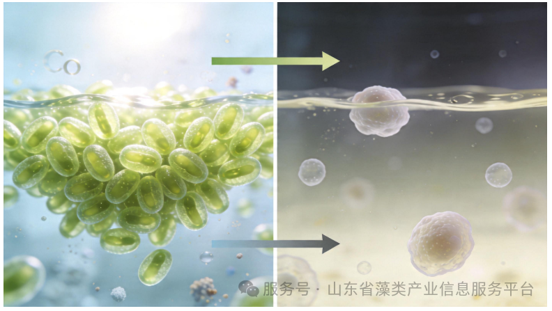
智药局消息,2026年4月透云生物旗下山西透云与深圳大学共建联合研究所,投500万启动衣藻格鲁肽产业化,这款藻产降糖药成本仅为传统工艺1/5,还能做成口服制剂。当你为注射降糖药的繁琐皱眉时,深圳大学的实验室里,一种直径只有10微米的单细胞绿藻,正在悄悄合成和司美格鲁肽作用相似的降糖蛋白。2026年4月,这种名为莱茵衣藻的微藻,从实验室的培养皿跳进了产业化流水线——山西透云生物与深圳大学共建联合研究所,砸下500万元启动资金,要把这种“藻产降糖药”变成货架上的商品。你或许会问:为什么放着成熟的大肠杆菌、哺乳动物细胞不用,偏偏要选一种微藻?这背后藏着生物制药行业一场静悄悄的成本革命。微藻工厂:比细胞培养省一半成本你可以把莱茵衣藻想象成一个自带太阳能板的微型工厂——它靠光合作用吃饭,只要有光、CO₂和简单的无机盐,就能24小时不停繁殖,48小时就能翻倍。而传统的哺乳动物细胞培养,不仅要昂贵的血清培养基,还要严格控制温度、氧气,像照顾婴儿一样精细。更关键的是,莱茵衣藻是真核生物,能像人体细胞一样给蛋白做“糖基化”“折叠”这类精细加工,让生产出的降糖蛋白和人体自身的蛋白结构几乎一致,降低过敏风险。深圳大学的团队花了27年打磨这个工厂:从1999年开始研究莱茵衣藻,发表了400多篇论文,建起了国内第一个药用微藻工程株库,终于把人源GLP-1受体激动剂的基因“焊”进了藻的基因组里——这就是他们的专利成果“衣藻格鲁肽”。直给数据:莱茵衣藻的培养成本仅为哺乳动物细胞的1/5蛋白表达量可达总可溶性蛋白的2%-3%整个生产过程的碳排放比传统工艺低60%但真实的机制比这个工厂类比更精确:研究人员用CRISPR/Cas9技术把目标基因插入莱茵衣藻的核基因组,再筛选出能稳定遗传、高效表达的工程株。这种藻株在光生物反应器里培养一周,就能收获足够的降糖蛋白,后续的纯化步骤也比细胞培养简单得多。产学研闭环:从专利到病床的最短路径这次合作的核心,是把“实验室成果”和“产业能力”拧成了一股绳。深圳大学出技术、出藻种,负责给企业的技术人员培训;山西透云出资金、出生产线,负责中试放大、临床试验报批和未来的销售。深圳大学不用再为“论文变产品”发愁,企业也不用从零开始搞研发——这种模式,把从专利到病床的时间至少缩短了3年。更聪明的是收益分配:深圳大学不拿一次性的技术转让费,而是拿产品销售额的分成。这意味着学校和企业成了真正的利益共同体,学校会持续跟进技术优化,企业也会更用心做市场。据透露,这种“远期收益”的比例通常在1%-5%,看起来不多,但如果这款口服降糖药能拿下10%的市场份额,就是每年上亿元的收入。不过,这条路也不是一帆风顺。莱茵衣藻的基因编辑效率还不够高,精准敲入一个基因的成功率只有不到10%;大规模培养时,光的均匀分布、CO₂的供应都是难题;而且,微藻生产的药物要通过临床试验,还得证明它和传统注射剂有一样的疗效和安全性。这些都是摆在联合研究所面前的硬骨头。绿色制药:下一个万亿赛道的敲门砖莱茵衣藻的野心,不止于一款降糖药。它就像一个通用的“蛋白生产平台”,只要换个基因,就能生产胰岛素、干扰素、疫苗抗原等各种生物药物。而且,它的细胞壁还能当“天然胶囊”,把蛋白包裹起来,做成口服制剂——这解决了肽类药物容易被胃酸降解的难题,未来或许能让糖尿病人不用再打针,直接吞一颗藻粉胶囊就行。全球微藻市场正在以每年6%的速度增长,预计2032年将达到73亿美元。而生物制药领域的微藻应用,才刚刚起步。美国能源部已经投入1880万美元支持微藻药物研发,欧洲的初创企业也在抢滩这个赛道。中国这次的产学研合作,相当于拿到了绿色制药赛道的一张入场券。但要真正跑赢,还要解决几个关键问题:比如开发更高效的基因递送技术,让编辑效率从10%提升到50%;优化光生物反应器的设计,让大规模培养的成本再降一半;还要建立微藻药物的质量标准,让监管部门和消费者都能放心。当我们谈论生物制药的未来时,我们总在说“精准医疗”“个性化治疗”,但往往忽略了“可及性”——让普通人用得起好药,才是最实在的进步。莱茵衣藻的意义,就在于它用光合作用的能量,把生物药物的成本打了下来。“微藻不是替代,而是重构。”这句话或许能概括这场革命的本质。它不是在旧的生产线上修修补补,而是用一种全新的思路,重新定义生物制药的生产方式。未来的某一天,当你拿起一颗降糖药,或许不会想到,它的前身是一滴池水里的微藻,正随着阳光的节奏,在光生物反应器里轻轻摇晃。信息来源:大圆镜
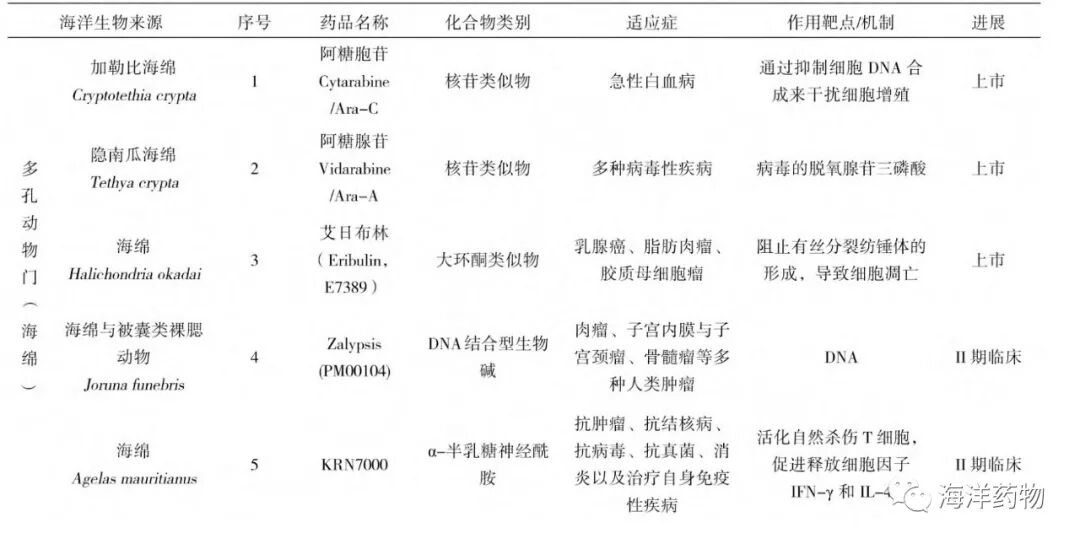
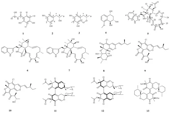
开发“蓝色药库”成为现今世界医药工业发展的重要方向,各制药强国均在不断加大投入,如美国国家研究委员会(National Research Council)和国立癌症研究所(National Cancer Institute)、日本海洋生物技术研究院(Japanese Marine Biotechnology Institute)及日本海洋科学技术中心(Japan Marine Science and Technology Center)、欧共体海洋科学和技术(Marine Sciences and Technology)等机构每年均投入上亿美元作为海洋药物开发研究的经费。目前国内外共有49种来自海洋的活性物质或其衍生物被批准上市或进入临床(见表1)。其中来自海洋多孔动物门(海绵)的活性物质14个(上市3个、II期5个,I期4个,折戟I期临床2个);来自于海洋软体动物门的活性物质10个(上市2个、III期1个、II期4个,I期3个);来自棘皮动物门的活性物质1个(III期);来自海洋尾索动物门的活性物质4个(上市2个、III期1个、折戟II期临床1个);来自节肢动物门的活性物质2个(均处于II期);来自海洋纽形动物门的活性物质1个(折戟II期临床);来自海洋藻类的活性物质7个(上市5个、III期1个,I期1个);来自海洋真菌、放线菌的活性物质4个(上市2个、III期2个);来自海洋脊椎动物(鱼类)的活性物质6个(目前上市3个,III期2个,II期1个)。这些活性物质的治疗范围涉及到众多疾病杂症领域,显示出独特疗效。本文整理了目前已成药或进入不同临床研究阶段的海洋活性物质的来源、特异结构、药理活性、作用机理等,以期能为医药工作者提供新的研究思路,推进海洋药物学科的发展,为人类的健康事业做出贡献。表1 来自海洋生物的药物和处于不同临床阶段的活性物质Table 1 An overview of marine drugs on the market and in clinical trials图1 来源于多孔动物门(海绵)的海洋药物 Fig.1 Marine drugs from Porifera (Marine sponge)图2 来源于多孔动物门(海绵)的海洋药物 Fig.2 Marine drugs from Porifera (Marine sponge)图3 来自软体动物门(海兔)的海洋药物 Fig.3 Marine drugs from Mollusca (Aplysia)图4 来自软体动物门的海洋药物 Fig.4 Marine drugs from Mollusca图5 来自棘皮动物门、节肢动物门和纽虫动物门的海洋药物 Fig.5 Marine drugs from Echinodermata、Arthropod and Nemertea图6 来自尾索动物门(海鞘)的海洋药物 Fig.6 Marine drugs from Urochordata(Pyrosomella verticilliata)图7 来源于藻类的海洋药物 Fig.7 Marine drugs from thallophytes图8 来源于海洋真菌的海洋药物 Fig.8 Marine drugs from marine fungi图9 来源于脊椎动物(鱼类)的海洋药物 Fig.9 Marine drugs from Vertebrate(Fish)来源:海洋药物版权声明:我们分享文章、图片来自网络,版权归原作者及原出处所有。此文出于学习分享、传播更多资讯之目的,无商业用途。若涉及作品内容、版权或其他问题,敬请原作者联系我们(邮箱:xianghaiwenyao@126.com),立即处理。

一株深海链霉菌的沉默基因簇被激活,产量跃升48倍后,新型聚醚PDM现身,并在糖尿病小鼠模型中展现促慢性伤口闭合能力,全球32项海洋药物管线八成押注抗肿瘤,PDM切入创伤修复真空地带,为1300亿产值目标下的“蓝色药库”提供了可复制的基因组挖掘范式。海经社获悉,中国科学院南海海洋研究所张长生研究员团队,联合中国科学院广州生物医药与健康研究院李英杰副研究员团队、中山大学附属第六医院骆衍新主任医师团队,从深海链霉菌中激活一个沉默的聚醚合成基因簇,经代谢工程将目标产物产量抬升48 倍,分离获得具有八氢萘骨架的新型聚醚离子载体 ——聚德卡林霉素(PDM)。糖尿病小鼠模型实验显示,该分子可显著加速慢性伤口闭合,成果已于 2026 年 9 月 9 日在线发表于Nature Communications(DOI: 10.1038/s41467-026-77444-w)。对产业界而言,这条消息的分量不在 “又一个海洋天然产物问世”。 48 倍产量:把 PDM 从 “可检测” 推到 “可放大”PDM 不是从发酵罐里碰运气筛出来的,团队先敲除菌株的主产物合成基因,逼迫其他代谢通路表达,在深海链霉菌 SCSIO 03032 中锁定处于沉默状态的pdm 基因簇;随后过表达正调控基因pdmRI,产量抬升48 倍,纯品才够支撑结构解析与活性评价。这道产量关卡的行业背景是:绝大多数海洋微生物次级代谢产物的基因簇在实验室条件下不表达或表达量极低,研究者长期面对 “知道有好东西、却拿不到手” 的死结。海洋天然产物结构复杂,分离纯化门槛高,一款海洋创新药从发现走到上市动辄十余年。48 倍的跃迁,实质是跨过工程化第一道门槛 —— 分子从“可检测” 进入 “可分离、可评价、可放大”的区间。张长生团队在该方向,此前已累计分离鉴定化合物236 个,其中新化合物 138 个。 切口选择:靶向糖尿病伤口的差异化定位糖尿病慢性伤口的核心病理,是持续炎症与氧化应激互相锁死的恶性循环,线粒体功能障碍让细胞无法完成正常的修复信号转导。PDM 作为聚醚离子载体,先短暂拉高线粒体内钠离子水平,触发活性氧爆发 —— 相当于给停滞的修复程序打一记启动脉冲 —— 随即激活Nrf2 介导的抗氧化通路,让线粒体回到稳态。“先激活、后保护” 的动态策略,与当前主流的抗菌敷料或单一生长因子制剂不在同一条逻辑线上。据 LP Information 数据,全球慢性伤口护理药物市场 2025 年约9.63 亿美元,预计 2032 年增至12.56 亿美元,年复合增速3.9%;更大的盘子在糖尿病足溃疡 —— 据 VPA Research 数据,该市场 2026 年约68 亿美元,预计 2034 年达119.5 亿美元,年复合增速7.3%。中国糖尿病足溃疡年发病率约8.1%,每年新发患者约300 万例。Regranex 等生长因子类产品长期不温不火的行情说明,这个赛道缺的不是又一层敷料,而是能改写愈合动力学的新机制分子。 1300 亿产业目标下的管线真空把 PDM 放进中国海洋生物医药的坐标系来看。2026 年 5 月,自然资源部等八部门印发《关于加快海洋药物和功能制品高质量发展的指导意见》,提出到 2030 年实现多项海洋创新药上市、产业增加值突破 1300 亿元,并明确支持山东以青岛为核心打造“蓝色药库”创新研发聚集区 —— 这是国家层面首部聚焦海洋生物医药的政策文件。中国海洋大学 2026 年 9 月发布的《蓝色药库发展报告(2026)》给出了行业横截面:全球海洋衍生药物市场规模预计从 2024 年的124 亿美元增至 2030 年的209 亿美元,年复合增速约9.1%;全球在研海洋创新药物管线共32 项,近八成扎堆抗肿瘤,青岛自主研发的免疫抗肿瘤药物BG136已完成 II 期临床、进入 III 期。管线高度集中于抗肿瘤,代谢性疾病、慢性创伤修复方向存在结构性空白。PDM 切入糖尿病伤口愈合,填的正是这个真空。更具范式价值的是其完整路径 ——“基因组挖掘 — 沉默基因簇激活 — 产量提升 — 体内活性验证”证明,深海微生物中大量未开发基因簇可以借合成生物学手段转化为可操作的药物先导物,不必再依赖大规模发酵加活性筛选的碰运气模式。 叩门之后:成药性考验在前从叩响到推开产业化之门,距离仍不短。当前研究止步于糖尿病小鼠模型,系统的药代动力学、毒理学与临床前安全性评价均待完成。聚醚离子载体这类分子有特有的成药性难题 ——细胞毒性窗口管理难度高,莫能菌素等同类化合物在兽药领域的应用经验提示,治疗窗口的界定将是临床开发的关键节点。往期推荐厦门《“十五五”海洋经济发展专项规划》的路线图:百亿生物医药与零碳航运双赛道起跑妆食同源到创新药:南极磷虾叩门“蓝色药库”,制度适配成关键变量成立一年资产奔300亿:青岛海洋集团的“三条鱼”与“蓝色药库”版图
Refers to Ali Bidaki, …, Qun Ren International Journal of Biological Macromolecules, Volume 311, Part 4, June 2025, Pages 143743 View PDF The authors regret that inadvertent errors were found in Fig. 2C and Fig. 5 A of the published article. The authors regret that an inadvertent error occurred during the analysis and preparation of the XRD spectra presented in Fig. 2C. Specifically, the spectra corresponding to samples e (Nio@CS-AL) and f (Nio-Bar@CS-AL) were incorrectly processed during the initial data analysis stage. The original data were subsequently re-examined, and the corrected XRD spectra are provided below. In addition, during the final assembly of Fig. 5 A, an incorrect histological image was inadvertently inserted for the “CS-AL, Short Time” sample, resulting in the duplication of another panel within the figure. The figure has now been corrected using the original histological image corresponding to the affected sample. The corrected Fig. 5 A is presented below. These errors resulted from an oversight during data processing and figure preparation. The authors confirm that these corrections do not affect the results, discussion, or conclusions of the study and have given their consent for the publication of this corrigendum. Download: Download high-res image (294KB) Download: Download full-size image Corrected Fig. 2C XRD analysis of a) Nio, b) Nio-Bar, c) Bar, d) CS-AL, e) Nio@CS-AL, and f) Nio-Bar@CS-AL). The spectra correctly depict the characteristic peaks for all components, with the Bar peaks marked with an asterisk (*) and the CS-AL peaks marked with circles (●). The corrected figure replaces the originally published version. Download: Download high-res image (1MB) Download: Download full-size image Corrected Fig. 5 A Microscopic analysis of the tissues after 7 (short time) and 28 (long time) days of treatment with bare scaffold CS-AL and loaded scaffold Nio-Bar@CS-AL (magnification: 40×). The corrected image (CS-AL, short Time) replaces the erroneously duplicated images in the original publication. The authors would like to apologies for any inconvenience caused.
Although hydrogel-based drug delivery shows promise for localized cancer therapy, effective combination strategies remain needed. Herein, a dual-drug …
Abstract Chitosan (CS) has emerged as a promising alternative against antimicrobial resistance due to its broad-spectrum antimicrobial properties. CS's antimicrobial activity is supposed to be influenced by its molecular weight, degree of deacetylation, chemical functionalization, and biological origin. Despite extensive research, the mechanisms underlying its bioactivity remain poorly understood, with hypotheses including membrane disruption, ion chelation, and intracellular interference. In this study, we systematically evaluated the antimicrobial efficacy and mechanisms of action of various CS samples having different molecular weight (from 2 to 300 kDa), different functionalization (carboxyl and lactate moieties) or different origin (from crustacean or fungal source). Through various chemical and microbiological assays, we identify the most effective structural features leading to the highest performance and its mechanism of action, corresponding to lactate-functionalised CS. Similarly, 7 kDa CS displays the best activity among pristine CS with different MWs. This systematic analysis offers valuable insights for the rational design of more efficient, sustainable CS-based antimicrobial therapies, contributing to developing alternative strategies against microbial infections. Keywords Chitosan ; Antimicrobial ; C. elegans ; Chitosan oligosaccharide lactate 1. Introduction Antimicrobial resistance represents a pressing global health challenge, accounting for approximately 5 million deaths annually worldwide [1]. The reduced development of new antimicrobials in the pipeline and the increase in the number of resistant species have only aggravated this scenario [1], [2], [3]. The discovery and production of effective antimicrobials is a process that demands substantial time and resources and often entails significant environmental implications and reduce long-term effectivity due to the rapid acquisition of antimicrobial resistant of these novel compounds [4]. Chitosan (CS), a chitin derivative composed of β-(1 → 4)-linked D-glucosamine units with varying degrees of N-acetylation [5], has demonstrated promising antimicrobial properties [6]. CS is a significant by-product recovered mainly from the seafood industry. Thereby, its repurpose contributes to a sustainable and circular economy [7], [8], [9]. Structurally, CS is a cationic polymer (in physiological pH range), whose physical properties heavily depend on its degree of deacetylation (DD), molecular weight (MW), and chemical functionalization [10], [11]. In this regard, CS with relatively large and broad MW distributions (MW > 100 kDa), typically characterized by high dispersity indices, together with a wide range of degrees of deacetylation (DD, ca. 75 to 90%). Moreover, very low-MW CS molecules (MW < 5 kDa) have been studied in the literature, most often produced by controlled depolymerization of high-MW precursors using chemical (acid, basic, or enzyme-catalysed hydrolysis) or physical (microwave assisted or gamma-radiation) methods. [12], [13], [14] However, less attention has been devoted to the synthesis and commercialization of intermediate-MW CS (MW = 5–10 kDa) due to the need of high controlled processed. Moreover, beyond average MW values, the control and reporting of MW dispersity (polydispersity index, Đ) has received limited consideration across both high- and low-MW CS systems, despite its critical influence on physicochemical properties, solubility, bioactivity, and reproducibility. Consequently, there remains a clear need for CS materials with well-defined MW, narrow dispersity, and precisely controlled DD, particularly in the intermediate-MW regime. The ability of CS to inhibit bacterial, fungal, and yeast growth has been previously reported revealing promising result for its implementation in biomedical applications [6], [15], [16]. Nevertheless, the mechanism of action of these biopolymers remains poorly elucidated. On the one hand, the main hypothesis involves the electrostatic interaction between the positively charged amino groups of the biopolymer with the negatively charged microbial membrane, as it happened with other positively charged macromolecules, as AMPs [15], [17]. This interaction would lead to membrane fluidization, intracellular content leaking, and hence, cell death. Pore formation by positively charged CS in microbial membranes has also been proposed as a potential mechanism of action [18], [19]. On the other hand, the ability of CS to scavenge divalent cations present in the bacterial membrane has been reported, as well as the effect on membrane destabilization in the osmotic balance of microbial cells [20]. Noteworthy, other mechanisms of action, such as enzyme deactivation, induction of metabolic changes, DNA transcription hindering, or efflux pump inhibition, have also been postulated [21], [22], [23]. Taken together, this mechanistic diversity, combined with the strong dependence of CS activity on structural parameters, provides a plausible explanation for the variability and apparent inconsistencies reported across different studies [24], [25]. Several factors influence the antimicrobial efficacy of CS, with MW and surface charge at physiological pH conditions being among the most significant, as they govern both CS solubility and its interactions with microbial cell membranes [26]. Low MW-CS have been hypothesized to penetrate microbial membranes more efficiently, enabling interactions with intracellular targets and potentially disrupting nutrient uptake as well as RNA and DNA replication and translation processes [27]. However, while some studies report enhanced activity of low MW-CS against Gram-positive bacteria, others observe comparable antimicrobial effects for higher-MW CS [26]. These apparently contradictory findings highlight the need for a more systematic evaluation of how MW modulate the antimicrobial activity of CS. The amine and hydroxyl groups of the glucosamine repeating unit in CS can be chemically modified or exploited to anchor functional moieties that provide additional properties while simultaneously affecting CS solubility [28]. Such chemical functionalization represents a powerful strategy to enhance antimicrobial activity by increasing interactions with microbial cells and improving affinity toward target structures [29]. For instance, functionalization with quaternary ammonium groups has been shown to improve the antimicrobial performance compared with the pristine CS [30], [31]. However, the introduction of cationic-groups is often associated with reduced biocompatibility, which can limit their practical applicability [32]. Other functionalization, such as N-lactate, have excelled in wound healing and haemostasis-related applications [33], [34], [35], and promising antimicrobial properties have been reported [36]. Yet, their antimicrobial activity has not been systematically investigated. In addition to deliberate chemical modification, structural variability in CS can also arise from its biological origin. CS can be obtained from various sources, including crustacean shells (shrimp, crab), fungal cell walls, and insect exoskeletons [5], [8]. While animal-derived CS typically exhibits a linear polymeric structure, fungal-derived CS often displays a branched architecture, in which β(1 → 4)-linked D-glucosamine backbones are substituted with β(1 → 6) linked glucan side chains at the C6 position [37]. According to the above discussion, the antimicrobial activity of CS is likely governed by the interplay of multiple structural and physicochemical factors rather than by a single parameter such as MW. Notably, the lack of a systematic and comparative studies in this field largely stems from the difficulty of evaluating CS samples that differ in MW, chemical functionalization, or biological origin sharing a common structural background. On this basis, this work investigates the antimicrobial activity and mechanism of action of a well-defined library of CS materials differing in MW, chemical functionalization, and origin. In particular, low-MW CS samples (CS7 and CS1) were produced by controlled depolymerization of the same high-MW precursor (CS300), thereby maintaining a constant DD. In addition, commercially available low-MW CS (CS1-c) and chemically modified derivatives, including lactate-functionalized (CSlac) and carboxymethylated CS (CScm), were examined. The inclusion of CSlac was motivated by the reported antimicrobial potential of lactate-containing compounds, [36] whereas the negatively charged groups in CScm were expected to attenuate antimicrobial activity and thus provide insight into the role of chemical functionalization. The antimicrobial performance of all CS samples was systematically evaluated, and the biocompatibility of the most active material was further assessed using Caenorhabditis elegans as a novel alternative method model following the EU 3R directives (Directive 2010/63/EU). Overall, this study advances the understanding of the structure-activity relationships governing CS antimicrobial behaviour and provides a rational framework for the design of more effective CS-based antimicrobial materials, contributing to the development of sustainable alternatives to combat antimicrobial resistance. 2. Materials and methods 2.1. Materials and reagents Commercial CSs were acquired form different sources; high MW CSs (CS300 and CS50) and, lactate functionalized CS (CSlac) were purchased form Sigma-Aldrich (St Louis, MO, USA), carboxymethyl functionalized CS (CScm) from Heppe Medical Chitosan (HMC+, Halle, Germany), low MW (CS1-c) from Qingdao Hehai Biotech Co. Ltd. (Qingdao, China). Information about their MW distribution and DD can be found in the main text (Table 1). Sodium chloride (NaCl), sodium hydroxide (NaOH), hydrogen peroxide (H2O2) (30%, v/v), Phosphate Buffered Saline (PBS) solution (0.1 M, pH 7.4), ethylendiaminetetraacetic acid disodium salt dihydrate (Na2EDTA·H2O), sodium acetate trihydrate and Propidium Iodide (PI) were purchased from Merck Sigma-Aldrich (St Louis, MO, USA). LIVE/DEAD BlackLight Bacterial viability kit and agarose were purchased from Invitrogen (Thermo Fisher, Waltham, Massachusetts, USA Bacterial DNA extraction was performed with a GeneJEt DNA purification kit (Thermo Fisher, Waltham, Massachusetts, USA). The GeneRuler™ 1 kb DNA ladder was purchased from Thermo Fisher Scientific (Waltham, Massachusetts, USA). European bacteriological agar, triptone, pentose, glucose and yeast extract were purchased from Condalab (Torrejón de Ardoz, Spain). Ethanol (EtOH, 96% v/v for clinical diagnosis), acetone (technical grade), and dimethyl formamide (DMF) were purchased from PanReac ApplicChem (ITW Reagents, Castellar del Vallès, Spain). Acetic Acid (Glacial 99.8% AGR ACS ISO Reag. Ph. Eur.) was supplied by Labkem (Premia de Dalt, Spain). All aqueous solutions were prepared with Milli-Q water (Milli-Q IQ 7000 Ultrapure Water Purification System, Millipore Burlington, Massachusetts, USA). All chemicals were used as received without further purification. Table 1. Summary of the different structural parameters (MW, dispersity Đ, deacetylation degree DD and functionalization) obtained from NMR and electrophoretic mobility for all the CS materials. Empty Cell MW (kDa) Đ DD (%) ζ-potential (pH 7) (mV) Functionalization CS300a, c 100-300b – >80b +3 ± 2 CS50c 30-50b – 80b +18 ± 2 CS7 7.5 ± 0.6 2.2 ± 0.3 75.8 +45 ± 5 CS1 1.7 ± 0.3 2.0 ± 0.2 76.6 +35 ± 1 CS1-cc 2.0 ± 0.2 1.7 ± 0.3 88.2 +7 ± 2 CSlacc 1075 ± 25 2.7 ± 0.2 62.1 +23 ± 3 N-lactate CScmc 790 ± 20 1.4 ± 0.1 23.5 −20 ± 4 N,O-carboxymethyl a Not measurable due to lack of solubility in aqueous media. b Information provided by the supplier. c Commercially acquired samples. 2.2. Experimental 2.2.1. Synthesis of CS7 and CS1 Low MW CSs (CS7 and CS1) were synthesized from CS300 via a microwave-assisted oxidative hydrolysis process with some modifications (See Supplementary Data). 2.2.2. NMR determination of molecular weight and dispersity NMR spectra were measured in a Bruker Avance III 500 spectrometer equipped with an inverse-detection TBI (1H/13P/BB) probe head. NMR samples of CS7, CS1, CS1-c, CSlac, and CScm were prepared by weighting 1 mg in 0.5 mL of D2O in oven-dried 5 mm NMR tubes. CS300 could not be analyzed by NMR due to its insolubility in deuterated water. CS50 was commercially acquired and used as received without further purification. Diffusion coefficients were determined via pulse-field gradient spin-echo (PGSE) diffusion NMR experiments that were conducted using the stimulated echo sequence within bipolar pair pulses [38]. Gradient pulses were applied using a smoothed rectangular shape (SMSQ) and were automatically adjusted in strength (G) throughout the experiments. The diffusion coefficients (D values) were extracted from the slope of the regression line plotting ln(I/I0) against G2. This regression approach linearly rises the inverse Laplace transformation based on the Stejskal-Tanner equation for SMSQ gradient pulses [39]. To check reproducibility and lack of convection, three different measurements with different diffusion times (Δ) were always carried out. The contribution of convection to the calculated D values seems to be negligible since it remains always constant under the three different Δ assayed. The conversion into MW values was achieved using previously established universal calibration approaches, which are independent of concentration and solvent viscosity [40], [41]. Molecular weight distributions and dispersity (Đ) were calculated as previously reported [42]. The degree of acetylation (DA) of all the CS samples was determined from their respective 1H NMR spectra recorded using a water-presaturation pulse sequence (noesygppr1d), and was based on the relative integrals of the N-acetyl methyl protons and the H2 proton (see Fig. 1) of glucosamine units, following established procedures reported in the literature [43]. The degree of deacetylation (DD) was calculated as DD (%) = 100 − DA (%). Download: Download high-res image (353KB) Download: Download full-size image Fig. 1. (A) Chemical structures of the investigated CS derivatives. Hydrogens atoms have been omitted for clarity. (B) Stacked 1H NMR spectra (D2O, 500 MHz) of the studied polymers: CS7 (magenta), CS1cm (teal), CS1 (blue), CS1lac (red), and CS1-c (black). (C) ζ-potential measurements of the studied polymers at different pH values (n = 3). Experimental data were fitted using a Boltzmann sigmoidal function (R2 > 0.95). (For interpretation of the references to colour in this figure legend, the reader is referred to the web version of this article.) 2.2.3. Protonation constant (pka) estimation by ζ-potential measurement The pka of the different CSs was determined by measuring the ζ-potential of aqueous solutions (1 mg/mL) at 25 °C using a Zetasizer ZS instrument (Malvern Panalytical, Malvern, United Kingdom) equipped with a HNe 633 nm laser and with a backscattering of 173 0 controlled by with a Malvern DTS 7.03 software at a constant 2.5 mS/cm conductivity. ζ-potential was calculated based on Henry's equation with a Smoluchowski approximation for Henry function. Obtained data were fitted following a Boltzmann function with a Pearson correlation coefficient (R2) > 0.95. The pka value was estimated as the minimum of the derivative of the fitting. Data were analyzed using OriginPro v.2024b (OriginLab Corporation, Northampton, MA, USA) software. 2.2.4. Fourier transformed infrared (ATR-FTIR) spectroscopy ATR-FTIR spectroscopic analysis was performed in a FT/IR 4700 (JASCO Co., Tokio, Japan) instrument. All CS samples were dried overnight at 60 °C before the measurement and pure samples were used for the measurements. 2.2.5. Thermogravimetric analysis (TGA) – differential scanning calorimetry (DSC) TGA-DSC analysis was performed in a NETZSCH -STA 449 F1 Jupiter instrument (Selb, Germany) from 30 °C to 800 °C at 5 °C/min under N2/O2 atmosphere. Samples were dried overnight at 60 °C prior analysis. 2.2.6. Minimal inhibitory concentration (MIC) determination MIC values for Staphylococcus (S.) aureus (ATCC 12600), Escherichia (E.) coli (K12), Pseudomonas (P.) aeruginosa (PA01), Salmonella (S.) tiphymurium (ATCC 14028) and Candida (C.) albicans (ATCC 10231) were obtained via the standard broth-dilution method in 96-well microlitre plates. Lysogeny Broth (LB) and Sabouraud media were used for bacterial cells and fungal cells, respectively. The MIC of each compound was evaluated using a 2-fold decreasing concentration of each sample between 60 and 0.5 mg/mL against a bacterial/fungal inoculum with an optical density (OD600) of 0.005. Note that depending on the microbial species, this OD600 corresponded to experimentally determined concentrations ranging from 107 to 109 colony forming units per mL (CFU/mL). Plates were incubated at 37 °C for 18 h. MIC values were determined as the lowest concentration where no turbidity was observed [44]. Simultaneously, the optical density at 600 nm (OD600) was measured with a SpectraMax iD3 (Molecular Devices, San José, CA, USA) plate reader. MIC reported values are based on three biological replicates. 2.2.7. Cell membrane ζ-potential measurements Surface charge of S. aureus (ATCC 12600), E. coli (K12), P. aeruginosa (PA01), S. tiphymurium (ATCC 14028) and C. albicans (ATCC 10231) in absence or presence of biopolymers was measured with a Zetasizer (ZS, Malvern Panalytical Ltd., Malvern, UKm) equipped with a HNe 633 nm laser and with a back-scattering of 173 0 using PBS (0.1 M, pH 7.4) as dispersant at 25 °C. Overnight cell inoculum of 107–109 CFU/mL in PBS were incubated with 1 mg/mL of each CS sample in PBS (0.1 M, pH 7.4) for 4 h at 37 °C under mild shaking. Then, samples were centrifuged (5000 rpm, 5 min), washed with fresh PBS and the ζ-potential measured. Depicted values (average ± SD) are based on three biological replicates. 2.2.8. Dye accumulation assays The integrity of the cell membrane in the presence of the CS's was evaluated following a procedure previously described by H. E. Smith & J. M. A. Blair [45]. Briefly, overnight inoculum of S. aureus (ATCC 12600), E. coli (K12), P. aeruginosa (PA01), S. tiphymurium (ATCC 14028) and C. albicans (ATCC 10231) were incubated for 4 h under mild shaking at 37 °C in PBS (0.1, pH 7.4, OD600 = 0.2). The buffer solution contained Propidium Iodide (PI, 5 mg/L for bacterial and fungal cultures) and the CS of choice at a final concentration 1 mg/mL. Then, the samples were centrifuged (5000 rpm, 5 min), washed with fresh PBS and finally suspended in PBS containing 5% glucose (w/v). 200 μL of each culture was transferred to a 96 well black microlitre tray (Corning, Amsterdam, the Netherlands) and fluorescence was measured (λexc = 575 nm, λem = 635 nm) with a SpectraMax iD3 plate reader (Molecular Devices, San José, CA, USA). Results (average ± SD) are based on three biological replicates. A PBS solution of Na2DTPA·2H2O (10%, w/v) was employed as positive control. 2.2.9. Fluorescence microscopy Bacterial viability experiments were performed using E. coli (K12) bacterial reference species and C. albicans (ATCC 10231). Overnight inoculate of microorganisms were washed and diluted in PBS buffer (0.1 M, pH 7.4) to a final concentration of 107–109 CFU/mL, and incubated with the different CS molecules at a concentration of 1 mg/mL for 4 h under magnetic stirring (150 rpm) at 37 °C. Then, cultures were centrifuged (6500 rpm, 3 min), washed with fresh PBS, and stained using the LIVE/DEAD BackLight bacterial viability test kit. Fluorescence imaging was performed in an Olympus IX73-DP75 (Olympus, Tokyo, Japan) fluorescence microscope. Image analysis was performed with OriginPro v.2024b (OriginLab Corporation, Northampton, MA, USA) software. Depicted results (average ± SD) are based on three technical replicas from three different biological replicates (N = 3, n = 3). 2.2.10. Gel electrophoresis E. coli (K12) DNA was extracted and purified using a GeneJET DNA purification kit (Thermo Fisher Scientific Inc., Waltham, USA) following the protocols established by the kit manufacturer. Gel electrophoresis was performed on a 1% agarose-TBE gel using 100 V for 90 min in 1xTBE buffer. [DNA] = 4.5 ng/mL. 2.2.11. Circular dichroism (CD) experiments CD measurements were performed in a J-815 CD Spectrometer (JASCO Co., Tokio, Japan) using Milli-Q water as a dispersant. Extracted E. coli DNA (40 ng/μL) was incubated for 10 min at room temperature with the desired CS in a 1:1 mass ratio. Depicted data were obtained after five accumulations. 2.2.12. Toxicity evaluation in C. elegans These experiments were performed with the N2 wild-type strain of Caenorhabditis (C.) elegans provided by the Caenorhabditis Genetics Centre (CGC, University of Minnesota). C. elegans survival, growth in and the reprotoxicity in the presence of CS7 or CSlac were studied following a previously reported methodology [46]. Early developmental-stage nematodes (L1) were exposed to CS7 or CSlac solutions for 72 h at 20 °C. The concentration ranges of CS7 or CSlac were determined from MIC values (0–15 mg/mL and 0–5 mg/mL, respectively). Experiments were performed in the presence or the absence (for survival studies) of heat-inactivated E. coli OP50 (OD600 = 0.1) in S-medium. Depicted results (average ± SD) are based on three biological replicates (15 ± 5 worms/replica) and the statistical significance was determined by 1-way ANOVA. 2.2.13. Statistical analysis Data are presented as mean ± standard deviation (SD) of at least three independent biological replicates. Statistical significance was determined by one-way or two-way ANOVA followed by a Tukey's HSD post-hoc test. ns p > 0.05; *: 0.01 < p ≤ 0.05; **: 0.001 < p ≤ 0.01; ***: p ≤ 0.001. Detailed p-values for comparisons across conditions are summarized in Tables S2 and S4. 3. Results and discussion 3.1. Chemical characterization of chitosan Parameters such as MW, DD, and chemical functionalization are supposed to play a crucial role in the antimicrobial activity of CS. To cover all these features, different CS molecules having different MW, dispersities, chemical functionalization and origin were studied (Fig. 1.A). Nuclear Magnetic Resonance (NMR), and diffusion NMR spectroscopy were employed to determine the chemical composition, molecular weight (MW) distribution and dispersity (Đ) of the different CS samples. Fig.S2 show the monomodal MW distribution of each type of CS assayed herein. First, a set of CS molecules with different MWs (CS1, CS7, CS50 and CS300) but showing the same DD were explored. Note that CS1 and CS7 were produced from CS300 by a microwave-assisted hydrolysis method with some modifications. [13] CS50 was commercially acquired and used as received without further purification. Then, and in order to further investigate the effect of the biopolymer origin on its antimicrobial activity, these samples were directly compared to a commercially available CS obtained from fungal origin (CS1-c) and CS molecules bearing different functional groups such as lactate (CSlac) and carboxymethyl (CScm). All CS samples present a narrow Đ besides the CScm (Table 1). 3.1.1. Structural characterization of CS CS7, and CS1 exhibited highly similar 1H NMR spectra, characterized by diagnostic resonances at δH 1.93, 2.75, and 4.42 ppm, which were assigned to the CH3 group of the N-acetyl-D-glucosamine unit, the H-2 proton of the D-glucosamine unit, and the anomeric H-1 protons of both units, respectively (Fig. 1B). As expected, all these CS samples displayed identical degrees of deacetylation (DD), as summarized in Table 1. These results confirm that the production of CS7 and CS1 from CS300 via microwave-assisted oxidative hydrolysis does not alter the chemical structure of the polymer backbone nor affect the DD of the resulting materials. MW of 7.5 ± 0.6 kDa, and 2.0 ± 0.2 kDa were determined for CS7 and CS1, respectively by NMR analysis. Furthermore, successful chemical functionalization of commercial CSlac and CScm was confirmed by 1H NMR spectroscopy (Fig. 1B). In the case of CSlac, new resonances appearing at δH 1.30 and 4.05 ppm were observed and assigned to the terminal CH3 and CH protons of the lactate moiety, respectively. The 1H NMR spectrum of CScm displayed an additional signal at ca. δH 3.8 ppm, which was attributed to the CH2 protons of the carboxymethyl group. Interestingly, an averaged MW of 1075 ± 25 kDa was determined by NMR analysis for CSlac, with only small fraction of the sample having the reported MW by the supplier. This discrepancy can be due to the different analytical techniques employed in the CSlac production and characterization, inherit calibration error, or batch-to-batch discrepancies. Complementary SEC-MALS analysis only showed a population with average MW of 40 kDa, also detected by DOSY (Fig. S1, Fig. S2 and Table S1). The guard columns and on-line filters prior the SEC column might trap hight MW CSlac, and, in that way, not send it in the chromatograph. Based on these results, we decided to used 1075 kDa as the average MW for CSlac. Determined MW for CScm (790 ± 20 kDa) is in good agreement with the commercially determined one. The presence of lactate and carboxymethyl functionalities in CSlac and CScm was further corroborated by 13C NMR spectroscopy (Fig. S3). The 13C NMR spectrum of CSlac showed additional resonances at δC 20–22 and 68.1 ppm, which were assigned to the methyl and methane carbons of the lactate moiety, respectively. A characteristic signal in the carbonyl region at δC 181.7 ppm corresponding to the lactate carboxylic carbon was also observed. In the case of CScm, the 13C NMR spectrum displayed an additional resonance at δC 69.5 ppm, attributed to the methylene carbon of the carboxymethyl substituent, along with a signal at δC 179.9 ppm associated with the carboxylic carbon. This confirms the successful carboxymethylation of the CS backbone. Finally, structural differences between the synthesized CS1 and the commercial CS1-c sample (fungal origin) were investigated by combined 1H and 13C NMR analysis in solution (Fig. S3). In the 1H NMR spectrum of CS1-c, an additional resonance was detected at δH 5.36 ppm, assigned to the H-1 of the glucan residue, which was not present in CS1. Another characteristic resonance at δH 3.28 was assigned to the H-2 proton of the same residue; typical of fungal-derived CS. [37] The difference in the integral intensity between H-2 from the glucan residue (δH 3.28) and H-2 from the glucosamine (δH 2.90 ppm) in the 1H NMR spectrum is 1:15. Establishing a degree of branching of approximately 6.7% (i.e., 1 glucan branch per 15 backbone units). Consistently, 13C NMR spectroscopy revealed extra signals in CS1-c beyond those corresponding to the CS backbone, notably resonances at δC 98.1 ppm (anomeric C1), 69–77 ppm (ring carbons C2–C5), and 60–62 ppm (C6), which appear in the same region as those of CS1. These features are in agreement with the reported branched architecture of fungal CS, consisting of a β-(1 → 4)-linked D-glucosamine backbone decorated with β-(1 → 6)-linked glucan chains/oligomers. [37]. The functionalization of CSlac and CScm was further confirmed by FT-IR spectroscopy (Fig. S4). Peaks corresponding to CO (1638 cm−1), CO (1363 cm−1), and CN (1105 cm−1) stretching bonds of the lactate moieties were identified in CSlac, [47] while vibrations of the terminal acid group (1591 cm−1 and 1410 cm−1) were found for CScm. The chemical differences between CS1 and CS1-c were also investigated by FT-IR (Fig. S4). The observed broadening of the vibration band centred at 3310 cm−1 in CS1-c can be associated with the higher density of hydroxyl groups from the glucan branching [37]. Similarly, the FT-IR spectrum of CS1-c presents a stronger vibration at 1510 cm−1 compared with CS1, which is associated with OH deformation vibration from the hydroxyl groups of the glucan branches. Thermogravimetric analysis coupled with differential scanning calorimetry (TGA-DSC) revealed a multi-step degradation process for both samples but with remarkable differences. On the one hand, CS1 exhibits a sharp mass loss around 648 °C, preceded by a gradual monotonic degradation. On the other hand, CS1-c shows earlier degradation events at approximately 375 °C and 610 °C (Fig. S5). The higher thermal stability of CS1 could be attributed to its greater packing efficiency of lineal polymers, in contrast to the lower packing density of the branched CS1-c, in line with reported literature [48]. 3.1.2. Physicochemical characterization of CS Finally, the ζ-potential of all the CS samples in aqueous solution was evaluated as a function of pH at a fixed conductivity of 2.5 mS/cm (Fig. 1C). The corresponding isoelectric points or apparent pKa values (Table 1) were estimated from the ζ-potential curves, acquired as described in the experimental section and Supplementary Data (Fig. S6). These parameters are particularly relevant, as the net charge of CS is expected to play a central role in its interaction with microbial membranes and, consequently, in its antimicrobial performance. A clear correlation between the MW and apparent pKa is observed for the unfunctionalized CS samples. Specifically, the pKa decreases with increasing MW, from 8.6 for CS1 to 6.4 for CS300. This behaviour can be rationalized by considering the increasing relative contribution of terminal sugar units in low-MW CS. Shorter polymer chains present a higher fraction of terminal units, which possess greater conformational freedom, and enhanced solvent accessibility compared to units embedded within long polymer chains. As a result, these terminal amino groups are more readily protonated as they experience reduced intramolecular electrostatic repulsion compared with amino groups embedded within long polymer chains. In contrast, in high-MW CSs, the higher local charge density and reduced accessibility of internal amino groups favour earlier deprotonation, leading to lower apparent pKa values. A markedly lower apparent pKa was observed for the fungal-derived CS1-c compared with CS1 of similar MW and DD. This difference can be attributed to the distinct macromolecular architecture of fungal CS, which contains β-(1 → 6)-linked glucan side chains attached to the CS backbone (see discussion above). The presence of these branched glucan units increases structural complexity and alters the intramolecular hydrogen-bonding network and polymer–polymer interactions in solution. As a consequence, the amino groups of the CS backbone in CS1-c experience a modified local microenvironment and reduced effective contribution to the electrokinetic potential compared with the linear CS1, which disfavours protonation from an electrokinetic standpoint. In addition, the more heterogeneous and branched polyelectrolyte architecture of CS1-c enhances collective electrostatic and interfacial effects, stabilizing the deprotonated state and resulting in a lower apparent pKa as observed from ζ-potential measurements. Accordingly, distinct antimicrobial behaviour can be anticipated for these two samples, despite their comparable MW and DD. ζ-potential measurements as a function of pH provide not only an estimation of the apparent pKa or isoelectric point, but also valuable information on the colloidal stability of the different CS samples in aqueous solution (Fig. 1C). In general, high absolute ζ-potential values are associated with enhanced electrostatic repulsion between particles and, therefore, greater dispersion stability, whereas values close to zero indicate a higher tendency toward aggregation. In contrast to the unfunctionalized CS samples, which exhibit a clear transition from positive to negative ζ-potential values and reach an isoelectric point within the investigated pH range, the chemically functionalized samples display markedly different electrokinetic behaviours. In particular, CSlac shows a ζ-potential profile similar in shape to that of unmodified CS, reaching near-neutral values at pH 7, consistent with the presence of both protonable amino groups and lactate functionalities that moderately modulate the net charge. By contrast, CScm exhibits a fundamentally different response, characterized by persistently negative ζ-potential values across the entire near-neutral pH range and the absence of an observable isoelectric point. This behaviour can be attributed to the high density of carboxymethyl groups, which are largely deprotonated above their intrinsic pKa (ca. 4.5) [49], thereby dominating the electrokinetic response and overpassing the contribution of protonated amino groups on the CS backbone. As a result, CScm remains negatively charged and electrostatically stabilized over a broad pH window, indicating enhanced colloidal stability under physiological and near-neutral conditions. 3.2. Antimicrobial activity of chitosan Previous studies have already demonstrated the antimicrobial properties of CS, CS derivatives biopolymers, and CS-based materials [27]. Efforts have also been made to correlate antibacterial performance with specific chemical and structural parameters of CS, such as MW or DD [6], [24]. However, these studies are generally limited to narrow sets of CS materials and often focus on individual parameters in isolation, rather than addressing the combined and interdependent effects of multiple variables, including MW, DD, dispersity, and chemical functionalization. Given the considerable potential of CS-based systems as antimicrobial agents, the present work investigates the antimicrobial properties and mechanisms of action of a comprehensive library of CS samples differing in MW, chemical functionality, and biological origin (animal or fungal). 3.2.1. Antimicrobial activity of linear CS First, the Minimum Inhibitory Concentration (MIC) of all samples against representative bacterial species (S. aureus, E. coli, P. aeruginosa, and S. tiphymurium) and fungus C. albicans was determined. MIC values were determined in Lysorgenic Broth (LB) for bacterial species and Sabouraud media for C. albicans (Table 2, Fig. S7–8). The highest-MW biopolymer, CS300, exhibited moderate to low antibacterial activity, with MIC values of 30 mg/mL against S. aureus and 60 mg/mL or higher against Gram-negative bacteria tested. Against C. albicans, CS300 showed moderate activity (MIC = 15 mg/mL). CS50 displayed a comparable antibacterial profile, although a slight improvement was observed against E. coli (MIC = 30 mg/mL). In contrast, a marked enhancement in activity was observed for lower-MW samples. Specifically, CS7 showed a 4-fold decrease in MIC values against S. aureus (7.5 mg/mL) and C. albicans (2.5 mg/mL), and an 8-fold decrease against E. coli (3.2 mg/mL). CS7 also showed improved activity against P. aeruginosa (MIC = 7.5 mg/mL), whereas its activity against S. typhimurium remained moderate (MIC = 30 mg/mL). Remarkably, CS1 showed even higher MIC values than CS7 across most strains. In contrast, the fungal-derived CS1-c displayed consistently lower activity than CS1 against all tested microorganisms, which can be attributed to its branched macromolecular architecture, in agreement with previous reports [50]. MIC values for C. albicans were also determined in LB media at neutral pH (7.4) (Table S2 and Fig. S8). As expected, most samples exhibited reduced antifungal activity under these conditions compared with assays performed in Sabouraud medium (pH 5.5), reflecting the lower degree of protonation of CS at neutral pH. Overall, these results highlight the strong influence of MW on the antimicrobial performance of non-functionalized CS, which is closely linked to its solubility and self-aggregation behaviour in aqueous solution [14]. In general, an increase in MW correlates with a progressive decrease in antimicrobial activity. Table 2. Minimum inhibitory concentration (MIC) values (mg/mL) for explored CS against representative bacterial (S. aureus, E. coli, P. aeruginosa, S. typhimurium) and fungal (C. albicans) species. MICs were determined by the 2-fold dilution method in LB (bacteria) and Sabouraud medium (fungus) at 37 °C under mild agitation (150 rpm). Data represent the mean of three biological replicates. Empty Cell Bacteria Fungi Gram+ Gram- S. aureus E. coli P. aeruginosa S. tiphymurium C. albicans CS300 30 60 > 60 > 60 15 CS50 30 30 > 60 > 60 15 CS7 7.5 3.2 7.5 30 2.5 CS1 15 15 30 30 7.5 CS1-c 30 30 30 >60 15 CSlac 3.2 0.9 3.2 15 0.2 CScm > 60 > 60 > 60 > 60 30 As discussed above, a peak in antimicrobial performance was observed for CS7, particularly against E. coli, P. aeruginosa, and C. albicans. At first glance, this observation may appear to contradict previous reports suggesting that antimicrobial activity generally decreases with increasing MW [24]. However, this apparent discrepancy highlights the existence of an optimal MW window in which multiple physicochemical factors are favourably balanced. The antimicrobial activity of CS mainly relies on electrostatic interactions between the positively charged amino groups and negatively charged components of the microbial cell envelope [6]. These interactions destabilize membrane integrity, ultimately leading to cell death. Noteworthy, the molecular conformation of CS in solution strongly influences the accessibility and effective exposure of these amino groups [14]. High-MW CS tends to form aggregates due to reduced solubility and increased chain entanglement, which limits amino-group exposure and diminishes antimicrobial efficacy, in line with the poor activity observed for CS300 and CS50 [24]. In contrast, CS7 appears to represent an optimal compromise between chain length and solubility. Its intermediate MW is sufficient to ensure multivalent interactions with microbial membranes, while still preventing extensive aggregation in solution. This combination maximizes the availability of protonated amino groups and enhances membrane interaction, resulting in superior antimicrobial and antifungal activity. Further reduction of MW, as in CS1, likely decreases the cooperative and multivalent character of these interactions, leading to a partial loss of activity. In addition to polymer-related factors, the composition and architecture of the microbial cell envelope play a crucial role in determining susceptibility to CS [26], [51]. Gram-positive bacteria such as S. aureus possess a relatively simple and permeable membrane, while Gram-negative species (E. coli, P. aeruginosa, and S. tiphymurium) feature an additional outer membrane that restricts the penetration of antimicrobial agents and confers intrinsic resistance [3]. Moreover, variations in membrane composition and surface charge among bacterial species and strains directly influence their interaction with positively charged polymers and peptides [17], [52], [53]. These differences likely contribute to the particularly pronounced activity of CS7 against E. coli and P. aeruginosa, as well as against C. albicans, whose cell envelope presents distinct negatively charged components susceptible to electrostatic disruption. As shown in Fig. 1.B, CS7 exhibit a higher positive ζ-potential than CS50 and CS300 at pH 7.4, which corresponds to the bacterial growth medium. This higher surface charge is expected to enhance both colloidal stability in aqueous solution and electrostatic interactions with negatively charged components of the microbial cell envelope, thereby contributing to its superior antimicrobial performance. Interestingly, CS1 displays a ζ-potential comparable to that of CS7 under the same conditions, despite showing higher MIC values. This apparent discrepancy suggests that surface charge alone is not sufficient to fully explain antimicrobial efficacy. This behaviour is consistent with previous reports and theoretical models indicating that low-molecular-weight polymers and oligomers interact with biological membranes through mechanisms distinct from those of longer chains [19]. In particular, smaller molecules may cross or insert into microbial membranes with limited membrane disruption, resulting in reduced membrane fluidization and weaker bactericidal effects. Instead, their activity may rely on interactions with intracellular targets, a mechanism that has been proposed for small cationic species but remains largely unexplored experimentally in the case of low-MW CS. CS1-c displayed a lower ζ-potential at pH 7.4 (Fig. 1.C, Table 1), which is consistent with its higher MIC values compared with linear CS1. In addition, as discussed above, 1H NMR analysis revealed clear compositional differences between fungal-derived CS1-c and the synthesized CS1. In particular, the presence of bulky β-(1 → 6)-linked glucan side chains increases the structural complexity of CS1-c, which can hinder effective biopolymer–cell interactions and reduce antimicrobial activity [37]. In the case of C. albicans, an overall increase in antifungal activity was observed for all unfunctionalized CS samples. The fungal cell wall is mainly composed of chitin, β-glucans, and mannoproteins [54]. The mannoproteins in the outer cell wall layer carry a high negative charge, which facilitates electrostatic interactions with cationic CS macromolecules. Moreover, direct interactions between exogenous CS and chitin present in the fungal cell wall have also been reported [55] The combined contribution of these interactions likely accounts for the enhanced antifungal activity observed for the unfunctionalized CS samples. Differences among individual CSs can therefore be rationalized using the same structure–charge–conformation arguments discussed for their antibacterial activity. 3.2.2. Antimicrobial activity of functionalized CS As summarized in Table 2, CSlac displays the lowest MIC values against all the microbial species explored. Besides its high MW (Table 1), CSlac displays a markedly enhanced antimicrobial performance respect to the rest of CSs. Specifically, a twofold decrease in MIC was observed for P. aeruginosa (MIC = 7.5 mg/mL) and S. typhimurium (MIC = 15 mg/mL), a fourfold decrease for S. aureus (MIC = 3.2 mg/mL), and a sixteen-fold decrease for E. coli (MIC = 0.9 mg/mL) was observed respect to CS7 (the second explored CS with highest activity). Most notably, the MIC value against C. albicans decreased by 64-fold (MIC = 0.2 mg/mL) compared with CS7. Based on previous reports demonstrating the ability of lactate-containing compounds to sequester membrane-associated divalent cations, [18] we hypothesize that CSlac may exert its antimicrobial activity, at least in part, through the scavenging of Ca2+ and Mg2+ ions from the bacterial membrane. Furthermore, this mechanistic is consistent with the pronounced improvement observed for CSlac against S. aureus, E. coli, and C. albicans. In contrast, CSlac showed only a modest enhancement in activity against P. aeruginosa compared with CS7. This behaviour can be explained by the well-known adaptive features of P. aeruginosa. In particular, P. aeruginosa secretes large amounts of lipopolysaccharides into the extracellular medium, which can interact with antibacterial polymers and prevent their direct contact with the bacterial outer membrane [56]. Moreover, several P. aeruginosa strains, including the PAO1 strain used in this study, harbour mutations that promote aminoarabinosylation of the lipid A region of the outer membrane's lipopolysaccharides. [57] This modification, driven by the arn operon, results in the covalent modification with a 4-amino-4-deoxy-L-arabinose molecule to the lipid A region's phosphate groups, protecting membrane divalent cations. Such adaptations, also associated with colistin resistance, diminish cation chelation by exogenous agents and could be a plausible explanation for the limited improvement in CSlac activity against P. aeruginosa [58]. Finally, CScm did not exhibit antibacterial activity against any of the bacterial species evaluated up to 60 mg/mL and showed only moderate antifungal activity against C. albicans (MIC = 15 mg/mL). This lack of activity is consistent with its strongly negative ζ-potential at physiological pH values, which prevents effective electrostatic interactions with microbial cell envelopes and underscores the critical role of surface charge in CS-based antimicrobial systems. 3.3. Mechanistic evaluation of chitosan's antimicrobial activity After evaluating the antimicrobial performance of the CS samples, a series of complementary microbiological and imaging studies was conducted to validate the proposed mechanistic interpretations. Specifically, microbial cell-CS interactions were assessed by ζ-potential measurements, while membrane and cell wall integrity were examined by dye accumulation assay and fluorescence microscopy. In addition, circular dichroism (CD) spectroscopy was applied to bacterial DNA to determine whether the different CS samples are able to effectively interact with intracellular macromolecules. 3.3.1. Cell membrane ζ-potential and membrane disruption As already mentioned, the interaction of cationic polymers with negatively charged microbial membranes leads to membrane fluidization, disruption, and intracellular content leak [53]. In order to evaluate whether the antimicrobial activity of CS results from membrane disruption, ζ-potential measurements of bacterial cells, dye accumulation assays, and fluorescence microscopy analyses were employed. First, the electrostatic interactions between the different CS biopolymers and the membrane of the different bacterial and fungal cells was monitored by ζ-potential measurements [59]. Microbial cells (106–107 CFU/mL) were incubated in PBS buffer containing 1 mg/mL of the biopolymer for 4 h at 37 °C. After isolation of microbial cells by centrifugation, the cell surface charge was measured in PBS and compared with untreated samples (Fig. 2A). Noteworthy, a clear correlation was observed between the antimicrobial activity of the CS biopolymers and the change in the surface charge of cells. In particular, the most active samples induced the strongest shift in the cells' surface charge toward neutral or even positive values (i.e., CSlac in E. coli and C. albicans). However, biopolymers with lower or no activity (i.e., CS300, CS50, and CScm) had little to no effect on the surface charge of the cells, suggesting that these compounds do not interact with the cells. Download: Download high-res image (348KB) Download: Download full-size image Fig. 2. (A) ζ-potential measurements of bacterial and fungal cells (106–107 CFU/mL) in PBS buffer (0.1 M, pH 7.4) before and after incubation with biopolymers (1 mg/mL) for 4 h at 37 °C. (B) Normalized propidium Iodide (PI) accumulation assays of bacterial and fungal cells (OD600 = 0.2) incubated in PBS buffer (0.1 M, pH 7.4) before and after incubation with each compound (1 mg/mL) for 4 h at 37 °C. EDTA PBS solution (10% w/v) was used as a positive control. Depicted values (average ± SD) are based on three biological replicas. Data has been normalized to the control experiment. For clarity, only the statistical significance between the control and the different CS conditions have been depicted. The rest of statistical comparisons can be found in Tables S3–5. Beyond electrostatic surface interactions, cationic polymers can induce different degrees of membrane perturbation, including pore formation, membrane fluidization, and structural damage, ultimately leading to the leakage of intracellular components [18]. To directly assess membrane integrity, cell membrane disruption was explored using dye (propidium iodide, PI) accumulation assays as reported previously [45]. Note that PI is only internalized within the cells if their membrane has been compromised. The fluorescence intensity of internalized PI was measured in microbial inoculate (OD600 = 0.2) incubated for 4 h in PBS containing each CS (1 mg/mL) and it was compared with a control microbial inoculum in PBS (Fig. 2B). The dye's intensity increased in those scenarios where the change in membrane ζ-potential was more pronounce, while membrane/cell wall damage was not observed in the case of the CS that did not alter cell surface charge. Interestingly, membrane damage by CS7 in P. aeruginosa and S. typhimurium was less pronounced than that observed for CSlac, even though both CS displayed comparable antibacterial activity. This discrepancy suggests that, in addition to membrane disruption, alternative or complementary mechanisms may contribute to the antimicrobial activity of CS7. Live/dead cell imaging of E. coli and C. albicans was performed to further elucidate their mechanisms of action. Microbial cells were incubated in PBS (0.1 M, pH 7.4) containing the studied CS (1 mg/mL) for 4 h at 37 °C. As a control for membrane permeability, cells were also treated with a 10% (w/v) EDTA solution in PBS (Fig. 3). The live/dead ratios (Fig. S9) obtained by fluorescence microscopy were in good agreement with the MIC results summarized in Table 2. Download: Download high-res image (565KB) Download: Download full-size image Fig. 3. Representative fluorescence microscopy of (A) E. coli and (B) C. albicans cells (106–107 CFU/mL) before and after incubation with biopolymers (1 mg/mL) in PBS buffer (0.1 M, pH 7.4) for 4 h at 37 °C. Green channel (SYTO-9) λexc = 460–495 nm, λem = 510–550 nm, red channel (PI) λexc 530–555 nm = nm, λem = 575–625 nm. Scale bar represents 20 μm. (For interpretation of the references to colour in this figure legend, the reader is referred to the web version of this article.) 3.3.2. Interaction between CS and bacterial DNA CS has been reported to interact with charged biomolecules within the bacterial cytoplasm once internalized [21], [22], [23]. In particular, CS-DNA interactions have been reported as an additional mechanism contributing to antibacterial activity [22]. Nevertheless, the influence of MW and chemical functionalization on these interactions has not been systematically explored. Accordingly, circular dichroism (CD) analysis of bacterial DNA (extracted from E. coli, Fig. S10) in the presence of the different CS biomaterials was performed (Fig. 4). The interaction between CS and DNA was inferred from: i) a decrease in the intensity of circular dichroism signals, associated with partial loss of DNA chirality upon complex formation, and ii) changes in the spectral profile, indicative of DNA conformational rearrangements induced by interactions with CS molecules [60]. Download: Download high-res image (268KB) Download: Download full-size image Fig. 4. (A) Circular dichroism (CD) spectra of E. coli DNA incubated with different CSs in PBS buffer (0.1 M, pH 7.4) at 37 °C for 1 h in a 1:1 mass ratio, and (B) calculated area under the curve (AUC) of each spectrum. Among the CS with different MW, CS7 exhibited the strongest interaction with bacterial DNA, whereas higher-MW CS (CS300 and CS50) showed weaker interactions. The well-known tendency of high-MW CSs to self-aggregate in solution could hinder their accessibility to DNA binding sites [14]. Interestingly, both CS1 and CS1-c interacted less efficiently with DNA than CS7, despite their lower MW. This behaviour may be related to the reduced number of available binding sites in very short CS chains, which limits multivalent interactions with the polyanionic DNA backbone. Indeed, the relationship between the number of binding sites and CS-DNA interaction strength has been previously described [61]. However, that study focused exclusively on variations in DD for already water-soluble CS and did not consider the aggregation behaviour of higher-MW samples in solution. Additionally, the branched architecture of CS1-c may further sterically hinder access to DNA binding sites, contributing to its reduced interaction. In contrast, functionalized CS (CSlac and CScm) exhibited weak interactions with bacterial DNA compared with unfunctionalized CS. The strong negative ζ-potential of CScm (−20 ± 4 mV at pH 7) would electrostatically disfavour interactions with the negatively charged DNA. Similarly, the presence of electron-rich lactate moieties in CSlac may reduce its affinity toward DNA, even though CSlac remains positively charged at pH 7.4. Although these ex-vivo experiment may present some limitations as the CS must cross the microbial membrane to reach intracellular components; the observed results indicate that for low-MW CS (CS1 and CS7), antimicrobial activity may preferentially involve interactions with intracellular components rather than direct membrane disruption, in contrast to CSlac, whose activity could be predominantly associated with membrane damage. 3.4. Biocompatibility screening of CS biopolymers in C. elegans The biosafety of CS has been extensively reported and constitutes one of its main advantages for biomedical applications. Both in vitro and in vivo toxicity assays on CS biopolymers with different MW and chemical functionalizations have generally shown negligible cytotoxicity at the in therapeutically relevant concentrations [62]. In the present work we evaluated the in vivo toxicity of the promising CS compounds, CSlac and CS7, in the model organism Caenorhabditis (C.) elegans. This nematode was chosen due to its experimental simplicity, ease of maintenance, and high degree of genetic homology with humans (> 60–80%) [63], [64]. Moreover, C. elegans has been widely employed to study the toxicity of biopolymers such as cellulose and CS [46], [65], as well as other antimicrobial polymers, nanoparticles and macromolecules [66], [67], [68]. CS7 and CSlac were assessed by first incubating young (L1) C. elegans with different concentration of each CS (within the range of their corresponding MIC values) in growth medium supplemented with inactivated E. coli OP50 as a food source. After 72 h of incubation, nematodes reached the egg-laying adult stage and their viability was assessed as previously described [46]. Survival rates were calculated from population counts after exposing C. elegans to different CS concentrations (Fig. 5). No significant decrease in survival was observed in the presence of CS7 or CSlac at concentrations up to 15 mg/mL and 5 mg/mL, respectively, when OP50 was present. These results indicate good biocompatibility of both samples under nutritionally supported conditions and support their potential for biomedical applications. Additional experiments were conducted in the absence of OP50 as acute treatment. Under these conditions, no significant toxicity was observed for CS7 across the tested concentration range. In contrast, a concentration-dependent decrease in viability was detected for CSlac at 5 mg/mL. This reduction in survival at high CSlac concentrations, exceeding the MIC values, may be attributed to an acute and toxic effect. Download: Download high-res image (589KB) Download: Download full-size image Fig. 5. Cytotoxicity of (A) CS7 and (B) CSlac on C. elegans in presence and the absence of inactivated OP50 E. coli. Depicted results (average ± SD) are based on three biological replicates. Statistical significance was determined by one-way ANOVA. ns p > 0.05, * p < 0.05, ** p < 0.01, *** p < 0.001. 4. Conclusions In this work, the antimicrobial activity of a series of CS biomaterials with different MW, chemical functionalities, and biological origins was explored via a range of microbiological and molecular biology approaches. The results indicate demonstrate that the antimicrobial activity of CS is not governed by a single parameter, but rather arises from the interplay of multiple, interconnected factors. On the one hand, MW plays a central role in determining antimicrobial performance. Unfunctionalized high-MW CS polymers tend to aggregate in aqueous media, which limits their accessibility and interaction with microbial membranes. In contrast, very low-MW polymers exhibited enhanced colloidal stability at physiological pH, facilitating their interaction with microbial cells. Notably, our data reveal that low-MW CS can strongly interact with bacterial DNA, suggesting that their antimicrobial mechanism extends beyond membrane disruption and may involve intracellular targets. On the other hand, chemical functionalization an effective means to modulate antimicrobial activity. In particular, the introduction of lactate groups in CSlac enhances CS-microbe interactions and promotes efficient membrane disruption, leading to markedly improved antimicrobial performance, besides its high MW. Conversely, carboxymethyl functionalization introduces a negative charge that can hinder electrostatic interactions with microbial membranes, resulting in reduced activity, as observed for CScm. Finally, the biological origin of CS was found to have significant impact on antimicrobial efficacy. Even at comparable MW and DD, fungal-derived CS (CS1-c) displays markedly lower antimicrobial activity compared to its animal-derived analogue (CS1). This behaviour is attributed to the presence of glucan side chains in fungal CS, which alter the macromolecular architecture and impair effective interactions with microbial cells. While the microbial strains evaluated in this work serve as established reference models, the results should not be directly generalized to all clinically relevant or multidrug-resistant pathogens. Furthermore, while the C. elegans assays provide robust preliminary in vivo biocompatibility data, further studies in mammalian models will be required to fully establish safety profiles for clinical translational applications. This work provides a comprehensive and systematic assessment of the key variables governing the antimicrobial activity of CS. Our findings demonstrate that CS activity emerges from a complex balance among MW, chemical functionality, molecular charge, and biological origin. These findings provide valuable mechanistic insight into the structure–activity relationships of CS derivatives, and support forward-looking design recommendations for next-generation CS-based antimicrobial polymers aimed at combating bacterial resistance (Fig. 6). In particular, our results suggest that the development of highly active CS materials should consider the use of lineal low-to-intermediate MW to improve colloidal stability and microbial accessibility, and the introduction of functional groups that enhance interactions with microbial membranes. Download: Download high-res image (420KB) Download: Download full-size image Fig. 6. Scheme summarizing the interplay between key macromolecular parameters of CS (i.e., Mw, degree of deacetylation, and linear vs. branched architecture), chemical functionalization, surface charge/colloidal behaviour, and their corresponding antimicrobial outcomes (membrane adhesion, cell disruption, and hypothesized intracellular effects). CRediT authorship contribution statement Asier R. Muguruza: Writing – review & editing, Writing – original draft, Methodology, Investigation, Conceptualization. Liqiong Li: Writing – review & editing, Writing – original draft, Investigation. Carlos López-Orts: Writing – review & editing, Methodology, Investigation. Francisco M. Arrabal-Campos: Writing – review & editing, Writing – original draft, Investigation. Rafael Contreras-Caceres: Writing – review & editing, Writing – original draft, Investigation. Anna Laromaine: Writing – review & editing, Writing – original draft, Supervision, Resources. Ignacio Fernández: Writing – review & editing, Writing – original draft, Supervision, Resources, Funding acquisition. Pablo Guardia: Writing – review & editing, Writing – original draft, Supervision, Resources, Funding acquisition, Conceptualization. Declaration of competing interest The authors declare that they have no known competing financial interests or personal relationships that could have influenced the work reported in this paper. Acknowledgments This work was supported by the State Research Agency of the Spanish Ministry of Science and Innovation under Grant Nos. CNS2022-135583, PID2021-122645OB-I00 and CPP2022-009967 by the Spanish Government (MCIN/AEI/10.13039/501100011033) and the European Union “Next Generation EU”/PRTR. The authors also acknowledge financial support from the Generalitat de Catalunya through AGAUR Grants No. 2021SGR00446 and from the “Severo Ochoa” Programme for Centers of Excellence in R&D funded by MCIN and FEDER (“A way of making Europe”) under Grant No. CEX2023-001263-S. Individual funding is acknowledged from the MCIN/AEI “Ramón y Cajal” Programme and the European Social Fund “FSE invierte en tu futuro” under Grant Nos. RYC2019-028414 and RYC2021-03447-I. Financial support from the Plan Propio de Investigación y Transferencia 2023 of the University of Almería is also acknowledged (ref. ARyC2023_01). L.L. acknowledges the Chinese Scholarship Council for her predoctoral fellowship (Grant No. 202206650005). C.L.O acknowledges the financial support from the Generalitat de Catalunya through the FI-STEP 2025 AGAUR Grant, as well as funding by the European Union as part of the European project NextGEM, under Grant Agreement Number 101016567. Data availability Data will be made available on request. References [1] C.J. Murray, K.S. Ikuta, F. Sharara, L. Swetschinski, G. Robles Aguilar, A. Gray, C. Han, C. Bisignano, P. Rao, E. Wool, S.C. Johnson, A.J. Browne, M.G. Chipeta, F. Fell, S. Hackett, G. Haines-Woodhouse, B.H. Kashef Hamadani, E.A.P. Kumaran, B. McManigal, R. Agarwal, S. Akech, S. Albertson, J. Amuasi, J. Andrews, A. Aravkin, E. Ashley, F. Bailey, S. Baker, B. Basnyat, A. Bekker, R. Bender, A. Bethou, J. Bielicki, S. Boonkasidecha, J. Bukosia, C. Carvalheiro, C. Castañeda-Orjuela, V. Chansamouth, S. Chaurasia, S. Chiurchiù, F. Chowdhury, A.J. Cook, B. Cooper, T.R. Cressey, E. Criollo-Mora, M. Cunningham, S. Darboe, N.P.J. Day, M. De Luca, K. Dokova, A. Dramowski, S.J. Dunachie, T. Eckmanns, D. Eibach, A. Emami, N. Feasey, N. Fisher-Pearson, K. Forrest, D. Garrett, P. Gastmeier, A.Z. Giref, R.C. Greer, V. Gupta, S. Haller, A. Haselbeck, S.I. Hay, M. Holm, S. Hopkins, K.C. Iregbu, J. Jacobs, D. Jarovsky, F. Javanmardi, M. Khorana, N. Kissoon, E. Kobeissi, T. Kostyanev, F. Krapp, R. Krumkamp, A. Kumar, H.H. Kyu, C. Lim, D. Limmathurotsakul, M.J. Loftus, M. Lunn, J. Ma, N. Mturi, T. Munera-Huertas, P. Musicha, M.M.M. ussi-Pinhata, T. Nakamura, R. Nanavati, S. Nangia, P. Newton, C. Ngoun, A. Novotney, D. Nwakanma, C.W. Obiero, A. Olivas-Martinez, P. Olliaro, E. Ooko, E. Ortiz-Brizuela, A.Y. Peleg, C. Perrone, N. Plakkal, A. Ponce-de-Leon, M. Raad, T. Ramdin, A. Riddell, T. Roberts, J.V. Robotham, A. Roca, K.E. Rudd, N. Russell, J. Schnall, J.A.G. Scott, M. Shivamallappa, J. Sifuentes-Osornio, N. Steenkeste, A.J. Stewardson, T. Stoeva, N. Tasak, A. Thaiprakong, G. Thwaites, C. Turner, P. Turner, H.R. van Doorn, S. Velaphi, A. Vongpradith, H. Vu, T. Walsh, S. Waner, T. Wangrangsimakul, T. Wozniak, P. Zheng, B. Sartorius, A.D. Lopez, A. Stergachis, C. Moore, C. Dolecek, M. Naghavi Global burden of bacterial antimicrobial resistance in 2019: a systematic analysis [2] R. Laxminarayan, A. Duse, C. Wattal, A.K.M. Zaidi, H.F.L. Wertheim, N. Smpradit, E. Vlieghe, G. Levy Hara, I.M. Gould, H. Goossens, C. Greko, A.D. So, M. Bigdeli, G. Tomson, W. Woodhouse, E. Ombaka, A. Quizhpe Peralta, F. Naz Qamar, F. Mir, S. Kariaki, Z.A. Bhutta, A. Coates, R. Bergstorm, G.D. Wright, E.D. Brown, O. Cars Antibiotic resistance-the need for global solutions [3] E.M. Darby, E. Trampari, P. Siasat, M.S. Gaya, I. Alav, M.A. Webber, J.M.A. Blair Molecular mechanisms of antibiotic resistance revisited [4] M.A.T. Blaskovich, M.A. Cooper Antibiotics re-booted — time to kick back against drug resistance check for updates [5] M. Rinaudo Chitin and chitosan: properties and applications [6] M. Kong, X.G. Chen, K. Xing, H.J. Park Antimicrobial properties of chitosan and mode of action: a state of the art review [7] K. Kurita Chitin and chitosan: functional biopolymers from marine crustaceans [8] V.P. Santos, N.S.S. Marques, P.C.S.V. Maia, M.A.B. de Lima, L. de O. Franco, G.M. de Campos-Takaki Seafood waste as attractive source of chitin and chitosan production and their applications [9] P.S. Bakshi, D. Selvakumar, K. Kadirvelu, N.S. Kumar Chitosan as an environment friendly biomaterial – a review on recent modifications and applications [10] C.K.S. Pillai, W. Paul, C.P. Sharma Chitin and chitosan polymers: chemistry, solubility and fiber formation [11] V. Zargar, M. Asghari, A. Dashti A review on chitin and chitosan polymers: structure, chemistry, solubility, derivatives, and applications [12] C. Gonçalves, N. Ferreira, L. Lourenço Production of low molecular weight chitosan and chitooligosaccharides (COS): a review [13] H. Jafari, C. Delporte, K.V. Bernaerts, G. De Leener, M. Luhmer, L. Nie, A. Shavandi Development of marine oligosaccharides for potential wound healing biomaterials engineering [14] O.E. Philippova, E.V. Volkov, N.L. Sitnikova, A.R. Khokhlov, J. Desbrieres, M. Rinaudo Two types of hydrophobic aggregates in aqueous solutions of chitosan and its hydrophobic derivative [15] D. Raafat, K. Von Bargen, A. Haas, H.G. Sahl Insights into the mode of action of chitosan as an antibacterial compound [16] R. Jayakumar, D. Menon, K. Manzoor, S.V. Nair, H. Tamura Biomedical applications of chitin and chitosan based nanomaterials - a short review [17] W. Jiang, J. Liu, A. Chen, T. Zhao, Z. Xu, L. Xu, Y. Du, X. Guan, J. Ding, Y. Liu, D. Sun Antimicrobial peptides as novel ecological approaches to caries prevention [18] B. Bertrand, R. Garduño-Juárez, C. Munoz-Garay Estimation of pore dimensions in lipid membranes induced by peptides and other biomolecules: a review [19] S.L. Regen Membrane-disrupting molecules as therapeutic agents: a cautionary note [20] D.H. Young, H. Kauss Release of calcium from suspension-cultured Glycine max cells by chitosan, other polycations, and polyamines in relation to effects on membrane permeability [21] A. Bozkir, O.M. Saka Chitosan nanoparticles for plasmid DNA delivery: effect of chitosan molecular structure on formulation and release characteristics Drug Deliv., 11 (2004), pp. 107-112 [22] A. Bozkir, O.M. Saka Chitosan–DNA nanoparticles: effect on DNA integrity, bacterial transformation and transfection efficiency [23] Sonia, Komal, S. Kukreti, M. Kaushik Exploring the DNA damaging potential of chitosan and citrate-reduced gold nanoparticles: physicochemical approach [24] M. Másson The quantitative molecular weight-antimicrobial activity relationship for chitosan polymers, oligomers, and derivatives [25] S. Gabriel Kou, L. Peters, M. Mucalo Chitosan: a review of molecular structure, bioactivities and interactions with the human body and micro-organisms [26] M. Nasaj, M. Chehelgerdi, B. Asghari, A. Ahmadieh-Yazdi, M. Asgari, S. Kabiri-Samani, E. Sharifi, M. Arabestani Factors influencing the antimicrobial mechanism of chitosan action and its derivatives: a review [27] H. Atay Yilmaz Antibacterial Activity of Chitosan-Based Systems [28] S.-L. Zou, X.-R. Liu, J.-H. Zhang, Y.-C. Xu, Q. Wang, L.-P. Xiao, R.-C. Sun Advanced functionalization strategies of chitin and chitosan toward sustainable nanocomposites [29] A. Plota-Pietrzak, M. Pęśko, R. Fotiadou, I.V. Pavlidis, A. Masek Strategies in chitin and chitosan functionalization – from the production of defined oligos to the synthesis of blends [30] Y.L. Qiu, Y. Li, G.L. Zhang, H. Hao, H.M. Hou, J. Bi Quaternary-ammonium chitosan, a promising packaging material in the food industry [31] W. Tan, Q. Li, F. Dong, Q. Chen, Z. Guo Preparation and characterization of novel cationic chitosan derivatives bearing quaternary ammonium and phosphonium salts and assessment of their antifungal properties [32] C.F. de Moura Junior, D. Ochi, E.D. Freitas, J. Kerwald, M.A. d'Ávila, M.M. Beppu Synthesis and characterization of n-phosphonium chitosan and its virucidal activity evaluation against coronavirus [33] Y. Jiang, X. Tang, T. Li, J. Ling, Y. Ge, Y. Yang Chitosan lactate particles for non-compression hemostasis on hepatic resection [34] Y. Lin, W. Lun, Y. Bai, Q. Che, H. Cao, J. Guo, Z. Su Chitosan lactate mitigates metabolic-associated fatty liver disease via TGR5-mediated anti-inflammatory mechanisms and the gut-hepatic-bile acid axis [35] M.R. Bazaz, H.P. Padhy, M.P. Dandekar Chitosan lactate improves repeated closed head injury-generated motor and neurological dysfunctions in mice by impacting microbiota gut-brain axis [36] L.A. Shelef Antimicrobial effects of lactates: a review [37] Q. Cheng, M.C.D. Widanage, J.R. Yarava, J.-P. Latgé, P. Wang, T. Wang Molecular architecture of chitin and chitosan-dominated cell walls in Zygomycetous fungal pathogens by solid-state NMR [38] D.H. Wu, A. Chen, C.S. Johnson An improved diffusion-ordered spectroscopy experiment incorporating bipolar-gradient pulses [39] D. Sinnaeve The Stejskal–Tanner equation generalized for any gradient shape—an overview of most pulse sequences measuring free diffusion [40] F.M. Arrabal-Campos, P. Oña-Burgos, I. Fernández Molecular weight prediction with no dependence on solvent viscosity. A quantitative pulse field gradient diffusion NMR approach [41] F.M. Arrabal-Campos, M. González-Lázaro, J.M. Pérez, J.A. Martínez Lao, I. Fernández Concentration-independent molecular weight determination of polymers via diffusion NMR: a universal approach across solvents [42] F.M. Arrabal-Campos, L.M. Aguilera-Sáez, I. Fernández Algebraic reconstruction technique for diffusion NMR experiments. Application to the molecular weight prediction of polymers [43] A. Hirai, H. Odani, A. Nakajima Determination of degree of deacetylation of chitosan by 1H NMR spectroscopy [44] I. Wiegand, K. Hilpert, R.E.W. Hancock Agar and broth dilution methods to determine the minimal inhibitory concentration (MIC) of antimicrobial substances [45] E.E. Whittle, S.W. Legood, I. Alav, P. Dulyayangkul, T.W. Overton, J.M.A. Blair Flow cytometric analysis of efflux by dye accumulation [46] A. Muñoz-Juan, A. Assié, A. Esteve-Codina, M. Gut, N. Benseny-Cases, B.S. Samuel, E. Dalfó, A. Laromaine Caenorhabditis elegans endorse bacterial nanocellulose fibers as functional dietary Fiber reducing lipid markers [47] M.F. Cervera, J. Heinämäki, N. de la Paz, O. López, S.L. Maunu, T. Virtanen, T. Hatanpää, O. Antikainen, A. Nogueira, J. Fundora, J. Yliruusi Effects of spray drying on physicochemical properties of chitosan acid salts [48] I. Farinha, P. Duarte, A. Pimentel, E. Plotnikova, B. Chagas, L. Mafra, C. Grandfils, F. Freitas, E. Fortunato, M.A.M. Reis Chitin-glucan complex production by Komagataella pastoris: downstream optimization and product characterization [49] S. Kalliola, E. Repo, V. Srivastava, F. Zhao, J.P. Heiskanen, J.A. Sirviö, H. Liimatainen, M. Sillanpää Carboxymethyl chitosan and its Hydrophobically modified derivative as pH-switchable emulsifiers [50] A. Khayrova, S. Lopatin, B. Shagdarova, O. Sinitsyna, A. Sinitsyn, V. Varlamov Evaluation of antibacterial and antifungal properties of low molecular weight chitosan extracted from Hermetia illucens relative to crab chitosan [51] Y. Xing, X. Wang, X. Guo, P. Yang, J. Yu, Y. Shui, C. Chen, X. Li, Q. Xu, L. Xu, X. Bi, X. Liu Comparison of antimicrobial activity of chitosan nanoparticles against bacteria and fungi [52] Y. Yang, Z. Cai, Z. Huang, X. Tang, X. Zhang Antimicrobial cationic polymers: from structural design to functional control [53] Z. Si, W. Zheng, D. Prananty, J. Li, C.H. Koh, E.T. Kang, K. Pethe, M.B. Chan-Park Polymers as advanced antibacterial and antibiofilm agents for direct and combination therapies [54] W.L. Chaffin, J.L. López-Ribot, M. Casanova, D. Gozalbo, J.P. Martínez Cell wall and secreted proteins of Candida albicans: identification, function, and expression [55] T. Pusztahelyi Chitin and chitin-related compounds in plant–fungal interactions [56] J.E. Cadieux, J. Kuzio, F.H. Milazzo, A.M. Kropinski Spontaneous release of lipopolysaccharide by Pseudomonas aeruginosa [57] A. Lo Sciuto, M. Cervoni, R. Stefanelli, C. Mancone, F. Imperi Effect of lipid a aminoarabinosylation on Pseudomonas aeruginosa colistin resistance and fitness [58] A. Lo Sciuto, F. Imperi Aminoarabinosylation of lipid a is critical for the development of colistin resistance in Pseudomonas aeruginosa Antimicrob. Agents Chemother., 62 (2018), Article e01820–17, 10.1128/AAC.01820-17 [59] S. Halder, K.K. Yadav, R. Sarkar, S. Mukherjee, P. Saha, S. Haldar, S. Karmakar, T. Sen Alteration of zeta potential and membrane permeability in bacteria: a study with cationic agents [60] N.C. Garbett, P.A. Ragazzon, J.O.B. Chaires Circular dichroism to determine binding mode and affinity of ligand-DNA interactions [61] L.M. Bravo-Anaya, J.F.A. Soltero, M. Rinaudo DNA/chitosan electrostatic complex [62] T. Kean, M. Thanou Biodegradation, biodistribution and toxicity of chitosan [63] E. Moreno-Arriola, N. Cárdenas-Rodríguez, E. Coballase-Urrutia, J. Pedraza-Chaverri, L. Carmona-Aparicio, D. Ortega-Cuellar Caenorhabditis elegans: a useful model for studying metabolic disorders in which oxidative stress is a contributing factor [64] L. Gonzalez-Moragas, A. Roig, A. Laromaine, C. Elegans as a tool for in vivo nanoparticle assessment [65] Z. Ma, A. Garrido-Maestu, C. Lee, J. Chon, D. Jeong, Y. Yue, K. Sung, Y. Park, K.C. Jeong Comprehensive in vitro and in vivo risk assessments of chitosan microparticles using human epithelial cells and Caenorhabditis elegans [66] S.H.O. Zarroug, J.S. Bajaman, F.N. Hamza, R.A. Saleem, H.K. Abdalla Caenorhabditis elegans as an in vivo model for the discovery and development of natural plant-based antimicrobial compounds [67] A. Desalermos, M. Muhammed, J. Glavis-Bloom, E. Mylonakis Using Caenorhabditis elegans for antimicrobial drug discovery [68] S.Y. Srinivasan, P.A. Illera, D. Kukhtar, N. Benseny-Cases, J. Cerón, J. Álvarez, R.I. Fonteriz, M. Montero, A. Laromaine Arrhythmic effects evaluated on Caenorhabditis elegans: the case of polypyrrole nanoparticles View Abstract
Article Menu Font Type: Arial Georgia Verdana Font Size: Aa Aa Aa Line Spacing: Column Width: Background: Open AccessSystematic Review by Valentina Lazzara 1,2,*, Andrea Perri 2,3, Alessandra Pandolfo 1,2, Paola Pinto 2,4, Giustino Carlino 2,5, Stefania La Grutta 2, Elisabetta Pace 2, Giusy Daniela Albano 2,† and Velia Malizia 2,† 1 Department of Economics, Business and Statistics (dSEAS), University of Palermo (UNIPA), 90128 Palermo, Italy 2 Institute of Translational Pharmacology (IFT), National Research Council (CNR), Via Ugo La Malfa 153, 90146 Palermo, Italy 3 Department of Earth and Marine Sciences (DiSTeM), University of Palermo (UNIPA), 90123 Palermo, Italy 4 PhD National Program in One Health Approaches to Infectious Diseases and Life Science Research, Department of Public Health, Experimental and Forensic Medicine, University of Pavia, 27100 Pavia, Italy 5 Department of Culture and Society, University of Palermo (UNIPA), 90128 Palermo, Italy * Author to whom correspondence should be addressed. † These authors contributed equally to this work. Submission received: 31 August 2026 / Revised: 30 September 2026 / Accepted: 8 October 2026 / Published: 13 October 2026 Abstract Marine-derived natural compounds have been investigated across respiratory diseases, but evidence remains fragmented across heterogeneous interventions, models, and outcomes. We mapped the evidence, examined biological convergence, and quantitatively synthesized comparable preclinical data. PubMed/MEDLINE, Scopus, and Web of Science were searched for studies published from 2000 to 2025; random-effects meta-analyses (Hedges’ g) were conducted for model–outcome combinations reported by at least three independent studies, with risk of bias and certainty assessed using design-specific tools and an adapted GRADE approach. Sixty-five studies were included (44 in vivo, 15 in vitro, 5 randomized trials, 1 non-randomized clinical study), with recurrent effects on inflammatory signalling, oxidative stress, immune regulation, tissue injury, and remodelling. Meta-analysis of 17 preclinical studies (60 comparisons, 11 outcomes) favoured marine-derived interventions for eosinophils, IgE-related outcomes, and type 2 cytokines in allergic airway models and for inflammatory/lung-injury markers in acute lung injury models, although heterogeneity was frequent and certainty was very low for all outcomes; clinical evidence was limited and heterogeneous. Marine-derived natural compounds show recurrent biological and directional convergence in selected preclinical respiratory models, but the evidence does not establish a therapeutic class effect or support direct clinical inference; translation requires standardized compound characterization, independent replication, and rigorous candidate prioritization. Graphical Abstract 1. Introduction Respiratory diseases encompass a heterogeneous group of conditions and remain a major cause of morbidity and mortality worldwide. Chronic respiratory diseases (CRDs), including chronic obstructive pulmonary disease (COPD), asthma, and interstitial lung diseases, accounted for approximately 55.2 million incident cases and 4.4 million deaths globally in 2021, with COPD responsible for the largest share of deaths, despite sustained declines in age-standardized rates over recent decades [1]. COPD remains a leading cause of death worldwide, while asthma is the most prevalent CRD and continues to show marked inequalities in disease burden and access to effective care [2,3,4]. Lower respiratory infections remain an important cause of acute morbidity and mortality, particularly among children and older adults [5], and environmental air pollution contributes substantially to respiratory disease burden [6]. Despite their clinical heterogeneity, many respiratory diseases involve interconnected pathological processes, including persistent or dysregulated inflammation, oxidative stress, immune dysfunction, epithelial and endothelial injury, and tissue remodeling [7,8,9]. Existing therapies have substantially improved outcomes in several respiratory conditions, but important unmet needs remain. In COPD, current and emerging treatment strategies increasingly target specific treatable traits, yet preventing disease progression remains challenging [7,10]. Similarly, antifibrotic therapies slow functional decline in progressive fibrosing interstitial lung diseases but do not reverse established fibrosis [11]. Long-term treatment of CRDs may also be affected by economic burden, adverse effects and suboptimal adherence [12,13]. These persistent gaps provide a rationale for investigating bioactive molecules capable of modulating multiple interacting components of respiratory pathobiology. Marine ecosystems represent a chemically diverse source of natural products generated under distinctive ecological and environmental pressures [14]. Macroalgae, microalgae, marine invertebrates, and marine microorganisms produce structurally diverse metabolites, including sulfated polysaccharides, carotenoids, polyphenols and phlorotannins, lipids and fatty-acid derivatives, peptides, pigments, alkaloids, and other specialized molecules [14,15,16]. Experimental research has attributed anti-inflammatory, antioxidant, immunomodulatory, and other biologically active properties to compounds from these sources [15,16]. Such activities are potentially relevant to respiratory tissues, which are continuously exposed to allergens, pathogens, pollutants, and other environmental stressors that can induce inflammatory and oxidative injury [8,9]. However, the term “marine-derived natural compounds” encompasses pharmacologically heterogeneous interventions, ranging from chemically defined molecules to enriched fractions, lipid extracts, polysaccharide preparations, crude extracts, and whole-organism or multicomponent preparations. Evidence of biological activity across this spectrum cannot therefore be assumed to indicate pharmacological equivalence or a common therapeutic class. The central problem is not the scarcity of positive experimental findings but the fragmentation and interpretability of the evidence. Studies are distributed across different respiratory disease contexts, compound classes, experimental systems, exposure protocols, doses, biological compartments, and outcomes. Much of the literature consists of small preclinical studies using short-term models and surrogate inflammatory or oxidative-stress markers. In addition, incomplete reporting of randomization, blinding, attrition, intervention characterization, and other safeguards against bias may limit confidence in reported effects. For complex natural products, variation in source organism, geographical origin, harvesting conditions, extraction procedures, molecular composition, and batch standardization creates an additional challenge for reproducibility and translation, consistent with broader challenges in marine natural-product discovery and development [14,16]. Consequently, it remains unclear whether the literature represents a collection of isolated positive experiments or whether recurrent biological effects converge across independent compounds and experimental models. This uncertainty is particularly relevant to translation. Mechanistic convergence across diverse studies may identify biologically recurrent pathways and inform candidate prioritization, but it does not establish clinical efficacy. Similarly, large effects in standardized animal models may reflect pharmacological activity while remaining vulnerable to model dependence, methodological limitations, small-study effects, and indirectness with respect to human disease. The limited and heterogeneous clinical literature further complicates assessment of whether preclinical findings have progressed along a coherent translational pathway. An integrated evaluation therefore requires not only mapping the breadth of the evidence but also distinguishing levels of evidence, assessing study-level risk of bias, determining when quantitative synthesis is justified, and evaluating the certainty and limits of the resulting estimates. Previous literature has largely described the chemical diversity and pharmacological potential of marine natural products or examined individual compounds and disease applications [14,15,16]. A cross-disease synthesis focused specifically on respiratory research is therefore needed to determine what the field has studied whether reported effects converge at the level of biological processes, which findings are sufficiently comparable for quantitative evaluation, and what the available evidence can and cannot support regarding therapeutic translation. Such an approach is especially important in a chemically heterogeneous field, in which repeated modulation of common inflammatory, or antioxidant pathways may indicate recurrent biological activity without demonstrating interchangeability among interventions. We therefore systematically reviewed preclinical and clinical studies of marine-derived natural compounds across major respiratory disease contexts. The objectives were to: (1) map the distribution of evidence across study designs, marine sources, compound classes, and respiratory applications; (2) characterize recurrent biological and mechanistic patterns across experimental systems; (3) quantitatively synthesize selected preclinical model–outcome combinations when studies were sufficiently comparable; (4) assess study-level risk of bias using design-specific tools; and (5) evaluate the certainty of the meta-analyzed preclinical evidence and define the current limits of therapeutic and translational inference. 2. Results 2.1. Study Selection The literature search identified a total of 342 records across PubMed/MEDLINE, Scopus, and Web of Science. After removal of 122 duplicate records, 220 records were screened by title and abstract, of which 139 were excluded. Full-text articles were sought for 81 records; two full texts could not be retrieved, and two additional duplicates were identified during full-text assessment. Therefore, 77 full-text articles were evaluated for eligibility. Twelve articles were excluded, leaving 65 unique studies for qualitative synthesis. The study selection process is summarized in the PRISMA flow diagram (Figure 1). Figure 1. PRISMA 2020 flow diagram of the study selection process. Adapted from [17]. 2.2. Study Characteristics and Evidence Map The 65 included studies were published between 2000 and 2025 and covered a broad range of marine-derived compounds, preparations, experimental systems, and respiratory disease contexts. Using the prespecified single-design classification hierarchy, the evidence base comprised 44 studies with an in vivo component, 15 pure in vitro studies, 5 randomized clinical trials, and 1 non-randomized clinical study. Studies combining in vitro and in vivo experiments were classified as in vivo for evidence mapping and risk-of-bias partitioning, while their mechanistic in vitro findings were retained in the narrative synthesis. The evidence base was therefore predominantly preclinical. Clinical evidence was limited and concentrated mainly in asthma and allergic airway disease, with heterogeneous interventions and endpoints. The most frequently investigated disease contexts were asthma and allergic airway diseases (28 studies), followed by acute lung injury/ARDS (14), pulmonary fibrosis and lung remodeling (8), respiratory infections (5), and pollution- or exposure-related airway injury (10). Marine sources included macroalgae, microalgae and cyanobacteria, marine invertebrates, marine-derived microorganisms, and marine lipid or polysaccharide preparations. The principal compound classes were sulfated polysaccharides, carotenoids, polyphenols and phlorotannins, fatty acids and lipid extracts, peptides, pigments, and other structurally diverse marine metabolites. The structural diversity of the low-molecular-weight compounds evaluated in the included studies, spanning carotenoids and apocarotenoids, terpenoids and sterols, polyketides and quinones, nitrogen-containing heterocycles, and fatty-acid derivatives is illustrated in Figure 2. Several of these molecules possess uncommon scaffolds, such as the polyether ladder of brevenal, the 2,2′-bipyridine core of collismycin C and pyrisulfoxin A, the 2-aminoimidazole–hydantoin framework of naamidine J, and the allenic, epoxide-bearing skeleton of fucoxanthin, and may therefore represent starting points for semi-synthetic optimization and structure–activity studies. Key characteristics of the included studies are summarized in Supplementary Table S1A–E. Figure 2. Chemical structures of representative low-molecular-weight marine-derived compounds evaluated in the included studies, grouped by structural class: (A) carotenoids and apocarotenoids; (B) terpenoids and sterols; (C) polyketides, quinones, and other oxygenated aromatics; (D) nitrogen-containing metabolites; and (E) fatty acids, lipids, and derivatives. The marine source is reported below each structure, and the corresponding references are as follows: fucoxanthin [18,19,20]; astaxanthin [21,22]; apo-9′-fucoxanthinone [23]; manoalide [24]; contignasterol [25]; fucosterol [26]; echinochrome A [27,28]; alternariol [29]; splenocin A [30]; talaroterphenyl A [31]; brevenal [32]; naamidine J [33]; collismycin C [34]; pyrisulfoxin A [34]; indole-6-carboxaldehyde [35]; EPA [36,37,38]; DHA [36,37,38,39]; oleamide [40]. Structures were drawn from PubChem records or from the original structural reports. * Phloroglucinol is shown as the monomeric building block of the phlorotannins trifuhalol A and eckmaxol [41,42]; † J9 is shown as the general scaffold of the semi-synthetic substituted-benzoate esters of DHA ethanolamine (Ar, substituted phenyl group) [43]; ‡ chimyl alcohol (1-O-hexadecylglycerol) is the main constituent of the squid-derived 1-O-alkylglycerol preparation, together with batyl alcohol [44]. Macromolecules (sulfated polysaccharides, laminarin, C-phycocyanin, and phycocyanin-derived peptides), saponin-rich extracts, and crude or multicomponent preparations are not depicted. 2.3. Marine-Derived Compounds in Asthma and Allergic Airway Diseases Asthma and allergic airway diseases represented the largest disease domain in the evidence base, comprising 28 studies. The evidence encompassed mechanistic cellular systems, predominantly OVA-based animal models, and a limited number of clinical studies. Despite substantial heterogeneity in marine sources and preparations, the preclinical findings clustered around four recurrent biological processes: suppression of type 2 inflammation, attenuation of oxidative stress, modulation of regulatory immune responses, and inhibition of pathways involved in airway remodeling. In cellular and ex vivo systems, structurally diverse marine-derived compounds affected several components of allergic airway inflammation. Oligo-fucoidan inhibited PDGF-induced airway smooth-muscle-cell proliferation through suppression of ERK1/2, Akt, and NF-κB signaling [45], whereas the sponge-derived metabolite manoalide reduced mast-cell inflammatory mediator release through inhibition of caspase-1, NF-κB, and MAPK activation [24]. Marine microbial metabolites also modulated adaptive immune responses: splenocins suppressed IL-5 and IL-13 production at low concentrations [30], while alternariol affected early T-cell activation, cytokine production, and inflammatory-cell migration [29]. Collectively, these studies suggested activity at multiple cellular levels relevant to allergic airway disease, although they used heterogeneous experimental systems and did not establish a common molecular target. Most in vivo evidence was generated in OVA-induced allergic airway models. Across sulfated polysaccharides, algal polyphenol-rich extracts, phlorotannins, carotenoids, marine pigments, and lipid-rich preparations, a recurrent pattern was observed: reductions in airway or bronchoalveolar eosinophilia, type 2 cytokines, IgE-related outcomes, mucus production, and, when measured, airway hyperresponsiveness. Fucoidan and several macroalgal extracts primarily suppressed IL-4, IL-5, IL-13, eosinophilic inflammation, and IgE responses [40,41,46,47,48,49,50]. Marine lipid preparation (Lyprinol, a green-lipped mussel extract) similarly reduced airway inflammation and improved lung function [51]. Echinochrome and fucoxanthin combined anti-inflammatory effects with restoration of antioxidant defences and reductions in oxidative-stress markers [18,27,28]. Other preparations appeared to act through broader immunoregulatory mechanisms. Sea-cucumber-derived extracts and lipid fractions increased IL-10, TGF-β, and Foxp3-positive regulatory T-cell responses while reducing Th2- and, in some models, Th17-associated inflammation [52,53]. In OVA models complicated by particulate-matter exposure, Sargassum horneri preparations attenuated mixed inflammatory responses through MAPK inhibition, Nrf2/HO-1 activation, and modulation of Th2/Th17-related signaling [54,55]. Additional experimental models extended these findings beyond conventional lower-airway OVA asthma. Contignasterol reduced allergen-induced plasma exudation in sensitized guinea-pig airways [25], suggesting an effect on allergen-associated microvascular leakage. Undaria pinnatifida extract attenuated an OVA-induced allergic rhinitis model, rebalancing Th1/Th2 and Th17/Treg responses and suppressing NF-κB/MAPK signaling [56]. In a combined allergic rhinitis–asthma syndrome (CARAS) model, the same extract additionally preserved epithelial-barrier integrity, downregulated IL-33, and enhanced Nrf2/HO-1-related antioxidant responses [57]. These studies broadened the biological contexts represented within the domain but remained preclinical. Overall, convergence across the animal studies was greater at the level of biological response than at the level of compound identity. Suppression of eosinophilic and type 2 inflammation was the most recurrent signal, accompanied in subsets of studies by antioxidant activity, Treg-associated immune modulation, protection of epithelial integrity, or inhibition of NF-κB, MAPK, and remodeling-related pathways. However, the evidence was heavily concentrated in short-term allergen-challenge models, particularly OVA-induced disease, and several tested interventions were crude extracts or incompletely standardized mixtures. The quantitative synthesis of selected OVA model–outcome combinations is reported separately in Section 2.9. Clinical evidence was limited to six studies and was heterogeneous in design, intervention, population, and outcome (Table 1). Three randomized studies evaluated marine lipid preparations in asthma. Lyprinol improved selected symptom, peak-flow, oxidative-stress, and exercise- or hyperpnea-induced bronchoconstriction outcomes in two small trials [36,37]. In the parallel-group trial (46 adults with steroid-naïve atopic asthma), 8 weeks of Lyprinol reduced daytime wheeze and exhaled H2O2 and increased morning PEF relative to an olive-oil placebo, without changes in FEV1 [36]. In the crossover trial (20 adults with hyperpnea-induced bronchoconstriction), 3 weeks of the Perna canaliculus lipid fraction PCSO-524 attenuated the maximum post-challenge fall in FEV1 (−8.4% vs. −22.5% with placebo) and reduced exhaled cysteinyl leukotrienes and 8-isoprostane, symptom scores and rescue-medication use [37]. In contrast, a multicenter placebo-controlled trial of salmon oil in type 2 eosinophilic asthma found no significant benefit for its primary composite exacerbation outcome or for FEV1 and FeNO [38]. In this exploratory trial (70 participants randomized, 66 analyzed; 6 g/day for 20 weeks), the median time to the first composite event was 37 versus 15 days (p = 0.347), 73% versus 82% of participants experienced at least one event, and ACQ-5 scores did not differ between groups [38]. A small uncontrolled study of squid-derived alkylglycerols reported improvements in asthma control, selected lung-function measures, inflammatory cytokines, and lipid mediators (0.4 g/day for 3 months, added to budesonide/formoterol in 19 obese patients with mild asthma; clinically relevant ACQ improvement in 17 of 19 patients) [44], but its open-label before–after design and serious risk of bias preclude causal interpretation. Two randomized studies of Spirulina in allergic rhinitis reported improvement in nasal symptoms or reduction in IL-4 production [58,59]. In the larger trial, 150 adults were randomized to 2 g/day of Spirulina or placebo for 6 months, with 129 completing the study and unequal completion between arms, and outcomes were largely self-reported symptom scores [58]; in the smaller crossover study (36 adults, 12 weeks), a 32% reduction in IL-4 production by stimulated peripheral blood mononuclear cells was observed only at 2 g/day, and no clinical endpoints were assessed [59]. Where reported, the interventions were well tolerated, with no significant side effects in the parallel-group Lyprinol trial [36] and no between-group differences in adverse events or withdrawals due to side effects in the salmon-oil trial [38]; however, none of the studies was designed or powered to assess safety. Across the six studies, sample sizes ranged from 19 to 150 participants and intervention durations from 3 weeks to 6 months; only two trials were prospectively registered [37,38], only one was judged at low risk of bias [37], and the trial with an exacerbation-based primary endpoint was neutral [38]. Overall, the clinical findings did not reproduce a single consistent efficacy signal and could not be quantitatively pooled. Table 1. Characteristics, main results, and risk of bias of the clinical studies included in the review. ACQ, Asthma Control Questionnaire; b.i.d., twice daily; DHA, docosahexaenoic acid; EBC, exhaled breath condensate; EPA, eicosapentaenoic acid; EVH, eucapnic voluntary hyperpnea; FeNO, fractional exhaled nitric oxide; FEV1, forced expiratory volume in 1 s; MCT, medium-chain triglycerides; PBMCs, peripheral blood mononuclear cells; PEF, peak expiratory flow; PHA, phytohaemagglutinin; PUFA, polyunsaturated fatty acids; RCT, randomized controlled trial; RoB 2, revised Cochrane risk-of-bias tool for randomized trials; ROBINS-I, Risk Of Bias In Non-randomized Studies of Interventions; VC, vital capacity. Domain-level risk-of-bias judgements are reported in Supplementary Tables S4 and S5. 2.4. Marine-Derived Compounds in Acute Lung Injury and ARDS The acute lung injury and ARDS domain comprised 14 studies and was almost entirely preclinical. Most studies used LPS-induced inflammatory models, whereas a smaller number investigated sepsis-associated, toxic, hyperoxic, or other forms of acute lung injury. Compared with asthma literature, the biological response was more consistently centered on macrophage activation, pro-inflammatory cytokine production, oxidative injury, pulmonary edema, and inflammasome signaling. Cellular studies identified several distinct upstream mechanisms. Naamidine J, a sponge-derived imidazole alkaloid, reduced macrophage inflammatory activation through the CSE1L–SP1 axis [33], whereas J9, a semi-synthetic derivative of docosahexaenoic acid ethanolamine (DHA-EA, N-docosahexaenoylethanolamine) obtained by esterifying a substituted benzoyl group to the terminal hydroxyl of DHA-EA prepared from DHA-rich micro-algal oil, suppressed NF-κB-dependent cytokine production through a Nur77-dependent mechanism [43]. Because J9 is a semi-synthetic derivative and not a native marine metabolite, its activity should not be attributed to a naturally occurring compound; however, it was retained because its DHA scaffold was obtained directly from a marine algal source. Although these compounds acted through different proximal targets, their effects were consistent with the broader in vivo evidence showing convergence on attenuation of innate inflammatory signaling. In LPS-induced animal models, sulfated polysaccharides, β-glucans, carotenoids, algal pigments, and marine extracts generally reduced inflammatory-cell recruitment, pulmonary edema, lung injury, and pro-inflammatory cytokine production. Fucoidan attenuated dendritic-cell activation and inflammatory responses [60], while laminarin modulated macrophage polarization through HIF-1α-related signaling [61]. Aerosolized algal polysaccharides combined suppression of TLR4/NF-κB signaling with activation of the Nrf2/HO-1 antioxidant response [62]. C-phycocyanin, fucoxanthin, Endarachne binghamiae extract, and sea-cucumber-derived preparations also reduced inflammatory mediators and histological injury, with recurrent involvement of NF-κB, MAPK, PI3K/AKT, TLR4/MyD88, and NLRP3-related pathways [19,63,64,65,66]. For combination products containing both marine and non-marine constituents, however, attribution of the observed effects to the marine component alone was not possible. A smaller group of studies used alternative acute-injury models. A marine probiotic attenuated inflammatory and oxidative injury in sepsis-associated lung injury [67], a brown-algal extract reduced oxidative stress and apoptosis in copper-induced toxic injury [68], and fucoidan showed protective effects in hyperoxia-induced neonatal lung injury [69]. These models broadened the biological contexts represented but were not directly comparable with the predominant LPS-induced ALI models. Across the domain, the most consistent preclinical pattern was attenuation of innate immune activation accompanied by reductions in cytokine release, oxidative stress, edema, and tissue injury. NF-κB suppression was the most recurrent signaling feature, frequently accompanied by MAPK inhibition, Nrf2/HO-1 activation, altered macrophage polarization, or NLRP3 inflammasome suppression. Quantitative synthesis was possible for selected LPS model–outcome combinations and is reported in Section 2.9. No clinical intervention studies in patients with ALI or ARDS were identified. 2.5. Marine-Derived Compounds in Pulmonary Fibrosis and Lung Remodeling The pulmonary fibrosis and lung-remodeling domain comprised eight studies and was entirely preclinical. Studies included fibroblast-based systems and animal models of bleomycin- or radiation-induced lung injury. Despite the limited evidence base, the findings converged on three related processes: suppression of fibroblast activation, attenuation of extracellular-matrix deposition, and reduction of oxidative and inflammatory signaling. In cellular models, talaroterphenyl A inhibited PDE4 activity and reduced expression of fibronectin, collagen I, and α-smooth-muscle actin in TGF-β1-stimulated lung fibroblasts [31]. Other marine-derived compounds acted primarily through antioxidant and cytoprotective pathways. Indole-6-carboxaldehyde and laminarin reduced reactive oxygen species and cellular injury while activating Nrf2/HO-1-related antioxidant responses [35,70], and a phycocyanin-derived peptide similarly modulated the Keap1–Nrf2–HO-1 axis in fibrosis- and remodeling-relevant cellular systems [71]. The in vivo studies extended these findings to experimental tissue remodeling. A polyphenol-rich Corallina officinalis extract improved antioxidant defenses and reduced bleomycin-induced fibrotic injury [72]. Fucoxanthin reduced fibroblast activation and extracellular-matrix markers through modulation of TGF-β/Smad and MAPK signaling [20], while Caulerpa okamurae extract affected both fibrotic signaling and NLRP3 inflammasome activation [73]. Astaxanthin reduced inflammatory and apoptotic responses in radiation-induced lung injury [21]. Overall, the domain showed a recurrent anti-fibrotic and cytoprotective signal centered on TGF-β/Smad-related fibroblast activation, extracellular-matrix deposition, and Nrf2-associated oxidative-stress responses. However, the small number of studies, diversity of preparations, and predominance of short-term experimental models limited assessment of reproducibility and durability. No clinical studies of marine-derived compounds for pulmonary fibrosis or lung remodeling were identified, and the available evidence did not support quantitative synthesis. 2.6. Marine-Derived Compounds in Respiratory Infections The respiratory-infection domain comprised five studies and was heterogeneous in pathogen, intervention, and experimental design, encompassing influenza, respiratory syncytial virus, SARS-CoV-2, and pneumococcal infection models. The evidence did not identify a single recurrent antiviral or antibacterial mechanism. In vitro evidence of direct antiviral activity was limited. HESA-A inhibited influenza A virus replication in infected epithelial cells, apparently by interfering with viral adsorption and penetration [74]. HESA-A was retained because it was reported as an herbal-marine preparation; however, the marine component was not taxonomically specified, and attribution of the observed activity to a defined marine constituent was therefore not possible. In animal models, the predominant pattern was modulation of host response rather than consistent direct suppression of pathogen replication. A combination of cordycepin, lactoferrin, and Sargassum fusiforme polysaccharides reduced viral load and lung injury in an RSV model and was associated with altered macrophage polarization [75]. Since this was a multicomponent formulation, the contribution of the marine-derived constituent could not be isolated. In H1N1 influenza, fucoidan reduced clinical disease severity and lung consolidation without significantly reducing viral titers, suggesting attenuation of infection-associated immunopathology rather than direct antiviral activity [76]. A solubilized sturgeon-oil nanoemulsion showed activity across experimental SARS-CoV-2, influenza, and RSV models [39], although interpretation was limited by the proprietary multicomponent formulation and high risk of bias. In bacterial infection, ascophyllan HS improved survival and reduced pulmonary bacterial burden in a pneumococcal model, with evidence of host immune activation [77]. Taken together, the infection studies suggested two potentially distinct patterns: direct interference with pathogen replication or entry in selected experimental systems and modulation of host inflammatory or immune responses in vivo. However, the small number of studies, diversity of pathogens and formulations, frequent use of multicomponent preparations, and absence of clinical trials prevented assessment of consistency across infection types or quantitative synthesis. 2.7. Marine-Derived Compounds in Pollution- and Exposure-Related Lung Injury The pollution- and exposure-related lung-injury domain comprised 10 studies, all preclinical. Studies focused on particulate-matter-induced inflammation, cigarette-smoke-related epithelial injury, and COPD-relevant experimental models. Studies combining allergen sensitization with particulate-matter exposure were classified within the asthma and allergic-airway-disease domain and are therefore discussed in Section 2.3. In particulate-matter models, the dominant biological pattern was protection against oxidative stress, inflammatory mediator production, and epithelial or endothelial barrier injury. Collismycin C and pyrisulfoxin A, isolated from a red alga-associated marine actinomycete, reduced PM2.5-induced endothelial hyperpermeability, bronchoalveolar leukocyte recruitment, and IL-6 and TNF-α production, with evidence of ROS scavenging, p38 MAPK inhibition, and PI3K/Akt1 activation [34]. In epithelial and macrophage systems, fucosterol [26], Sargassum horneri extracts [54,55], Sargassum swartzii sulfated polysaccharides [78], the phlorotannin eckmaxol from Ecklonia maxima [42], and a Prasiola japonica fraction [79] attenuated particulate-matter-induced inflammatory and oxidative responses through modulation of Nrf2-related antioxidant defenses, NF-κB signaling, MAPK activation, and TLR-associated pathways. Cigarette-smoke-related studies were fewer but addressed mechanisms relevant to COPD pathobiology. Apo-9′-fucoxanthinone protected bronchial epithelial cells from cigarette-smoke-extract-induced cytotoxicity by reducing mitochondrial ROS generation, apoptosis, and DNA damage [23]. Brevenal modulated inflammatory cytokine production and macrophage activation in LPS-stimulated respiratory cell systems [32]. In the only chronic in vivo exposure model within this domain, dietary astaxanthin reduced inflammatory-cell recruitment and emphysematous lung damage after 12 weeks of cigarette-smoke exposure, accompanied by activation of the Nrf2–ARE/HO-1 antioxidant pathway [22]. Overall, the pollution- and exposure-related evidence suggested a recurrent cytoprotective profile involving antioxidant activation, NF-κB/MAPK inhibition, TLR-pathway modulation, and protection of epithelial or endothelial integrity. However, the domain remained heterogeneous in exposure type, model duration, tested preparation, and outcome selection. Except for one chronic cigarette-smoke model, most studies used acute or short-term exposure systems, limiting inference regarding chronic disease modification in COPD or long-term pollution-related respiratory disease. 2.8. Risk of Bias Risk of bias was assessed for all 65 unique included studies using the design-specific tools defined in Section 4.8. Overall, methodological limitations were common across the evidence base. Among the 44 studies with an in vivo component assessed with SYRCLE, none was rated as low risk of bias, 35 were rated as unclear risk, and 9 were rated as high risk. Among the 15 pure in vitro studies assessed with OHAT, none was rated as low risk, 13 were rated as unclear risk, and 2 were rated as high risk. Among the 5 randomized clinical trials assessed with RoB 2, 1 was rated as low risk and 4 as having some concerns. The single non-randomized clinical study assessed with ROBINS-I was rated as having a serious risk of bias. Overall, 1 of 65 studies was rated as low risk of bias, 52 as unclear risk or having some concerns, and 12 as high or serious risk of bias. In the animal studies, the predominant limitation was insufficient reporting of procedures intended to reduce selection, performance, and detection bias, including sequence generation, allocation concealment, random housing, and blinding. Allocation concealment was generally rated as unclear when unreported and was not, by itself, considered sufficient to classify a study as high risk of bias. High-risk judgements were instead driven by confirmed threats to internal validity, including major or unexplained attrition, internal inconsistencies, missing ethics approval, inadequately characterized test materials, use of a non-validated disease model, or serious commercial conflicts of interest. In the pure in vitro evidence base, unclear ratings mainly reflected incomplete reporting of blinding, selective-outcome-reporting safeguards, or conflicts of interest, whereas high-risk judgements were associated with inadequate characterization of the tested material or other major design limitations. Among the clinical studies, the randomized trials were generally limited by concerns related to the randomization process, deviations from intended interventions, or selective reporting. The uncontrolled before–after study was rated as having serious risk of bias, primarily because of confounding and the absence of a concurrent control group. Full domain-level judgements and study-specific justifications are reported in Supplementary Tables S2–S5. 2.9. Quantitative Synthesis Quantitative synthesis was feasible only for selected preclinical model–outcome combinations. Overall, 17 preclinical studies contributed 60 comparisons across 11 outcomes, mainly from OVA-induced allergic airway disease and LPS-induced acute lung injury models. Clinical outcomes were not meta-analyzed because the clinical studies differed substantially in intervention, population, comparator, duration, and endpoint. Positive Hedges’ g values indicated a reduction in the pathological marker or injury-related outcome in the marine-derived intervention group relative to the corresponding disease-model control. In OVA-induced allergic airway disease models, marine-derived interventions were associated with reductions in BALF eosinophils and type 2 inflammatory markers, including IL-4, IL-5, IL-13, and IgE-related outcomes. Pooled analyses showed reductions in BALF eosinophils (Hedges’ g 2.23, 95% CI 1.29–3.16), IL-4 (1.90, 0.89–2.90), IL-5 (2.18, 0.99–3.37), IL-13 (1.63, 0.82–2.43), OVA-specific IgE (1.46, 0.82–2.11), and total IgE (1.22, 0.21–2.23). The pooled effects were large and statistically significant across the selected outcomes, but heterogeneity was substantial for several estimates (I2 approximately 64–84%), and the number of contributing studies was small for some outcomes (e.g., total IgE, k = 3). These findings therefore indicated a consistent direction of effect across the pooled OVA model–outcome combinations rather than a precise estimate of the magnitude expected for any individual compound. In LPS-induced acute lung injury models, pooled analyses showed reductions in TNF-α (Hedges’ g 4.45, 95% CI 2.25–6.65), IL-1β (3.62, 0.41–6.84), IL-6 (3.06, 0.98–5.15), myeloperoxidase activity (2.13, 1.31–2.96), and the lung wet-to-dry weight ratio (2.85, 1.87–3.83). Between-study heterogeneity was substantial for most outcomes, with I2 values of approximately 45–88% but was absent for myeloperoxidase activity (I2 = 0%) and lower for the wet-to-dry weight ratio (I2 = 44%). Across both disease-model clusters, the pooled estimates consistently favored the marine-derived interventions. Post hoc stratified analyses indicated that the direction of effect was preserved across compound classes, whereas its magnitude was not (Supplementary Table S8). In the OVA cluster, all class-specific estimates favored the intervention, but significant between-class differences were detected for BALF eosinophils (p < 0.001), IL-13 (p = 0.023), IL-5 (p = 0.049), and OVA-specific IgE (p = 0.030). For BALF eosinophils, for example, pooled effects ranged from g = 1.25 (95% CI 0.35–2.14; k = 3) for polyphenol- or phlorotannin-rich preparations to g = 7.49 (4.74–10.25; k = 1) for echinochrome A, whereas the lipid extract Lyprinol showed a non-significant effect (g = 0.58, −0.49 to 1.65). In the LPS cluster, estimates for polysaccharides (IL-6: g = 2.25, 0.56–3.94; TNF-α: 3.13, 1.87–4.40; wet-to-dry ratio: 2.76, 0.73–4.80) and for C-phycocyanin (wet-to-dry ratio: 2.88, 0.62–5.13; myeloperoxidase: 2.43, 1.38–3.48) were directionally concordant, without significant between-class differences (p = 0.28–0.89), although the C-phycocyanin estimates for cytokines were highly heterogeneous and imprecise (k = 2). Stratification by preparation type did not reveal significant differences between isolated compounds or purified constituents and crude or multicomponent preparations for any testable outcome (p ≥ 0.13). In leave-one-out analyses, all pooled estimates retained a positive direction and statistical significance was preserved for 9 of 11 outcomes; IL-1β lost significance when either the C-phycocyanin study of Leung et al. [63] or the algal-polysaccharide study of Zhang et al. [62] was omitted, as did total IgE when either the fucoidan study of Maruyama et al. [46] or the more recent echinochrome study [28] was omitted. Excluding studies at high risk of bias did not materially change the estimates, except those for IL-1β (g = 4.30, −2.82 to 11.41; k = 2). Small-study effects were explored using Egger’s regression test for outcomes with at least four contributing studies. Signals compatible with small-study effects were detected for several pooled outcomes; however, given the small number of studies within each meta-analysis, these results were considered exploratory and were not interpreted as definitive evidence of publication bias. The large, standardized effect sizes should likewise be interpreted cautiously because the pooled analyses combined structurally diverse compounds and preparations. Several outcomes showed substantial heterogeneity, and all contributing evidence was preclinical. Complete study-level data, pooled estimates, heterogeneity statistics, Egger-test results, and forest plots are reported in Supplementary Table S6 and Supplementary Figures S1–S11. 2.10. Certainty of Preclinical Meta-Analytic Evidence The certainty of evidence was rated as very low for all 11 meta-analyzed outcomes. Downgrading reflected recurrent methodological limitations in the contributing studies, indirectness arising from the use of animal models and surrogate biomarker outcomes, imprecision related to the small number of studies contributing to several pooled estimates, inconsistency for outcomes with substantial between-study heterogeneity, and suspected publication bias or small-study effects where applicable. Thus, although the pooled estimates were generally large, statistically significant, and consistent in direction, confidence in the magnitude and reproducibility of these effects was very limited. Quantitative synthesis supports the presence of a recurrent preclinical pharmacological signal across selected allergic-airway and acute-lung-injury models but does not provide confirmatory evidence of efficacy and cannot support direct clinical inference. Confirmation will require standardized and adequately characterized interventions, prospectively defined experimental protocols, appropriate randomization and blinding, transparent reporting of attrition and all prespecified outcomes, adequate sample sizes, and independent replication. The outcome-level GRADE assessments and reasons for downgrading are reported in Supplementary Table S7. GRADE certainty was not formally assigned to the clinical evidence because the small number of studies and the heterogeneity of interventions, populations, and outcomes precluded pooled outcome-level assessment; the interpretability of the clinical findings should therefore be considered in conjunction with the RoB 2 and ROBINS-I assessments reported in Section 2.8. 3. Discussion 3.1. Principal Findings and Interpretation of the Evidence This systematic review maps a broad but methodologically uneven body of evidence that points to biological activity of marine-derived natural compounds across experimental models of respiratory disease [7,8,9]. What emerges is a pattern rather than a proven treatment. No single compound class reached established efficacy in a defined respiratory condition; instead, structurally diverse marine-derived interventions were repeatedly reported to act on the same limited set of pathological processes that recur across respiratory diseases, i.e., inflammatory activation, oxidative stress, dysregulated immune responses, tissue injury, and remodeling. This activity was documented across a wide range of marine sources and chemical classes, from sulfated polysaccharides [46,60], carotenoids [18,22], and polyphenols and phlorotannins [41,42] to lipid-derived products [44,51], peptides [71], and microbial metabolites [29,34]. Despite such chemical heterogeneity, the reported effects converged on a shared set of interconnected inflammatory, antioxidant, immune, and tissue-remodeling pathways rather than dispersing across unrelated mechanisms. Within the subset of preclinical studies comparable enough to be pooled, the direction of effect remained favorable across all 11 meta-analyzed outcomes (Section 2.9). These observations are consistent with a recurrent preclinical pharmacological signal, yet they do not establish a general therapeutic effect of “marine-derived compounds” as a single intervention class. The evidence base remains dominated by small preclinical studies, heterogeneous preparations, surrogate outcomes, incompletely reported experimental procedures, and a limited number of clinical investigations. The central interpretation of this review is therefore one of biological convergence without therapeutic confirmation. 3.2. Mechanistic Convergence Versus Pharmacological Class Effect One of the most consistent patterns across the included studies was the convergence of structurally different compounds on a relatively restricted network of biological processes. Across different experimental systems, marine-derived interventions repeatedly suppressed NF-κB- and MAPK-related inflammatory signaling, activated Nrf2/HO-1-dependent antioxidant responses, modulated type 2 and other immune pathways, attenuated inflammasome-related activity, and interfered with TGF-β/Smad-mediated remodeling (Section 2.3, Section 2.4, Section 2.5, Section 2.6 and Section 2.7). Each of these axes has an established role in respiratory pathobiology [80,81,82,83,84,85], which makes their recurrence across structurally unrelated marine compounds biologically coherent. These processes are, moreover, tightly interconnected: oxidative stress, innate immune activation, cytokine release, epithelial dysfunction, inflammatory-cell recruitment, and tissue remodeling frequently amplify one another [7,8,9]. Modulation of a single node may therefore propagate across several downstream outcomes as a plausible reading of the preclinical evidence rather than a causal mechanism confirmed across compounds. Mechanistic convergence, however, should not be interpreted as pharmacological equivalence. The interventions included in this review ranged from purified molecules with defined chemical structures to complex extracts, lipid mixtures, polysaccharide preparations, and incompletely characterized biological products. Compounds acting on the same signaling pathway can still differ markedly in potency, selectivity, bioavailability, metabolism, tissue distribution, toxicity, and manufacturability [81]. Part of the apparent convergence may also be an artefact of how the evidence was generated. NF-κB, for instance, is targeted by a broad range of dietary phytochemicals, including curcumin, resveratrol, quercetin, and epigallocatechin gallate, so its repeated inhibition is a common feature of natural-product pharmacology rather than a distinctive property of marine compounds [86]. Its recurrence may further reflect the predominance of LPS- and OVA-driven models in which this pathway is deliberately activated, rather than a mechanistic signature specific to marine sources. Therefore, the evidence supports convergence at the level of biological pathways while leaving a class effect unproven. A productive next step would move beyond confirming that further marine products lower familiar inflammatory markers and instead identify which specific compounds, preparations, and molecular features produce reproducible effects under standardized experimental conditions. 3.3. Scope and Limits of the Quantitative Synthesis The meta-analysis, comprising 17 preclinical studies contributing 60 comparisons across 11 outcomes, extends the narrative evidence in an important way. In OVA-induced allergic airway disease models, marine-derived interventions reduced BALF eosinophilia, IgE-related outcomes, and the type 2 cytokines IL-4, IL-5, and IL-13. In LPS-induced acute lung injury models, pooled effects favored the interventions for TNF-α, IL-1β, IL-6, myeloperoxidase activity, and pulmonary oedema measured by the lung wet-to-dry weight ratio. The consistency in direction across all 11 outcomes suggests that the recurrent anti-inflammatory signal observed narratively was not confined to isolated positive experiments. The quantitative findings are best read as evidence of directional convergence. Standardized mean differences were often large, yet large effects in small preclinical studies are particularly vulnerable to methodological bias, selective reporting, model homogeneity, and small-study effects, as well documented in the in vivo literature [87], and compounded by incomplete reporting of experimental procedures [88]. Several pooled estimates also showed substantial between-study heterogeneity. Moreover, structurally different compounds and preparations were combined within shared model–outcome frameworks. This approach was appropriate for testing whether a broad cross-compound signal existed, but the resulting estimates should not be interpreted as the expected effect size of any individual intervention. The post hoc stratified analyses support this reading: the direction of effect was consistent across chemical classes, but its magnitude differed significantly between classes for several allergic-airway outcomes, and class-specific estimates frequently rested on one or two studies. The pooled estimates should therefore be regarded as a summary of directional consistency across heterogeneous interventions rather than as evidence that these compounds are interchangeable or share a common effect size. Compound-specific quantitative synthesis will require independent replication of the same, adequately characterized intervention in comparable models. The meta-analysis therefore addresses a specific question: When different marine-derived interventions are tested in comparable preclinical respiratory models, do their effects converge in the same direction? Within the limits of the available evidence, the pooled results indicate directional consistency. Interchangeability of the interventions, reproducibility of the pooled effect magnitude, comparative superiority among compounds, and translation to humans all remain open questions, and treatment effects observed in animal experiments have frequently diverged from those subsequently measured in clinical trials [89]. The certainty assessment reinforces this reading. All 11 meta-analysed outcomes were rated as very low certainty because of recurrent risk-of-bias concerns, indirectness arising from animal models and surrogate outcomes [90], inconsistency or imprecision for several estimates, and suspected small-study effects where applicable. Statistical significance and effect magnitude are thus distinct dimensions from certainty of evidence: here, large pooled effects coexist with very low confidence in efficacy. 3.4. Why the Evidence Remains Translationally Immature Translation depends less on whether biological activity is present than on whether it has been demonstrated with sufficient reliability, comparability, and translational relevance. On all three counts the current evidence falls short. Only 1 of 65 studies was rated at low risk of bias, against 52 rated unclear risk or some concerns and 12 rated high or serious risk. In animal studies, random sequence generation, allocation concealment, random housing, investigator blinding, and random outcome assessment were frequently unreported, a pattern consistent with long-standing deficiencies in the in vivo literature [87] and with persistent gaps in adherence to reporting standards [88]. High-risk judgements were additionally driven by unexplained attrition, internal inconsistencies, missing ethics approval, insufficient characterization of test materials, non-validated disease models, and serious commercial conflicts of interest. Incomplete reporting is not proof that appropriate safeguards were absent. Yet when randomization, allocation procedures, blinding, attrition, or prespecified outcomes cannot be reconstructed from the published report, confidence in the estimated intervention effect is inevitably reduced, and this is a concern amplified in a literature characterized by small experimental groups and large reported effects. Model dependence compounds this problem. The quantitative evidence was generated almost entirely from two highly standardized systems, OVA-induced allergic airway disease and LPS-induced acute lung injury, which are valuable for mechanistic and proof-of-concept research yet reproduce selected components of human disease rather than its full biological and clinical complexity [91]. OVA models predominantly capture experimentally induced type 2 inflammation, whereas severe human asthma encompasses type 2-low and mixed inflammatory endotypes that such models do not represent [92]. LPS models generate acute innate inflammatory responses that differ substantially from the clinical and biological heterogeneity of human ARDS [93]. The fibrosis evidence illustrates the same constraint in vitro, where anti-fibrotic activity was largely demonstrated against supraphysiological exogenous TGF-β1 stimulation, a reductionist system that does not reproduce the chronic, multicellular, self-amplifying remodeling of human pulmonary fibrosis [83,94]. Reproducibility within these systems therefore leaves the problem of indirectness unresolved. Intervention identity poses further difficulty. Natural products are particularly sensitive to variation in species, geographical origin, harvesting conditions, extraction procedures, purification, molecular-weight distribution, formulation, and storage [95]. For complex extracts and polysaccharide-rich preparations, nominally similar interventions may therefore represent materially different exposures [96]. Without chemical characterization, batch standardization, and pharmacokinetic information, replication across laboratories and progression towards clinical development remain difficult. Pharmacokinetics and pulmonary delivery represent a final, and arguably decisive, gap. Many of the compounds identified here (i.e., carotenoids, phycobiliproteins, and sulfated polysaccharides) were administered systemically in preclinical models, yet their oral bioavailability and lung-tissue exposure are frequently limited and rarely quantified [97,98]. The translational cost of this omission is illustrated by a small randomized, placebo-controlled crossover trial of sulforaphane, a widely studied Nrf2 activator, in which supplementation in healthy volunteers neither induced antioxidant gene expression nor protected against ozone-induced neutrophilic airway inflammation [99], despite the well-established role of this pathway in respiratory disease [81,82]. Demonstrable target engagement in the lung, rather than systemic administration alone, is therefore likely to be a prerequisite for clinical translation and a requirement that may depend on inhaled or otherwise optimized formulations [100]. 3.5. From Biological Activity to Candidate Prioritization The current evidence base reports biological activity for many compounds and preparations but provides limited basis for clearly prioritizing development candidates. Continued accumulation of isolated positive studies is unlikely, by itself, to advance the field, consistent with the broader disconnect documented between preclinical findings and clinical translation [89]. A more selective translational framework is required. Candidate prioritization should integrate at least five dimensions: reproducibility across independent laboratories; adequate chemical characterization and batch standardization; evidence of activity across more than one complementary experimental system; plausible exposure at pharmacologically relevant concentrations; and an acceptable preliminary safety profile, in line with established principles for natural-product drug development [95]. Additional value should be assigned to compounds supported by mechanistic coherence across in vitro and in vivo studies, dose–response relationships, active comparators, and evidence that the observed effect is not restricted to a single surrogate biomarker. Under such a framework, priority follows the completeness of the chain from compound identity to target engagement, biological effect, reproducibility, exposure, and safety. The size of the effect reported in a single experiment carries comparatively little weight in this ranking. This shift is particularly important for complex extracts. Such preparations may offer multi-target activity, but their development requires rigorous compositional profiling and identification of the constituents responsible for the reported biological effects. Purified compounds provide greater pharmacological precision but may lose potential synergistic effects present in the original biological matrix [101,102]. The appropriate development pathway may therefore differ by intervention type, but the requirement for reproducible identity and exposure is common to both. 3.6. Clinical Evidence and the Translational Gap Clinical evidence remains insufficient to determine whether the preclinical signals identified in this review translate into meaningful respiratory benefit. The available human studies were few and heterogeneous with respect to compound, formulation, population, treatment duration, comparator, and outcome selection [36,37,38,44]. Most focused on asthma or allergic airway disease, and no sufficiently homogeneous clinical outcome could be pooled. Some studies reported favorable changes in symptoms, pulmonary function, oxidative-stress markers, or immune outcomes, but these findings were not consistent across interventions. Recent clinical evidence also included neutral findings for clinically relevant outcomes [38], illustrating that strong biological plausibility and favorable preclinical results do not guarantee therapeutic efficacy. Several features of the clinical evidence further limit its interpretability (Table 1). The interventions were nutritional supplements or lipid preparations rather than pharmaceutical-grade formulations, and the delivered doses of the putative active constituents were modest, approximately 72 mg EPA plus 48 mg DHA per day in the PCSO-524 trial and at least 120 mg EPA plus 180 mg DHA per day in the salmon-oil trial, with no link to pharmacokinetic or target-engagement data [37,38]. Favorable findings clustered in short-term studies using physiological, symptom-based, or biomarker endpoints such as hyperpnea-induced bronchoconstriction, exhaled mediators, or ex vivo cytokine production [36,37,59]; the one trial with an exacerbation-based primary endpoint was neutral [38]. Most studies lacked prospective registration, some relied on completer analyses or self-reported symptom scores, and product manufacturers were frequently involved in supply or funding [37,38,44]. All were conducted in adults with asthma or allergic rhinitis, so no clinical evidence exists for children or for COPD, pulmonary fibrosis, ALI/ARDS, or respiratory infections. Safety data were sparse and not systematically collected: no signal of harm emerged, but the available studies cannot exclude uncommon adverse events or interactions with standard inhaled therapy. The gap between preclinical and clinical evidence reflects discontinuity across the translational pathway. Compounds tested clinically were not always those supported by the strongest or most reproducible preclinical evidence, preclinical dose–exposure relationships were rarely linked to human pharmacokinetics, and outcome selection often differed substantially between experimental and clinical studies. Future early-phase clinical development should therefore be preceded by explicit candidate selection and should incorporate pharmacokinetics, target engagement, safety, and mechanistically aligned biomarkers. Proof-of-concept trials should use standardized formulations and prospectively defined outcomes that can test whether the biological mechanisms suggested preclinically are also engaged in humans. Larger efficacy trials should follow only when this translational chain has been demonstrated. 3.7. Strengths and Limitations This review has several strengths. It integrates in vitro, in vivo, and clinical evidence across the major respiratory applications investigated for marine-derived natural compounds while preserving the distinction between experimental and human evidence. The broad qualitative synthesis was complemented by quantitative pooling only when studies shared a sufficiently comparable disease model and outcome. Risk of bias was assessed using design-specific tools, and certainty was evaluated separately for each meta-analyzed preclinical outcome. This approach allowed statistical significance, methodological quality, and certainty of evidence to be interpreted as distinct dimensions. Several limitations should also be acknowledged. First, the diversity of compounds, preparations, models, doses, routes of administration, and outcomes limited quantitative synthesis to a subset of the evidence base. Second, the meta-analysis combined structurally different interventions within common model–outcome frameworks. This was intended to evaluate cross-compound convergence and should not be interpreted as evidence of pharmacological interchangeability. Post hoc stratification by chemical class and preparation type partly addressed this issue, but it was not prespecified and was limited by the small number of studies per class. Third, the highest-dose eligible comparison was selected according to the prespecified analytical strategy. This avoided non-independence from multiple treatment arms but may preferentially represent maximal experimental efficacy rather than the most clinically relevant exposure. Fourth, most pooled outcomes were based on small numbers of studies, several showed substantial heterogeneity, and small-study effects were suspected for multiple outcomes. Egger’s regression was only exploratory because the number of studies per meta-analysis was limited. Fifth, poor reporting frequently resulted in unclear risk-of-bias judgements; although this does not prove that appropriate methods were not used, it reduces confidence in the reported effects. Sixth, the restriction to English-language publications and the exclusion of grey literature may have increased susceptibility to publication bias. Finally, clinical heterogeneity precluded quantitative synthesis and formal outcome-level certainty assessment of the human evidence. 3.8. Research Priorities and Conclusions The next phase of research should focus less on expanding the catalogue of marine compounds reported to have anti-inflammatory activity and more on establishing a reproducible translational pipeline. Preclinical studies should be prospectively planned, adequately powered, randomized, blinded where feasible, and reported in accordance with current experimental standards [88]. Intervention identity should be supported by chemical characterization, batch information, and, where relevant, pharmacokinetic or tissue-exposure data [95]. Replication by independent groups should become a prerequisite for candidate advancement. Quantitative synthesis should also evolve from broad cross-compound comparisons towards compound-specific or chemically coherent meta-analyses as the evidence base matures. Harmonization of core outcomes within major respiratory models would substantially improve comparability and reduce selective reporting in future studies. For compounds progressing towards clinical investigation, preclinical models should be selected to test specific translational hypotheses rather than to reproduce additional demonstrations of generic anti-inflammatory activity. Sustainability and biodiversity considerations must also be integrated into the development framework for marine-derived therapeutics. The low natural abundance of many bioactive marine metabolites is a recognized constraint for pharmaceutical development [103], and wild harvest at the scale required would be environmentally unsustainable and risk depleting the biodiversity that constitutes the field’s core asset. Biotechnological solutions such as heterologous biosynthesis, marine aquaculture, and fermentation-based production of marine-derived compounds should be actively developed in parallel with pharmacological research to ensure scalable and ecologically responsible supply chains [103]. In conclusion, marine-derived natural compounds represent a chemically diverse source of compounds and preparations with reported biological activity on inflammatory, oxidative, immune, and tissue-injury pathways relevant to respiratory disease. The consistency of direction across the meta-analyzed preclinical outcomes is compatible with a recurrent cross-study pharmacological signal. However, this signal is accompanied by pervasive methodological uncertainty, substantial indirectness, heterogeneous interventions, suspected small-study effects, and very low certainty of evidence. The current literature therefore supports cautious prioritization of selected candidates for rigorous evaluation, not a general claim of therapeutic efficacy for marine-derived compounds. Progress towards possible respiratory therapeutics will depend on moving from repeated demonstrations of biological activity to standardized compounds, reproducible experimental evidence, explicit candidate-selection criteria, and mechanistically continuous clinical development. 4. Methods 4.1. Search Strategy and Information Sources The present systematic review was registered in PROSPERO (CRD420261396319) and conducted in accordance with the PRISMA 2020 guidelines [17]; the completed PRISMA 2020 checklist is provided as Supplementary Material. The review title was subsequently refined relative to the PROSPERO-registered protocol to more accurately reflect the final scope of the manuscript; the review question, eligibility criteria, and methodology remained unchanged. A comprehensive literature search was performed in PubMed/MEDLINE, Scopus, and Web of Science for studies published from January 2000 to December 2025. The search strategy combined controlled vocabulary, where available, and free-text terms related to marine-derived natural compounds and respiratory diseases or respiratory-relevant experimental models, including asthma, chronic obstructive pulmonary disease, acute lung injury, interstitial lung diseases, pulmonary fibrosis, respiratory infections, and pollution- or exposure-related lung injury. Only full-text articles published in English were considered eligible. The complete database-specific search strategies are reported in the Supplementary Material. 4.2. Eligibility Criteria Eligibility criteria were defined a priori and are summarized in Table 2. Studies were eligible if they investigated natural compounds, extracts, fractions, or preparations directly derived from marine organisms, including macroalgae, microalgae or cyanobacteria, marine invertebrates, marine microorganisms, or marine-derived biological materials. For this review, “marine-derived natural compounds” was used as an operational umbrella term encompassing chemically defined molecules, enriched fractions, lipid extracts, polysaccharide preparations, and crude or whole-organism preparations, provided that the marine origin of the tested material was explicit. Consistent with this definition, semi-synthetic derivatives prepared directly from an isolated marine-derived natural product were considered eligible (one study [43]), whereas synthetic analogues without a direct marine-derived precursor were excluded. Table 2. Eligibility criteria. Eligible study designs included original in vitro studies using respiratory-relevant cells or disease-relevant stimulated systems, original in vivo animal studies, randomized or non-randomized clinical intervention studies, and studies combining in vitro and in vivo experiments. Eligible models had to address respiratory diseases or airway/lung pathobiology, including allergic airway disease, asthma, acute lung injury or ARDS, pulmonary fibrosis or lung remodeling, respiratory infection, COPD-related injury, cigarette-smoke exposure, particulate-matter exposure, or other environmentally induced airway or lung injury models. Studies were required to report quantitative respiratory-relevant outcomes, including inflammatory, oxidative-stress, immune, remodeling, histological, lung-function, pathogen-related, or clinical symptom endpoints. Studies were excluded if they evaluated synthetic derivatives without a direct marine-derived natural compound or preparation, non-respiratory disease models, purely in silico analyses, reviews, editorials, conference abstracts, case reports, case series, or antimicrobial studies that did not include a respiratory disease model or respiratory-relevant outcome. Articles for which the full text was unavailable were also excluded. 4.3. Study Selection After removal of duplicate records, titles and abstracts were independently screened by two reviewers against the predefined eligibility criteria. Records considered potentially eligible by either reviewer underwent full-text assessment to confirm eligibility. Disagreements at either stage were resolved by discussion and consensus; when consensus could not be reached, a third reviewer was consulted. Reasons for exclusion at the full-text stage were recorded and are summarized in the PRISMA flow diagram. 4.4. Data Extraction Data were independently extracted by two reviewers using a standardized extraction form. Extracted information included first author; year; country; study design; marine source organism or biological material; compound or preparation tested; chemical class, degree of characterization or standardization when reported; experimental or clinical mode; population or animal species; sample size; dose, route, timing and duration of administration; comparator or disease-model control; biological compartment; outcomes assessed; and main findings. For clinical studies, additional extracted data included participant characteristics, intervention duration, control condition, clinical endpoints, and safety information when available. For studies eligible for quantitative synthesis, numerical outcome data were extracted from text, tables, or figures when sufficient information was available to compute standardized mean differences. Studies were grouped by respiratory disease context and model type. 4.5. Study Classification and Evidence Mapping Each included study was assigned to one primary design category to avoid double counting across evidence strata. The prespecified hierarchy for design classification was clinical human study > in vivo study > pure in vitro study. Thus, studies combining in vitro and in vivo experiments were classified as in vivo for the risk-of-bias partitioning and evidence mapping, while their mechanistic in vitro findings were retained in the narrative synthesis. Using this hierarchy, the 65 unique included studies comprised 44 studies with an in vivo component, 15 pure in vitro studies, 5 randomized clinical trials, and 1 non-randomized clinical study. Studies were then mapped according to respiratory disease context: asthma and allergic airway diseases, acute lung injury and ARDS, pulmonary fibrosis and lung remodeling, respiratory infections, and pollution- and exposure-related lung injury. 4.6. Narrative Synthesis Given the expected heterogeneity in marine sources, compound classes, preparation methods, experimental models, doses, routes of administration, and reported outcomes, the evidence was first synthesized narratively. The synthesis was organized by respiratory disease context and by level of evidence, distinguishing pure in vitro studies, in vivo models, and clinical studies. Particular attention was given to recurrent mechanistic pathways, including inflammatory signaling, oxidative-stress regulation, immune modulation, epithelial or endothelial injury, and tissue remodeling. Mechanistic convergence across models was interpreted as pharmacological plausibility rather than evidence of clinical efficacy. Clinical studies were summarized separately because interventions, populations, and endpoints were too heterogeneous for quantitative pooling. 4.7. Quantitative Synthesis (Meta-Analysis) Quantitative synthesis was restricted to preclinical model–outcome combinations for which at least three independent studies reported sufficiently comparable outcomes. Clinical studies were not meta-analyzed because of the small number of trials and the non-overlapping interventions, populations, comparators, and endpoints. For each eligible comparison, the standardized mean difference was calculated as Hedges’ g (bias-corrected) [104] comparing the marine-derived compound group with the corresponding disease-model control group. To harmonize interpretation across outcomes, effect sizes were coded so that a positive Hedges’ g indicated a reduction of the pathological marker or injury-related outcome by the marine-derived intervention relative to the disease-model control. When multiple doses were reported within the same study, the highest-dose group was selected for the primary analysis to avoid including multiple correlated comparisons from the same experiment and to capture the maximal pharmacological signal. When studies reported outcomes in multiple biological compartments, data were extracted from the compartment most consistently reported across studies within the same model–outcome cluster. Random-effects meta-analyses were performed using the Der Simonian-Laird estimator [105]. Between-study heterogeneity was assessed using Cochran’s Q, I2, and τ2. Small-study effects were explored using Egger’s regression test [106] when at least four studies contributed to a given pooled estimate. Because the number of studies per outcome was small, Egger’s test was interpreted as an exploratory signal rather than definitive evidence of publication bias. Dispersion measures reported as standard error of the mean were converted to standard deviations. Studies that did not report, or allow derivation of, an appropriate measure of dispersion were excluded from quantitative synthesis. Additional post hoc analyses, not prespecified in the registered protocol, were performed to examine whether pooled estimates were driven by particular types of intervention. Each pooled outcome was stratified by the chemical class of the tested intervention (polysaccharides; polyphenol- or phlorotannin-rich preparations; carotenoids; phycobiliproteins; naphthoquinone pigments; marine lipid extracts; other crude seaweed extracts) and, separately, by preparation type (isolated compound or purified constituent versus crude or multicomponent preparation). Random-effects estimates were computed within each subgroup, and between-subgroup differences were assessed with the Q-test for subgroup differences. The robustness of the pooled estimates was further explored with leave-one-out analyses and by excluding studies rated at high risk of bias. Compound-specific meta-analyses were not feasible, as no single compound or preparation had been evaluated in at least three independent studies for the same model–outcome combination; where two studies were available, within-compound estimates were reported descriptively. Per-study data, pooled estimates, and forest plots are reported in Supplementary Table S6 and Supplementary Figures S1–S11. The post hoc analyses are reported in Supplementary Table S8. Statistical analyses were performed using Python 3.11 (Python Software Foundation, Wilmington, DE, USA). 4.8. Risk-of-Bias Assessment Risk of bias was assessed independently by two reviewers using tools appropriate to the study design. Randomized clinical trials were assessed with the revised Cochrane Risk of Bias tool for randomized trials (RoB 2) [107]. The single non-randomized clinical intervention study was assessed with ROBINS-I [108], because its uncontrolled before–after design could not be appropriately evaluated using RoB 2. Studies with an in vivo component were assessed with the SYRCLE risk-of-bias tool [109], and pure in vitro studies were assessed with the OHAT/NTP risk-of-bias tool [110]. To avoid double counting, risk-of-bias tool assignment followed the prespecified study-classification hierarchy: clinical human studies first, then in vivo animal studies, and finally pure in vitro studies. Accordingly, 5 randomized clinical trials were assessed using RoB 2, 1 non-randomized clinical study with ROBINS-I, 44 studies with an in vivo component using SYRCLE, and 15 pure in vitro studies with OHAT. For studies combining in vitro and in vivo experiments, tool assignment was based on the in vivo component, while relevant in vitro mechanistic findings were retained in the narrative synthesis. RoB 2 and ROBINS-I overall judgements followed the standard algorithms for each tool. Because SYRCLE and OHAT do not prescribe a single fixed algorithm for deriving overall judgements, explicit decision rules were prespecified. For SYRCLE, a study was rated as high risk of bias if any domain critical to internal validity was judged high risk, including known baseline imbalance, detection bias for a subjective primary outcome, major or unexplained attrition, missing ethics approval, major internal inconsistency, non-validated disease model, or serious undisclosed commercial conflict of interest. Allocation concealment rated as unclear when unreported was not, by itself, considered sufficient to classify a study as high risk. For OHAT, studies were rated as high risk when the test material was inadequately characterized, when the design was confounded, or when a material commercial conflict of interest directly affected interpretation. Studies not meeting criteria for low or high risk were rated as unclear or some concerns, depending on the tool. Disagreements were resolved by consensus and, when necessary, by a third reviewer. Full domain-level judgements are reported in Supplementary Tables S2–S5. 4.9. Certainty of Evidence The certainty of the quantitative preclinical evidence was assessed for each meta-analyzed outcome using a GRADE approach adapted for preclinical evidence synthesis. Certainty was downgraded for risk of bias, inconsistency, indirectness, imprecision, and suspected publication bias. Indirectness was considered inherent when evidence came from animal models or surrogate biomarker outcomes rather than clinical respiratory endpoints. Imprecision was considered when the number of contributing studies was small, or confidence intervals were wide. Suspected publication bias was informed by small-study effects and by the broader predominance of small, single-laboratory preclinical studies. GRADE certainty was applied only to outcomes included in the preclinical meta-analysis. Clinical studies were not assigned pooled GRADE certainty ratings because of the limited number of studies, heterogeneity of interventions and outcome measures, and the absence of outcome-level pooling. The certainty and interpretability of clinical findings were therefore considered in relation to the corresponding RoB 2 or ROBINS-I judgements. Supplementary Materials The following supporting information can be downloaded at https://www.mdpi.com/article/10.3390/md24100358/s1: Supplementary Methods and Overview (database-specific search strategies and risk-of-bias methods); PRISMA 2020 Checklist; Table S1: Characteristics of included studies, by disease category, with Tables S1A–S1E (asthma and allergic airway diseases; acute lung injury and ARDS; pulmonary fibrosis and lung remodeling; respiratory infections; pollution- and exposure-related lung injury); Table S2: SYRCLE risk of bias, in vivo animal studies; Table S3: OHAT risk of bias, in vitro studies; Table S4: Cochrane RoB 2, randomised controlled trials; Table S5: ROBINS-I, non-randomised clinical study; Table S6: Pooled random-effects estimates; Table S7: GRADE certainty of evidence (Summary of Findings); Table S8: Post hoc analyses, with Table S8A (pooled preclinical outcomes stratified by chemical class and by preparation type) and Table S8B (leave-one-out and risk-of-bias sensitivity analyses); Figures S1–S11: Forest plots of the pooled preclinical outcomes (S1–S5, acute lung injury, LPS model: IL-1β, IL-6, lung wet/dry ratio, MPO, TNF-α; S6–S11, asthma, OVA model: BALF eosinophils, IL-13, IL-4, IL-5, OVA-specific IgE, total IgE). References [18,19,20,21,22,23,24,25,26,27,28,29,30,31,32,33,34,35,36,37,38,39,40,41,42,43,44,45,46,47,48,49,50,51,52,53,54,55,56,57,58,59,60,61,62,63,64,65,66,67,68,69,70,71,72,73,74,75,76,77,78,79,111,112,113] are cited in the Supplementary Materials. Author Contributions Conceptualization, V.L.; methodology, V.L.; formal analysis, V.L., A.P. (Andrea Perri), A.P. (Alessandra Pandolfo) and G.C.; software, A.P. (Andrea Perri), A.P. (Alessandra Pandolfo) and G.C.; investigation, P.P.; data curation, V.L. and P.P.; writing—original draft preparation, V.L., A.P. (Andrea Perri), A.P. (Alessandra Pandolfo) and G.C.; writing—review and editing, P.P., S.L.G., E.P., G.D.A. and V.M.; supervision, S.L.G., E.P., G.D.A. and V.M. All authors have read and agreed to the published version of the manuscript. Funding This research received no external funding. Institutional Review Board Statement Not applicable. Informed Consent Statement Not applicable. Data Availability Statement The original contributions presented in this study are included in the article/Supplementary Material. Further inquiries can be directed to the corresponding author. Conflicts of Interest The authors declare no conflicts of interest. References Ren, C.; Li, J.; Zheng, B.; Li, R.; Cao, N.; Zhang, Y.; Guan, L.; Tian, J.; Shi, D.; Zhao, L.; et al. Global, regional, and national burden and attributable risk factors of chronic respiratory diseases from 1990 to 2021 and projections to 2030. Intern. Emerg. Med. 2025, 20, 2443–2454. [Google Scholar] [CrossRef] [Scilit] [PubMed] World Health Organization; Regional Office for Europe. Chronic Respiratory Diseases and Health Equity by 2050: A Spotlight on Chronic Obstructive Pulmonary Disease and Asthma in the WHO European Region; WHO Regional Office for Europe: Copenhagen, Denmark, 2025; Available online: https://iris.who.int (accessed on 13 May 2026). De Oca, M.M.; Perez-Padilla, R.; Celli, B.; Aaron, S.D.; Wehrmeister, F.C.; Amaral, A.F.S.; Mannino, D.; Zheng, J.; Salvi, S.; Obaseki, D.; et al. The global burden of COPD: Epidemiology and effect of prevention strategies. Lancet Respir. Med. 2025, 13, 709–724. [Google Scholar] [CrossRef] [Scilit] [PubMed] Lu, W.; Aarsand, R.; Schotte, K.; Han, J.; Lebedeva, E.; Tsoy, E.; Maglakelidze, N.; Soriano, J.B.; Bill, W.; Halpin, D.M.G.; et al. Tobacco and COPD: Presenting the World Health Organization (WHO) Tobacco Knowledge Summary. Respir. Res. 2024, 25, 338. [Google Scholar] [CrossRef] [Scilit] [PubMed] Jiang, Q.-Q.; Zhang, X.-Y.; Yu, X.; Liu, Y.-D.; Pan, W.; Xue, J. Global, regional, and national trends in the disease burden and inequality of lower respiratory infections, 1990 to 2021: A systematic analysis of the 2021 global burden of disease study. Medicine 2025, 104, e44280. [Google Scholar] [CrossRef] [Scilit] [PubMed] Wu, Y.; Song, P.; Lin, S.; Peng, L.; Li, Y.; Deng, Y.; Deng, X.; Lou, W.; Yang, S.; Zheng, Y.; et al. Global Burden of Respiratory Diseases Attributable to Ambient Particulate Matter Pollution: Findings from the Global Burden of Disease Study 2019. Front. Public Health 2021, 9, 740800. [Google Scholar] [CrossRef] [Scilit] [PubMed] Brewer, K.D.; Santo, N.V.; Samanta, A.; Nag, R.; Trotsyuk, A.A.; Rajadas, J. Advances in Therapeutics for Chronic Lung Diseases: From Standard Therapies to Emerging Breakthroughs. J. Clin. Med. 2025, 14, 3118. [Google Scholar] [CrossRef] [Scilit] [PubMed] Eckhardt, C.M.; Wu, H. Environmental Exposures and Lung Aging: Molecular Mechanisms and Implications for Improving Respiratory Health. Curr. Environ. Health Rep. 2021, 8, 281–293. [Google Scholar] [CrossRef] [Scilit] [PubMed] Berry, S.B.; Haack, A.J.; Theberge, A.B.; Brighenti, S.; Svensson, M. Host and Pathogen Communication in the Respiratory Tract: Mechanisms and Models of a Complex Signaling Microenvironment. Front. Med. 2020, 7, 537. [Google Scholar] [CrossRef] [Scilit] [PubMed] Cazzola, M.; McDonald, V.M.; Stolz, D.; Rogliani, P.; Matera, M.G. Emerging Therapeutics in COPD: Mapping Innovation to Treatable Traits. Lung 2025, 203, 92. [Google Scholar] [CrossRef] [Scilit] [PubMed] Denis, A.; Tsiri, P.; Guiot, J.; Tzouvelekis, A. A new era in the treatment of progressive fibrosing interstitial lung diseases. Breathe 2025, 21, 240259. [Google Scholar] [CrossRef] [Scilit] [PubMed] Czira, A.; Banks, V.; Requena, G.; Wood, R.; Tritton, T.; Wild, R.; Compton, C.; Ismaila, A. Treatment pathways, economic burden and clinical outcomes in new users of inhaled corticosteroid/long-acting B2-agonist dual therapy with chronic obstructive pulmonary disease in a primary care setting in England: A retrospective cohort study. BMJ Open 2024, 14, e072361. [Google Scholar] [CrossRef] [Scilit] [PubMed] Patel, S.; Huang, M.; Miliara, S. Understanding Treatment Adherence in Chronic Diseases: Challenges, Consequences, and Strategies for Improvement. J. Clin. Med. 2025, 14, 6034. [Google Scholar] [CrossRef] [Scilit] [PubMed] Carroll, A.R.; Copp, B.R.; Grkovic, T.; Keyzers, R.A.; Prinsep, M.R. Marine natural products. Nat. Prod. Rep. 2024, 41, 162–207. [Google Scholar] [CrossRef] [PubMed] Karthikeyan, A.; Joseph, A.; Nair, B.G. Promising bioactive compounds from the marine environment and their potential effects on various diseases. J. Genet. Eng. Biotechnol. 2022, 20, 14. [Google Scholar] [CrossRef] [Scilit] [PubMed] Kalasariya, H.S.; Maya-Ramírez, C.E.; Cotas, J.; Pereira, L. Marine-Based Drug Candidates: Cutting-Edge Innovations and Strategies in Modern Drug Discovery. In Natural Product Drugs; Srivastava, V., Ed.; Royal Society of Chemistry: Cambridge, UK, 2025; pp. 148–213. [Google Scholar] [CrossRef] [Scilit] Page, M.J.; McKenzie, J.E.; Bossuyt, P.M.; Boutron, I.; Hoffmann, T.C.; Mulrow, C.D.; Shamseer, L.; Tetzlaff, J.M.; Akl, E.A.; Brennan, S.E.; et al. The PRISMA 2020 statement: An updated guideline for reporting systematic reviews. BMJ 2021, 372, n71. [Google Scholar] [CrossRef] [Scilit] [PubMed] Wu, S.-J.; Liou, C.-J.; Chen, Y.-L.; Cheng, S.-C.; Huang, W.-C. Fucoxanthin Ameliorates Oxidative Stress and Airway Inflammation in Tracheal Epithelial Cells and Asthmatic Mice. Cells 2021, 10, 1311. [Google Scholar] [CrossRef] [Scilit] [PubMed] Li, X.; Huang, R.; Liu, K.; Li, M.; Luo, H.; Cui, L.; Huang, L.; Luo, L. Fucoxanthin attenuates LPS-induced acute lung injury via inhibition of the TLR4/MyD88 signaling axis. Aging 2021, 13, 2655–2667. [Google Scholar] [CrossRef] [Scilit] [PubMed] Ma, S.Y.; Park, W.S.; Lee, D.-S.; Choi, G.; Yim, M.-J.; Lee, J.M.; Jung, W.-K.; Park, S.G.; Seo, S.-K.; Park, S.J.; et al. Fucoxanthin inhibits profibrotic protein expression in vitro and attenuates bleomycin-induced lung fibrosis in vivo. Eur. J. Pharmacol. 2017, 811, 199–207. [Google Scholar] [CrossRef] [Scilit] [PubMed] Li, J.; She, X.; Ding, Z.; Yao, L.; Yang, Y.; Tang, H.; Liu, H.; Zhao, D.; Li, B. Astaxanthin protects the radiation-induced lung injury in C57BL/6 female mice. Radiat. Prot. Dosim. 2023, 199, 2096–2103. [Google Scholar] [CrossRef] [Scilit] [PubMed] Kubo, H.; Asai, K.; Kojima, K.; Sugitani, A.; Kyomoto, Y.; Okamoto, A.; Yamada, K.; Ijiri, N.; Watanabe, T.; Hirata, K.; et al. Astaxanthin Suppresses Cigarette Smoke-Induced Emphysema through Nrf2 Activation in Mice. Mar. Drugs 2019, 17, 673. [Google Scholar] [CrossRef] [Scilit] [PubMed] Jang, J.-H.; Lee, J.-H.; Chand, H.; Lee, J.-S.; Lin, Y.; Weathington, N.; Mallampalli, R.; Jeon, Y.-J.; Nyunoya, T. APO-9′-Fucoxanthinone Extracted from Undariopsis peteseniana Protects Oxidative Stress-Mediated Apoptosis in Cigarette Smoke-Exposed Human Airway Epithelial Cells. Mar. Drugs 2016, 14, 140. [Google Scholar] [CrossRef] [Scilit] [PubMed] Yeom, J.-H.; Kim, H.-Y.; Lim, J.-H.; Yoon, K.W.; Kim, H.-M.; Jeong, H.-J. A calcium channel blocker, manoalide exerts an anti-allergic inflammatory effect through attenuating NF-κB activity. Immunopharmacol. Immunotoxicol. 2021, 43, 799–805. [Google Scholar] [CrossRef] [Scilit] [PubMed] Coulson, F.R.; O’Donnell, S.R. The effects of contignasterol (IZP-94,005) on allergen-induced plasma protein exudation in the tracheobronchial airways of sensitized guinea-pigs in vivo. Inflamm. Res. 2000, 49, 123–127. [Google Scholar] [CrossRef] [Scilit] [PubMed] Fernando, I.P.S.; Jayawardena, T.U.; Kim, H.-S.; Lee, W.W.; Vaas, A.P.J.P.; De Silva, H.I.C.; Abayaweera, G.S.; Nanayakkara, C.M.; Abeytunga, D.T.U.; Lee, D.-S.; et al. Beijing urban particulate matter-induced injury and inflammation in human lung epithelial cells and the protective effects of fucosterol from Sargassum binderi (Sonder ex J. Agardh). Environ. Res. 2019, 172, 150–158. [Google Scholar] [CrossRef] [Scilit] [PubMed] Abdelmawgood, I.A.; Mahana, N.A.; Badr, A.M.; Mohamed, A.S.; Al Shawoush, A.M.; Atia, T.; Abdelrazak, A.E.; Sakr, H.I. Echinochrome Ameliorates Physiological, Immunological, and Histopathological Alterations Induced by Ovalbumin in Asthmatic Mice by Modulating the Keap1/Nrf2 Signaling Pathway. Mar. Drugs 2023, 21, 455. [Google Scholar] [CrossRef] [Scilit] [PubMed] Abdelmawgood, I.A.; Mahana, N.A.; Badr, A.M.; Mohamed, A.S. Echinochrome exhibits anti-asthmatic activity through the suppression of airway inflammation, oxidative stress, and histopathological alterations in ovalbumin-induced asthma in BALB/c mice. Naunyn Schmiedeberg’s Arch. Pharmacol. 2024, 397, 1803–1815. [Google Scholar] [CrossRef] [Scilit] [PubMed] Liu, C.; Gu, F.; Zou, Z.; Wang, F.; Li, D.; Song, J.; Hong, Y.; Wu, X.; Yang, X.; Liu, W.-H.; et al. Marine-Derived Alternariol Suppresses Inflammation by Regulating T Cell Activation and Migration. Mar. Drugs 2025, 23, 133. [Google Scholar] [CrossRef] [Scilit] [PubMed] Strangman, W.K.; Kwon, H.C.; Broide, D.; Jensen, P.R.; Fenical, W. Potent Inhibitors of Pro-Inflammatory Cytokine Production Produced by a Marine-Derived Bacterium. J. Med. Chem. 2009, 52, 2317–2327. [Google Scholar] [CrossRef] [Scilit] [PubMed] Cai, J.; Zhou, Q.; Qi, X.; Zhang, F.; Yang, J.; Chen, C.; Zhang, K.; Chen, Z.; Luo, H.-B.; Liu, Y.; et al. Discovery of Oxidized p-Terphenyls as Phosphodiesterase 4 Inhibitors from Marine-Derived Fungi. J. Nat. Prod. 2024, 87, 1808–1816. [Google Scholar] [CrossRef] [Scilit] [PubMed] Keeler, D.M.; Grandal, M.K.; McCall, J.R. Brevenal, a Marine Natural Product, is Anti-Inflammatory and an Immunomodulator of Macrophage and Lung Epithelial Cells. Mar. Drugs 2019, 17, 184. [Google Scholar] [CrossRef] [Scilit] [PubMed] Gao, C.-L.; Song, J.-Q.; Yang, Z.-N.; Wang, H.; Wu, X.-Y.; Shao, C.; Dai, H.-X.; Chen, K.; Guo, Y.-W.; Pang, T.; et al. Chemoproteomics of Marine Natural Product Naamidine J Unveils CSE1L as a Therapeutic Target in Acute Lung Injury. J. Am. Chem. Soc. 2024, 146, 28384–28397. [Google Scholar] [CrossRef] [Scilit] [PubMed] Choi, H.; Lee, W.; Kim, E.; Ku, S.-K.; Bae, J.-S. Inhibitory effects of collismycin C and pyrisulfoxin A on particulate matter-induced pulmonary injury. Phytomedicine 2019, 62, 152939. [Google Scholar] [CrossRef] [Scilit] [PubMed] Kim, S.O.; Choi, Y. Indole-6-Carboxaldehyde Isolated from Sargassum thunbergii (Mertens) Kuntze Prevents Oxidative Stress-Induced Cellular Damage in V79-4 Chinese Hamster Lung Fibroblasts through the Activation of the Nrf2/HO-1 Signaling Pathway. Cell. Physiol. Biochem. 2020, 54, 959–974. [Google Scholar] [CrossRef] [Scilit] [PubMed] Emelyanov, A.; Fedoseev, G.; Krasnoschekova, O.; Abulimity, A.; Trendeleva, T.; Barnes, P.J. Treatment of asthma with lipid extract of New Zealand green-lipped mussel: A randomised clinical trial. Eur. Respir. J. 2002, 20, 596–600. [Google Scholar] [CrossRef] [Scilit] [PubMed] Mickleborough, T.D.; Vaughn, C.L.; Shei, R.-J.; Davis, E.M.; Wilhite, D.P. Marine lipid fraction PCSO-524TM (lyprinol®/omega XL®) of the New Zealand green lipped mussel attenuates hyperpnea-induced bronchoconstriction in asthma. Respir. Med. 2013, 107, 1152–1163. [Google Scholar] [CrossRef] [Scilit] [PubMed] Mølsæter, K.; Roth, K.; Myklebust, T.Å.; Hermansen, E.; Singh, D.; Currie, C.; Hoff, D.A.L. The Efficacy of Daily Salmon Oil for Adult Type 2 Asthma: An Exploratory Randomized Double-Blind Trial. Mar. Drugs 2025, 23, 328. [Google Scholar] [CrossRef] [Scilit] [PubMed] Park, S.O.; Uyangaa, E.; Lee, Y.-K.; Yun, S.-H.; Yu, M.; Kim, H.J.; Cho, H.W.; Byeon, H.W.; Lee, C.-K.; Eo, S.K. Exploring the Preventive Potential of Solubilized Sturgeon Oil on Acute Infection with Respiratory Viruses. Mar. Drugs 2025, 23, 112. [Google Scholar] [CrossRef] [Scilit] [PubMed] Lee, H.L.; Ju, Y.H.; Kim, I.Y.; Choi, H.J.; Heo, Y.M.; Na, H.R.; Heo, H.J. Codium fragile Extract Ameliorates Respiratory Function by Controlling Allergic Inflammation in Ovalbumin-Induced Bronchial Disorders in Mice. Mar. Drugs 2025, 23, 221. [Google Scholar] [CrossRef] [Scilit] [PubMed] Kim, J.; Lee, S.H.; Zhang, S.; Bong, S.-K.; Kim, A.T.; Lee, H.; Liu, X.; Kim, S.M.; Kim, S.-N. Anti-Allergic Inflammatory Effect of Agarum cribrosum and Its Phlorotannin Component, Trifuhalol A, against the Ovalbumin-Induced Allergic Asthma Model. Curr. Issues Mol. Biol. 2023, 45, 8882–8893. [Google Scholar] [CrossRef] [Scilit] [PubMed] Nagahawatta, D.P.; Liyanage, N.M.; Jayawardhana, H.H.A.C.K.; Jayawardena, T.U.; Lee, H.-G.; Heo, M.-S.; Jeon, Y.-J. Eckmaxol Isolated from Ecklonia maxima Attenuates Particulate-Matter-Induced Inflammation in MH-S Lung Macrophage. Mar. Drugs 2022, 20, 766. [Google Scholar] [CrossRef] [Scilit] [PubMed] Fang, H.; Li, M.; Wang, X.; Chen, W.; He, F.; Zhang, Y.; Guo, K.; Jin, W.; Li, B.; Fang, M. Discovery of new DHA ethanolamine derivatives as potential anti-inflammatory agents targeting Nur77. Bioorg. Chem. 2023, 141, 106887. [Google Scholar] [CrossRef] [Scilit] [PubMed] Denisenko, Y.; Novgorodtseva, T.; Antonyuk, M.; Yurenko, A.; Gvozdenko, T.; Kasyanov, S.; Ermolenko, E.; Sultanov, R. 1-O-alkyl-glycerols from Squid Berryteuthis magister Reduce Inflammation and Modify Fatty Acid and Plasmalogen Metabolism in Asthma Associated with Obesity. Mar. Drugs 2023, 21, 351. [Google Scholar] [CrossRef] [Scilit] [PubMed] Yang, C.-H.; Tsao, C.-F.; Ko, W.-S.; Chiou, Y.-L. The Oligo Fucoidan Inhibits Platelet-Derived Growth Factor-Stimulated Proliferation of Airway Smooth Muscle Cells. Mar. Drugs 2016, 14, 15. [Google Scholar] [CrossRef] [Scilit] [PubMed] Maruyama, H.; Tamauchi, H.; Hashimoto, M.; Nakano, T. Suppression of Th2 Immune Responses by Mekabu Fucoidan from Undaria pinnatifida Sporophylls. Int. Arch. Allergy Immunol. 2005, 137, 289–294. [Google Scholar] [CrossRef] [Scilit] [PubMed] Lee, D.-S.; Park, W.S.; Heo, S.-J.; Cha, S.-H.; Kim, D.; Jeon, Y.-J.; Park, S.-G.; Seo, S.-K.; Choi, J.S.; Park, S.-J.; et al. Polyopes affinis alleviates airway inflammation in a murine model of allergic asthma. J. Biosci. 2011, 36, 869–877. [Google Scholar] [CrossRef] [Scilit] [PubMed] Park, W.; Lee, K.; Chun, J.; Urm, S.; Lee, D.; Lee, D.; Park, S.; Seo, S.; Heo, S.; Qian, Z.; et al. Investigation of the Antiasthmatic Properties of Ethanol Extract of Callophyllis japonica in Mice. Trop. J. Pharm. Res. 2014, 12, 981. [Google Scholar] [CrossRef] [Scilit] Kim, S.-K.; Lee, D.-Y.; Jung, W.-K.; Kim, J.-H.; Choi, I.; Park, S.-G.; Seo, S.-K.; Lee, S.-W.; Lee, C.M.; Yea, S.S.; et al. Effects of Ecklonia cava ethanolic extracts on airway hyperresponsiveness and inflammation in a murine asthma model: Role of suppressor of cytokine signaling. Biomed. Pharmacother. 2008, 62, 289–296. [Google Scholar] [CrossRef] [Scilit] [PubMed] Abu Bakar, N.; Anyanji, V.U.; Mustapha, N.M.; Lim, S.-L.; Mohamed, S. Seaweed (Eucheuma cottonii) reduced inflammation, mucin synthesis, eosinophil infiltration and MMP-9 expressions in asthma-induced rats compared to Loratadine. J. Funct. Foods 2015, 19, 710–722. [Google Scholar] [CrossRef] [Scilit] Wood, L.G.; Hazlewood, L.C.; Foster, P.S.; Hansbro, P.M. Lyprinol reduces inflammation and improves lung function in a mouse model of allergic airways disease. Clin. Exp. Allergy 2010, 40, 1785–1793. [Google Scholar] [CrossRef] [Scilit] [PubMed] Lee, D.-I.; Park, M.-K.; Kang, S.A.; Choi, J.-H.; Kang, S.-J.; Lee, J.-Y.; Yu, H.S. Preventive Intra Oral Treatment of Sea Cucumber Ameliorate OVA-Induced Allergic Airway Inflammation. Am. J. Chin. Med. 2016, 44, 1663–1674. [Google Scholar] [CrossRef] [Scilit] [PubMed] Lee, D.-I.; Kang, S.A.; Md, A.; Jeong, U.-C.; Jin, F.; Kang, S.-J.; Lee, J.-Y.; Yu, H.S. Sea Cucumber Lipid-Soluble Extra Fraction Prevents Ovalbumin-Induced Allergic Airway Inflammation. J. Med. Food 2018, 21, 21–29. [Google Scholar] [CrossRef] [Scilit] [PubMed] Sanjeewa, K.K.A.; Jayawardena, T.U.; Lee, H.G.; Herath, K.H.I.N.M.; Jee, Y.; Jeon, Y.-J. The protective effect of Sargassum horneri against particulate matter-induced inflammation in lung tissues of an in vivo mouse asthma model. Food Funct. 2019, 10, 7995–8004. [Google Scholar] [CrossRef] [Scilit] [PubMed] Herath, K.H.I.N.M.; Kim, H.J.; Lee, J.H.; Je, J.G.; Yu, H.-S.; Jeon, Y.-J.; Kim, H.J.; Jee, Y. Sargassum horneri (Turner) C. Agardh containing polyphenols attenuates particulate matter-induced inflammatory response by blocking TLR-mediated MYD88-dependent MAPK signaling pathway in MLE-12 cells. J. Ethnopharmacol. 2021, 265, 113340. [Google Scholar] [CrossRef] [Scilit] [PubMed] Yu, Z.N.; Fan, Y.J.; Nguyen, T.V.; Piao, C.H.; Lee, B.; Lee, S.; Shin, H.S.; Song, C.H.; Chai, O.H. Undaria pinnatifida ameliorates nasal inflammation by inhibiting eosinophil and mast cell activation and modulating the NF-κB/MAPKs signaling pathway. Immun. Inflamm. Dis. 2024, 12, e1215. [Google Scholar] [CrossRef] [Scilit] [PubMed] Yu, Z.N.; Fan, Y.J.; Nguyen, T.V.; Piao, C.H.; Lee, B.-H.; Lee, S.-Y.; Shin, H.S.; Kim, T.-G.; Song, C.H.; Chai, O.H. Undaria pinnatifida extract attenuates combined allergic rhinitis and asthma syndrome by the modulation of epithelial cell dysfunction and oxidative stress. Acta Biochim. Biophys. Sin. 2025, 57, 792–804. [Google Scholar] [CrossRef] [Scilit] [PubMed] Cingi, C.; Conk-Dalay, M.; Cakli, H.; Bal, C. The effects of spirulina on allergic rhinitis. Eur. Arch. Otorhinolaryngol. 2008, 265, 1219–1223. [Google Scholar] [CrossRef] [Scilit] [PubMed] Mao, T.K.; Water, J.V.D.; Gershwin, M.E. Effects of a Spirulina-Based Dietary Supplement on Cytokine Production from Allergic Rhinitis Patients. J. Med. Food 2005, 8, 27–30. [Google Scholar] [CrossRef] [Scilit] [PubMed] Zhang, W.; Lee, P.C.W.; Jin, J.-O. Anti-Inflammatory Effect of Fucoidan from Costaria costata Inhibited Lipopolysaccharide-Induced Inflammation in Mice. Mar. Drugs 2024, 22, 401. [Google Scholar] [CrossRef] [Scilit] [PubMed] Zeng, L.; Zhang, J.; Song, R.; Dong, X.; Wei, Z.; Li, X.; Zeng, X.; Yao, J. Laminarin Alleviates Acute Lung Injury Induced by LPS Through Inhibition of M1 Macrophage Polarisation. J. Cell. Mol. Med. 2025, 29, e70440. [Google Scholar] [CrossRef] [Scilit] [PubMed] Zhang, X.; Lv, D.; Li, B.; Ding, Y. Inhaled aerosolized algal polysaccharides: A novel and reliable strategy for treating pneumonia through inflammation and oxidative stress inhibition. Int. Immunopharmacol. 2024, 137, 112532. [Google Scholar] [CrossRef] [Scilit] [PubMed] Leung, P.; Lee, H.-H.; Kung, Y.-C.; Tsai, M.-F.; Chou, T.-C. Therapeutic Effect of C-Phycocyanin Extracted from Blue Green Algae in a Rat Model of Acute Lung Injury Induced by Lipopolysaccharide. Evid. Based Complement. Altern. Med. 2013, 2013, 916590. [Google Scholar] [CrossRef] [Scilit] [PubMed] Zhang, L.; Kong, D.; Huang, J.; Wang, Q.; Shao, L. The Therapeutic Effect and the Possible Mechanism of C-Phycocyanin in Lipopolysaccharide and Seawater-Induced Acute Lung Injury. Drug Des. Devel. Ther. 2022, 16, 1025–1040. [Google Scholar] [CrossRef] [Scilit] [PubMed] Lee, S.-H.; Lee, S.-S.; Lee, G.-Y.; Han, S.-Y.; Kim, D.-S.; Lee, B.-H.; Yoo, Y.-C. Endarachne binghamiae Extract Ameliorates Inflammatory Responses in Macrophages Through Regulation of MAPK, NF-kB and PI3K/AKT Pathways, and Prevents Acute Lung Injury in Mice. Life 2025, 15, 88. [Google Scholar] [CrossRef] [Scilit] [PubMed] Fagbohun, O.F.; Thilakarathna, W.P.D.W.; Zhou, J.; Lehmann, C.; Jiao, G.; Rupasinghe, H.P.V. Sea Cucumber and Blueberry Extracts Suppress Inflammation and Reduce Acute Lung Injury through the Regulation of NF-κB/MAPK/JNK Signaling Pathway in Lipopolysaccharide-Treated C57BL/6 Mice. Molecules 2024, 29, 1511. [Google Scholar] [CrossRef] [Scilit] [PubMed] Abuohashish, H.M.; Zaghloul, E.H.; El Sharkawy, A.S.; Abbas, E.M.; Ahmed, M.M.; Al-Rejaie, S.S. Pharmacological Effects of Marine-Derived Enterococcus faecium EA9 against Acute Lung Injury and Inflammation in Cecal Ligated and Punctured Septic Rats. BioMed. Res. Int. 2021, 2021, 5801700. [Google Scholar] [CrossRef] [Scilit] [PubMed] Alharbi, R.M. Hydroclathrus clathratus as anti-damaging agent against lung injury in male albino rats. Beni-Suef Univ. J. Basic. Appl. Sci. 2020, 9, 21. [Google Scholar] [CrossRef] [Scilit] Zhang, Y.; Du, H.; Yu, X.; Zhu, J. Fucoidan attenuates hyperoxia-induced lung injury in newborn rats by mediating lung fibroblasts differentiate into myofibroblasts. Ann. Transl. Med. 2020, 8, 1501. [Google Scholar] [CrossRef] [Scilit] [PubMed] Liu, X.; Liu, H.; Zhai, Y.; Li, Y.; Zhu, X.; Zhang, W. Laminarin protects against hydrogen peroxide-induced oxidative damage in MRC-5 cells possibly via regulating NRF2. PeerJ 2017, 5, e3642. [Google Scholar] [CrossRef] [Scilit] [PubMed] Liu, R.-Z.; Li, W.-J.; Zhang, J.-J.; Liu, Z.-Y.; Li, Y.; Liu, C.; Qin, S. The Inhibitory Effect of Phycocyanin Peptide on Pulmonary Fibrosis in vitro. Mar. Drugs 2022, 20, 696. [Google Scholar] [CrossRef] [Scilit] [PubMed] Dhaouafi, J.; Abidi, A.; Nedjar, N.; Romdhani, M.; Tounsi, H.; Sebai, H.; Balti, R. Protective Effect of Tunisian Red Seaweed (Corallina officinalis) Against Bleomycin-Induced Pulmonary Fibrosis and Oxidative Stress in Rats. Dose-Response 2023, 21, 15593258231179906. [Google Scholar] [CrossRef] [Scilit] [PubMed] Seo, S.H.; Truong, T.M.T.; Hyeon, H.; Ham, Y.-M.; Jung, Y.-H.; Kim, H.-J.; Kim, D.-S.; Kang, I. Caulerpa okamurae extract alleviates pulmonary fibrosis in vitro and in vivo by modulating Tgfβ/SMAD/MAPK signaling and NLRP3 inflammasome activation. J. Funct. Foods 2025, 127, 106734. [Google Scholar] [CrossRef] [Scilit] Mehrbod, P.; Ideris, A.; Omar, A.R.; Hair-Bejo, M. Prophylactic effect of herbal-marine compound (HESA-A) on influenza A virus infectivity. BMC Complement. Altern. Med. 2014, 14, 131. [Google Scholar] [CrossRef] [Scilit] [PubMed] Fu, X.; Huang, P.; Zhang, Y.; Li, Y.; Hu, S. Cordycepin, lactoferrin, and Sargassum fusiforme polysaccharides protects against RSV via M2-like macrophage polarization. Front. Immunol. 2025, 16, 1576069. [Google Scholar] [CrossRef] [Scilit] [PubMed] Richards, C.; Williams, N.A.; Fitton, J.H.; Stringer, D.N.; Karpiniec, S.S.; Park, A.Y. Oral Fucoidan Attenuates Lung Pathology and Clinical Signs in a Severe Influenza A Mouse Model. Mar. Drugs 2020, 18, 246. [Google Scholar] [CrossRef] [Scilit] [PubMed] Okimura, T.; Jiang, Z.; Komatsubara, H.; Hirasaka, K.; Oda, T. Therapeutic effects of an orally administered edible seaweed-derived polysaccharide preparation, ascophyllan HS, on a Streptococcus pneumoniae infection mouse model. Int. J. Biol. Macromol. 2020, 154, 1116–1122. [Google Scholar] [CrossRef] [Scilit] [PubMed] Liyanage, N.M.; Nagahawatta, D.P.; Jayawardhana, H.H.A.C.K.; Jayawardena, T.U.; Kim, Y.-S.; Lee, H.-G.; Park, Y.-J.; Jeon, Y.-J. Therapeutic effect of Sargassum swartzii against urban particulate matter-induced lung inflammation via toll-like receptor-mediated NF-κB pathway inhibition. Algal Res. 2023, 71, 103045. [Google Scholar] [CrossRef] [Scilit] Park, S.H.; Kim, J.H.; Song, M.; Lee, H.P.; Yoon, J.H.; Kim, D.S.; Jang, S.G.; Kim, D.S.; Cho, J.Y. Chloroform Fraction of Prasiola japonica Ethanolic Extract Alleviates UPM 1648a-Induced Lung Injury by Suppressing NF-κB Signaling. Foods 2022, 12, 88. [Google Scholar] [CrossRef] [Scilit] [PubMed] Guo, Q.; Jin, Y.; Chen, X.; Ye, X.; Shen, X.; Lin, M.; Zeng, C.; Zhou, T.; Zhang, J. NF-κB in biology and targeted therapy: New insights and translational implications. Signal Transduct. Target. Ther. 2024, 9, 53. [Google Scholar] [CrossRef] [Scilit] [PubMed] Wang, Y.; Wei, J.; Deng, H.; Zheng, L.; Yang, H.; Lv, X. The Role of Nrf2 in Pulmonary Fibrosis: Molecular Mechanisms and Treatment Approaches. Antioxidants 2022, 11, 1685. [Google Scholar] [CrossRef] [Scilit] [PubMed] Audousset, C.; McGovern, T.; Martin, J.G. Role of Nrf2 in Disease: Novel Molecular Mechanisms and Therapeutic Approaches—Pulmonary Disease/Asthma. Front. Physiol. 2021, 12, 727806. [Google Scholar] [CrossRef] [Scilit] [PubMed] Fernandez, I.E.; Eickelberg, O. The Impact of TGF-β on Lung Fibrosis: From Targeting to Biomarkers. Proc. Am. Thorac. Soc. 2012, 9, 111–116. [Google Scholar] [CrossRef] [Scilit] [PubMed] Colunga Biancatelli, R.M.L.; Solopov, P.A.; Catravas, J.D. The Inflammasome NLR Family Pyrin Domain-Containing Protein 3 (NLRP3) as a Novel Therapeutic Target for Idiopathic Pulmonary Fibrosis. Am. J. Pathol. 2022, 192, 837–846. [Google Scholar] [CrossRef] [Scilit] [PubMed] Leszczyńska, K.; Jakubczyk, D.; Górska, S. The NLRP3 inflammasome as a new target in respiratory disorders treatment. Front. Immunol. 2022, 13, 1006654. [Google Scholar] [CrossRef] [Scilit] [PubMed] Sharif, M.; Mutiullah, I.; Kausar, K.; Khan, R.; Irfan, M.; Habtemariam, S.; Khan, K.; Quispe, C.; Cho, W.C.; Sharifi-Rad, J.; et al. Natural Dietary Compounds Targeting NF-κB Signaling in Cancer: Implications for Nutrition-Based Prevention and Therapy. Food Front. 2026, 7, e70214. [Google Scholar] [CrossRef] [Scilit] Sena, E.S.; van der Worp, H.B.; Bath, P.M.W.; Howells, D.W.; Macleod, M.R. Publication Bias in Reports of Animal Stroke Studies Leads to Major Overstatement of Efficacy. PLoS Biol. 2010, 8, e1000344. [Google Scholar] [CrossRef] [Scilit] [PubMed] Percie Du Sert, N.; Hurst, V.; Ahluwalia, A.; Alam, S.; Avey, M.T.; Baker, M.; Browne, W.J.; Clark, A.; Cuthill, I.C.; Dirnagl, U.; et al. The ARRIVE guidelines 2.0: Updated guidelines for reporting animal research. PLoS Biol. 2020, 18, e3000410. [Google Scholar] [CrossRef] [Scilit] [PubMed] Perel, P.; Roberts, I.; Sena, E.; Wheble, P.; Briscoe, C.; Sandercock, P.; Macleod, M.; E Mignini, L.; Jayaram, P.; Khan, K.S. Comparison of treatment effects between animal experiments and clinical trials: Systematic review. BMJ 2007, 334, 197. [Google Scholar] [CrossRef] [Scilit] [PubMed] Hooijmans, C.R.; De Vries, R.B.M.; Ritskes-Hoitinga, M.; Rovers, M.M.; Leeflang, M.M.; IntHout, J.; Wever, K.E.; Hooft, L.; de Beer, H.; Kuijpers, T.; et al. Facilitating healthcare decisions by assessing the certainty in the evidence from preclinical animal studies. PLoS ONE 2018, 13, e0187271. [Google Scholar] [CrossRef] [Scilit] [PubMed] Feng, T.; Cao, J.; Ma, X.; Wang, X.; Guo, X.; Yan, N.; Fan, C.; Bao, S.; Fan, J. Animal models of chronic obstructive pulmonary disease: A systematic review. Front. Med. 2024, 11, 1474870. [Google Scholar] [CrossRef] [Scilit] [PubMed] Israel, E.; Denlinger, L.C.; Bacharier, L.B.; LaVange, L.M.; Moore, W.C.; Peters, M.C.; Georas, S.N.; Wright, R.J.; Mauger, D.T.; Noel, P.; et al. PrecISE: Precision Medicine in Severe Asthma: An adaptive platform trial with biomarker ascertainment. J. Allergy Clin. Immunol. 2021, 147, 1594–1601. [Google Scholar] [CrossRef] [Scilit] [PubMed] Matthay, M.A.; Zemans, R.L.; Zimmerman, G.A.; Arabi, Y.M.; Beitler, J.R.; Mercat, A.; Herridge, M.; Randolph, A.G.; Calfee, C.S. Acute respiratory distress syndrome. Nat. Rev. Dis. Primers 2019, 5, 18. [Google Scholar] [CrossRef] [Scilit] [PubMed] Redente, E.F.; Black, B.P.; Backos, D.S.; Bahadur, A.N.; Humphries, S.M.; Lynch, D.A.; Tuder, R.M.; Zemans, R.L.; Riches, D.W.H. Persistent, Progressive Pulmonary Fibrosis and Epithelial Remodeling in Mice. Am. J. Respir. Cell Mol. Biol. 2021, 64, 669–676. [Google Scholar] [CrossRef] [Scilit] [PubMed] Atanasov, A.G.; Zotchev, S.B.; Dirsch, V.M.; the International Natural Product Sciences Taskforce; Supuran, C.T. Natural products in drug discovery: Advances and opportunities. Nat. Rev. Drug Discov. 2021, 20, 200–216. [Google Scholar] [CrossRef] [Scilit] [PubMed] Jim, E.L.; Jim, E.L.; Surya, R.; Permatasari, H.K.; Nurkolis, F. Marine-Derived Polymers–Polysaccharides as Promising Natural Therapeutics for Atherosclerotic Cardiovascular Disease. Mar. Drugs 2025, 23, 325. [Google Scholar] [CrossRef] [Scilit] [PubMed] Mohibbullah, M.; Haque, M.N.; Sohag, A.A.M.; Hossain, M.T.; Zahan, M.S.; Uddin, M.J.; Hannan, A.; Moon, I.S.; Choi, J.-S. A Systematic Review on Marine Algae-Derived Fucoxanthin: An Update of Pharmacological Insights. Mar. Drugs 2022, 20, 279. [Google Scholar] [CrossRef] [Scilit] [PubMed] El-Sherbiny, G.M.; Kalaba, M.H. Marine Carotenoids: A Critical Review of Bioactivities, Bioavailability, and Therapeutic Potential. BioMed. Res. Int. 2025, 2025, 4147524. [Google Scholar] [CrossRef] [Scilit] [PubMed] Duran, C.G.; Burbank, A.J.; Mills, K.H.; Duckworth, H.R.; Aleman, M.M.; Kesic, M.J.; Peden, D.B.; Pan, Y.; Zhou, H.; Hernandez, M.L. A proof-of-concept clinical study examining the NRF2 activator sulforaphane against neutrophilic airway inflammation. Respir. Res. 2016, 17, 89. [Google Scholar] [CrossRef] [Scilit] [PubMed] Yang, W.; Liu, H.; Liu, X.; Cao, Y.; Wang, X.; Wang, X.; Cao, W.; Guo, C.; Chen, D. Biomimetic fucoidan nanoparticles with regulation of macrophage polarization for targeted therapy of acute lung injury. Carbohydr. Polym. 2025, 351, 122967. [Google Scholar] [CrossRef] [Scilit] [PubMed] Caesar, L.K.; Cech, N.B. Synergy and antagonism in natural product extracts: When 1 + 1 does not equal 2. Nat. Prod. Rep. 2019, 36, 869–888. [Google Scholar] [CrossRef] [Scilit] [PubMed] Ebrahimi, B.; Baroutian, S.; Li, J.; Zhang, B.; Ying, T.; Lu, J. Combination of marine bioactive compounds and extracts for the prevention and treatment of chronic diseases. Front. Nutr. 2023, 9, 1047026. [Google Scholar] [CrossRef] [Scilit] [PubMed] Ahmad, M.; Tahir, M.; Hong, Z.; Zia, M.A.; Rafeeq, H.; Ahmad, M.S.; Rehman, S.u.; Sun, J. Plant and marine-derived natural products: Sustainable pathways for future drug discovery and therapeutic development. Front. Pharmacol. 2025, 15, 1497668. [Google Scholar] [CrossRef] [Scilit] [PubMed] Hedges, L.V. Distribution Theory for Glass’s Estimator of Effect size and Related Estimators. J. Educ. Stat. 1981, 6, 107–128. [Google Scholar] [CrossRef] [Scilit] DerSimonian, R.; Laird, N. Meta-analysis in clinical trials. Control. Clin. Trials 1986, 7, 177–188. [Google Scholar] [CrossRef] [Scilit] [PubMed] Egger, M.; Smith, G.D.; Schneider, M.; Minder, C. Bias in meta-analysis detected by a simple, graphical test. BMJ 1997, 315, 629–634. [Google Scholar] [CrossRef] [Scilit] [PubMed] Sterne, J.A.C.; Savović, J.; Page, M.J.; Elbers, R.G.; Blencowe, N.S.; Boutron, I.; Cates, C.J.; Cheng, H.Y.; Corbett, M.S.; Eldridge, S.M.; et al. RoB 2: A revised tool for assessing risk of bias in randomised trials. BMJ 2019, 366, l4898. [Google Scholar] [CrossRef] [Scilit] [PubMed] Sterne, J.A.; Hernán, M.A.; Reeves, B.C.; Savović, J.; Berkman, N.D.; Viswanathan, M.; Henry, D.; Altman, D.G.; Ansari, M.T.; Boutron, I.; et al. ROBINS-I: A tool for assessing risk of bias in non-randomised studies of interventions. BMJ 2016, 355, i4919. [Google Scholar] [CrossRef] [Scilit] [PubMed] Hooijmans, C.R.; Rovers, M.M.; De Vries, R.B.; Leenaars, M.; Ritskes-Hoitinga, M.; Langendam, M.W. SYRCLE’s risk of bias tool for animal studies. BMC Med. Res. Methodol. 2014, 14, 43. [Google Scholar] [CrossRef] [Scilit] [PubMed] National Toxicology Program (NTP). OHAT Risk of Bias Rating Tool for Human and Animal Studies; National Toxicology Program: Research Triangle Park, NC, USA, 2015. Available online: https://ntp.niehs.nih.gov/sites/default/files/ntp/ohat/pubs/riskofbiastool_508.pdf (accessed on 11 May 2026). Nie, M.; Wang, Y.; Lu, Y.; Yuan, Y.; Liu, Y.; Li, X. Protective effects of fucoidan against hyperoxic lung injury via the ERK signaling pathway. Mol. Med. Rep. 2018, 17, 1813–1818. [Google Scholar] [CrossRef] [Scilit] [PubMed] Herath, K.H.I.N.M.; Kim, H.J.; Mihindukulasooriya, S.P.; Kim, A.; Kim, H.J.; Jeon, Y.-J.; Jee, Y. Sargassum horneri extract containing mojabanchromanol attenuates the particulate matter exacerbated allergic asthma through reduction of Th2 and Th17 response in mice. Environ. Pollut. 2020, 265, 114094. [Google Scholar] [CrossRef] [Scilit] [PubMed] Sanjeewa, K.K.A.; Jayawardena, T.U.; Kim, S.-Y.; Lee, H.G.; Je, J.-G.; Jee, Y.; Jeon, Y.-J. Sargassum horneri (Turner) inhibit urban particulate matter-induced inflammation in MH-S lung macrophages via blocking TLRs mediated NF-κB and MAPK activation. J. Ethnopharmacol. 2020, 249, 112363. [Google Scholar] [CrossRef] [Scilit] [PubMed] Figure 1. PRISMA 2020 flow diagram of the study selection process. Adapted from [17]. Figure 2. Chemical structures of representative low-molecular-weight marine-derived compounds evaluated in the included studies, grouped by structural class: (A) carotenoids and apocarotenoids; (B) terpenoids and sterols; (C) polyketides, quinones, and other oxygenated aromatics; (D) nitrogen-containing metabolites; and (E) fatty acids, lipids, and derivatives. The marine source is reported below each structure, and the corresponding references are as follows: fucoxanthin [18,19,20]; astaxanthin [21,22]; apo-9′-fucoxanthinone [23]; manoalide [24]; contignasterol [25]; fucosterol [26]; echinochrome A [27,28]; alternariol [29]; splenocin A [30]; talaroterphenyl A [31]; brevenal [32]; naamidine J [33]; collismycin C [34]; pyrisulfoxin A [34]; indole-6-carboxaldehyde [35]; EPA [36,37,38]; DHA [36,37,38,39]; oleamide [40]. Structures were drawn from PubChem records or from the original structural reports. * Phloroglucinol is shown as the monomeric building block of the phlorotannins trifuhalol A and eckmaxol [41,42]; † J9 is shown as the general scaffold of the semi-synthetic substituted-benzoate esters of DHA ethanolamine (Ar, substituted phenyl group) [43]; ‡ chimyl alcohol (1-O-hexadecylglycerol) is the main constituent of the squid-derived 1-O-alkylglycerol preparation, together with batyl alcohol [44]. Macromolecules (sulfated polysaccharides, laminarin, C-phycocyanin, and phycocyanin-derived peptides), saponin-rich extracts, and crude or multicomponent preparations are not depicted. Table 1. Characteristics, main results, and risk of bias of the clinical studies included in the review. Study Design and Population Sample Size Intervention (Daily Dose) and Comparator Duration Outcomes Assessed Main Results Overall Risk of Bias Emelyanov 2002 [36] Double-blind, placebo-controlled, parallel-group RCT; adults with steroid-naïve atopic asthma 46 randomized (23/23); all completed Lipid extract of Perna canaliculus (Lyprinol), 2 capsules b.i.d. (each 50 mg n − 3 PUFA + 100 mg olive oil) vs. olive-oil placebo 8 weeks Daytime wheeze, morning PEF, exhaled H2O2, β2-agonist use, FEV1 Reduced daytime wheeze (p = 0.026) and exhaled H2O2 (p = 0.022); increased morning PEF (+47.0 vs. −33.4 L/min); reduced β2-agonist use; FEV1 unchanged; no significant side effects Some concerns (RoB 2) Mickleborough 2013 [37] Double-blind, placebo-controlled, randomized crossover (2-week washout); adults with asthma and hyperpnea-induced bronchoconstriction 20 PCSO-524 (P. canaliculus lipid fraction), 8 capsules/day (~72 mg EPA + 48 mg DHA) vs. placebo 3 weeks per period Maximum post-EVH fall in FEV1; EBC cysteinyl leukotrienes and 8-isoprostane; urinary mediators; FeNO; symptoms; rescue medication Maximum post-EVH FEV1 fall −8.4% vs. −22.5% with placebo (p < 0.05); reduced EBC cysteinyl leukotrienes and 8-isoprostane; improved symptoms and reduced rescue-medication use Low (RoB 2) Mølsæter 2025 [38] Double-blind, placebo-controlled, multicentre exploratory RCT; adults with type 2 (eosinophilic) asthma 70 randomized; 66 analyzed (33/33) Unrefined salmon oil (Salmo salar; OmeGO), 6 g/day (≥120 mg EPA, ≥180 mg DHA) vs. MCT-oil placebo 20 weeks Composite of moderate/severe exacerbations (primary); ACQ-5; FEV1; FeNO; adverse events No effect on the primary outcome (median time to first event 37 vs. 15 days, p = 0.347; ≥1 event 73% vs. 82%); no differences in ACQ-5, FEV1, or FeNO; adverse events similar between groups Some concerns (RoB 2) Denisenko 2023 [44] Open-label, uncontrolled before–after study; obese adults with mild asthma on budesonide/formoterol (healthy volunteers as untreated reference) 19 patients (+16 healthy volunteers) 1-O-alkylglycerols from the squid Berryteuthis magister, 0.4 g/day (0.2 g b.i.d.) added to usual therapy; no concurrent control 3 months ACQ; spirometry (FEV1, FEV1/VC); plasma fatty acids and plasmalogens; cytokines; adipokines; oxylipins Increased FEV1 and FEV1/VC; ACQ improved in 17/19 patients; reduced TNF-α, IL-4, IL-17a, TXB2, and LTB4; increased n − 3 PUFA and plasmalogens Serious (ROBINS-I) Cingi 2008 [58] Double-blind, placebo-controlled, parallel-group RCT; adults with allergic rhinitis 150 randomized; 129 completed (85/44) Spirulina platensis, 2 g/day vs. placebo 6 months Nasal symptoms (discharge, sneezing, congestion, itching); physical findings; patient-rated satisfaction and effectiveness Improvement in all symptoms and physical findings vs. placebo (p < 0.001) Some concerns (RoB 2) Mao 2005 [59] Double-blind, placebo-controlled, randomized crossover; adults with allergic rhinitis 36 enrolled Spirulina-based supplement, 1 g/day or 2 g/day vs. placebo 12 weeks IL-4, IFN-γ, and IL-2 production by PHA-stimulated PBMCs (no clinical endpoints) IL-4 reduced by 32% with 2 g/day (p = 0.0082); no effect with 1 g/day; IFN-γ and IL-2 unchanged Some concerns (RoB 2) Table 2. Eligibility criteria. Domain Criteria Study characteristics Original in vitro studies with disease-relevant cell lines or primary cultures under stimulated conditions, e.g., LPS, OVA, TGF-β, or cigarette smoke exposure; original in vivo studies; clinical studies; respiratory diseases or airway/lung models involving upper or lower respiratory tract pathology; quantitative data required. Intervention Natural compounds directly derived from marine organisms (e.g., macroalgae, microalgae, marine invertebrates, marine microorganisms); extracts or purified marine-derived molecules. Outcomes Respiratory-related endpoints including inflammation, oxidative stress, immune modulation, lung injury or remodeling, histopathology, pulmonary function, or clinical symptom measures. Exclusions Synthetic derivatives not directly marine-derived; non-respiratory studies; reviews/editorials/conference abstracts; case reports and case series; exclusively in silico or antimicrobial studies without respiratory models; unavailable full text. Disclaimer/Publisher’s Note: The statements, opinions and data contained in all publications are solely those of the individual author(s) and contributor(s) and not of MDPI and/or the editor(s). MDPI and/or the editor(s) disclaim responsibility for any injury to people or property resulting from any ideas, methods, instructions or products referred to in the content. © 2026 by the authors. Licensee MDPI, Basel, Switzerland. This article is an open access article distributed under the terms and conditions of the Creative Commons Attribution (CC BY) license. Share and Cite MDPI and ACS Style Lazzara, V.; Perri, A.; Pandolfo, A.; Pinto, P.; Carlino, G.; Grutta, S.L.; Pace, E.; Albano, G.D.; Malizia, V. Marine-Derived Natural Compounds in Respiratory Diseases: A Systematic Review of Preclinical Mechanisms, Clinical Evidence, and Translational Gaps. Mar. Drugs 2026, 24, 358. https://doi.org/10.3390/md24100358 AMA Style Lazzara V, Perri A, Pandolfo A, Pinto P, Carlino G, Grutta SL, Pace E, Albano GD, Malizia V. Marine-Derived Natural Compounds in Respiratory Diseases: A Systematic Review of Preclinical Mechanisms, Clinical Evidence, and Translational Gaps. Marine Drugs. 2026; 24(10):358. https://doi.org/10.3390/md24100358 Chicago/Turabian Style Lazzara, Valentina, Andrea Perri, Alessandra Pandolfo, Paola Pinto, Giustino Carlino, Stefania La Grutta, Elisabetta Pace, Giusy Daniela Albano, and Velia Malizia. 2026. "Marine-Derived Natural Compounds in Respiratory Diseases: A Systematic Review of Preclinical Mechanisms, Clinical Evidence, and Translational Gaps" Marine Drugs 24, no. 10: 358. https://doi.org/10.3390/md24100358 APA Style Lazzara, V., Perri, A., Pandolfo, A., Pinto, P., Carlino, G., Grutta, S. L., Pace, E., Albano, G. D., & Malizia, V. (2026). Marine-Derived Natural Compounds in Respiratory Diseases: A Systematic Review of Preclinical Mechanisms, Clinical Evidence, and Translational Gaps. Marine Drugs, 24(10), 358. https://doi.org/10.3390/md24100358 Note that from the first issue of 2016, this journal uses article numbers instead of page numbers. See further details here. Article Metrics Article Access Statistics For more information on the journal statistics, click here. Multiple requests from the same IP address are counted as one view.
1. Introduction Breast cancer remains the most diagnosed cancer among women worldwide, accounting for 24.5% of all new cases and 15.5% of cancer-related deaths in women. Notably, China reports the highest incidence, comprising approximately 18.4% of global breast cancer cases [1]. Compared to other subtypes, TNBC predominantly affects younger women and is characterized by higher malignancy and mortality rates [2]. Currently, chemotherapeutic agents such as taxanes and anthracyclines remain the mainstay of TNBC treatment; however, the development of acquired drug resistance severely limits their clinical efficacy [3]. Consequently, novel therapeutic strategies are urgently needed. In recent years, targeted approaches focusing on PARP, CDK4/6, PI3K, and AKT inhibitors have emerged as prominent areas of research. Despite these advances, their efficacy in TNBC remains suboptimal, underscoring the importance of identifying new molecular targets for innovative drug discovery in this subtype [4], [5], [6]. Emerging evidence indicates that the pathogenesis and progression of TNBC are closely associated with the aberrant activation of PIM1 kinase [7], [8], [9]. PIM1 is a constitutively active serine/threonine kinase that, while not essential for normal cellular function, plays a pivotal role in the development and unfavorable prognosis of several malignancies, including TNBC [7], [8], hepatocellular carcinoma [10], prostate cancer [11], and colorectal cancer [12]. In TNBC, PIM1 gene amplification and overexpression significantly promote cell proliferation by cooperating with c-MYC to inhibit mitochondria-mediated apoptosis, as evidenced by reduced proliferation and bcl-2 expression upon PIM1 knockdown in most tested cell lines [7]. PIM1 kinase inhibitors such as SGI-1776 (Fig. 1) has demonstrated the ability to suppress transcriptional level of MYC, restore the function of cell cycle inhibitor p27, and inhibit tumor growth in both patient-derived xenograft (PDX) models and MYC-driven transgenic mouse models of breast cancer [8]. These findings collectively establish PIM1 as a critical factor promoting TNBC tumorigenesis through MYC signaling pathways. Download: Download high-res image (427KB) Download: Download full-size image Fig. 1. Representative PIM1 inhibitors from the literature. Currently, PIM1 has emerged as a promising kinase drug target for several refractory hematological malignancies and solid tumors. Despite extensive efforts, no PIM1 inhibitor has yet been approved for clinical use, primarily due to limitations in efficacy and concerns regarding adverse effects [13], [14]. Several PIM1 kinase inhibitors (Fig. 1), such as LGH-447 (phase I), TP-3654 (phase II), AZD1208 (phase I) and INCB053914 (phase II), have entered clinical trials. Although studies have provided encouraging evidence for the efficacy and relative safety of PIM kinase inhibitors, important challenges remain [14]. Notably, the first-generation PIM inhibitor SGI-1776 was withdrawn from early clinical development due to cardiac toxicity. Hematological toxicities, particularly thrombocytopenia and anemia, have also posed major barriers [15]. It has been reported that administration at higher doses can lead to fatal outcomes (NCT01456689), and poor tolerability has led to the early termination of some trials (NCT01456689, NCT02144038). Therefore, there is an urgent need to identify and develop novel lead compounds as PIM kinase inhibitors, with a specific focus on improving efficacy and minimizing side effects. Preclinical studies indicate that the antitumor efficacy of PIM1 inhibitors is significantly enhanced when combined with chemotherapeutic agents or other targeted therapies; however, monotherapy targeting PIM1 alone has shown limited clinical benefit [13]. This limitation is generally attributed to functional redundancy between PIM1 kinase and the PI3K/AKT/mTOR signaling pathway, whereby PIM1 inhibition frequently elicits compensatory activation of the PI3K/AKT/mTOR cascade [9]. This functional interplay indicates that simultaneous inhibition of PIM1 and PI3K/AKT/mTOR-related kinases can synergistically enhance antitumor activity. Accordingly, developing multi-kinase inhibitors that retain potent PIM1 activity while modulating selected kinases connected to PI3K/AKT/mTOR signaling may represent a pragmatic strategy for TNBC lead discovery. In this study, virtual screening was employed in conjunction with biological activity assays to identify a series of indolocarbazole derivatives with PIM1 inhibitory activity. Indolocarbazoles represent a prominent class of marine-derived natural products, with staurosporine serving as a prototypical compound (Fig. 2). To date, over 150 natural indolocarbazoles, predominantly in glycosidic or glycosidic conjugate forms, have been isolated from bacteria and marine invertebrates. Numerous natural indolocarbazoles were purified from diverse marine actinomycetes [16], [17], [18], [19], [20]. These molecules are structurally characterized as ATP-competitive protein kinase inhibitors with broad inhibitory activity against kinases such as FLT3, TRKA, PKCδ, and PKCε [21], [22], [23], [24]. Notably, several indolocarbazole derivatives, such as midostaurin, lestaurtinib, enzastaurin, and edotecarin (Fig. 2), have been approved or are currently in clinical development, highlighting the drug-like potential and value of the indolocarbazole scaffold. The substituent pattern on indolocarbazole cores substantially influences target selectivity and biological activity. Structural modifications, particularly glycosylation, ring closure between the sugar and aglycone moieties, and introduction of various substituents (e.g., hydroxyl groups), have been shown to markedly affect both kinase inhibition and selectivity profiles [20], [25]. For example, the clinical antitumor agents lestaurtinib and midostaurin primarily target FLT3 kinase [26], [27]; the anti-Parkinson's disease compound CEP1347 (Fig. 2) targets MLK3 kinase, while edotecarin acts primarily as a topoisomerase I inhibitor [28], [29]. These findings highlight the structural versatility of the indolocarbazole framework in modulating diverse signaling pathways. Download: Download high-res image (688KB) Download: Download full-size image Fig. 2. Reported indolocarbazole derivatives. Nevertheless, most indolocarbazole-based inhibitors exhibit strong inhibitory activity toward PKC isoforms, and excessive PKC inhibition has been implicated in off-target toxicity and poor tolerability in preclinical and clinical studies [20], [30]. To overcome these challenges, we sought to optimize indolocarbazole derivatives to reduce undesired PKC inhibition while retaining potent PIM1 activity and a controlled multi-kinase profile relevant to TNBC survival signaling. Building on our prior isolation and synthesis of novel indolocarbazole analogs, we first used molecular dynamics (MD) simulations to map key interactions between the hit compounds and the PIM1 active site. Then integrated a discrete variational auto-encoder with an improved NSGA-III genetic algorithm to perform multi-objective, AI-assisted molecular evolution aimed at maximizing inhibitory potency against PIM1, retaining activity toward kinases within the PI3K/AKT/mTOR pathway, and minimizing off-target inhibition of PKCδ/ε/η to improve safety. This workflow furnished P19, a PIM1-potent indolocarbazole derivative that showed improved PKC-sparing properties relative to N6 while retaining a broader multi-kinase activity profile. 2. Results and discussion 2.1. Discovery of the lead compound A virtual screening campaign was conducted against the PIM1 kinase (PDB: 1XWS) using the Schrödinger Glide flexible docking protocol, applying it to a library of 1056 compounds previously isolated, synthesized, or derived by our research group (Fig. 3A). Following redocking and manual inspection of the docking poses, 51 indolocarbazole compounds were selected for further investigation. Of these, 18 compounds (N1-N18) originated from the secondary metabolites of marine-derived Streptomyces species, while 33 compounds (N19-N51) were obtained from synthetic derivatives. All 51 compounds were subsequently evaluated for their inhibitory activity against PIM1 kinase using the HTRF®KinEASE™ kinase assay kit, with TP-3654 (IC50 = 31.1 nM) serving as a positive control (Table 1). Remarkably, 16 of these compounds, including N6 (IC50 = 5.4 nM), exhibited potent PIM1 kinase inhibitory activity, with seven compounds (N4, N6, N12, N26, N46, N48, and N50) demonstrated activity comparable to or exceeding that of TP-3654. Based on structure-activity relationship (SAR) analysis and comparison with MD simulation results, N6 was selected as the lead compound for further structural optimization. Download: Download high-res image (1MB) Download: Download full-size image Fig. 3. (A) Identification of the lead compound by virtual screening and in vitro kinase assays. (B) Induced fit docking model of N6 in complex with PIM1 (PDB: 1XWS). (C) Representative binding conformations of N6 with PIM1 observed at 10 ns (green) and 30 ns (yellow) during the MD simulations. (D) A schematic of detailed ligand atom interactions with the protein residues. Interactions that occur more than 30.0% of the simulation time. (For interpretation of the references to color in this figure legend, the reader is referred to the web version of this article.) Table 1. Structures and PIM1 inhibitory activities of compounds N1-N51.a No. R1 R2 R3 R4R5 IC50 No. R1 R2 R3 R4R5 IC50 N1 OCH3 H 3-OH H, H >1000 N27 H 9-OH 3-OH H, H >1000 N2 H 9-OH H H, H >1000 N28 H 9-OCH3 3-OCH3 H, H >1000 N3 OH H H H, H >1000 N29 C=O 9-OCH3 3-OH H, H 534.8 N4 H H H 13.5 N30 H 9-OH 3-OCH3 H, H >1000 N5 OH H H >1000 N31 H 9-OCH3 3-OH H, H >1000 N6 C=O H H 5.4 N32 C=O H 3-CH3 H, H >1000 N7 H H H >1000 N33 H 9-CH3 H H, H >1000 N8 H H H 703.0 N34 H H 3-CH3 H, H >1000 N9 H H 3-OH >1000 N35 C=O H 3-t-Bu H, H >1000 N10 H H 3-OH >1000 N36 H 9-t-Bu H H, H >1000 N11 H H 3-OH 149.1 N37 H H 3-t-Bu H, H >1000 N12 H H H 15.6 N38 C=O H 1-OCH3 H, H >1000 N13 H H H >1000 N39 C=O H 1-OH H, H >1000 N14 H H H >1000 N40 H 11-OH H H, H >1000 N15 H 9-OH H >1000 N41 H H 1-OH H, H >1000 N16 H H H >1000 N42 C=O H 4-OCH3 H, H 166.1 N17 OCH3 H H >1000 N43 C=O H 2-OH H, H 319.8 N18 b C=O H H 670.9 N44 H 10-OH H H, H >1000 N20 C=O H 3-OCH3 H, H >1000 N45 H H 2-OH H, H >1000 N21 C=O H 3-OH H, H 86.2 N46 C=O H H 31.2 N22 H H 3-OH H, H >1000 N47 H H H >1000 N23 H 9-OCH3 H H, H >1000 N48 C=O H H 3.1 N24 H H 3-OCH3 H, H >1000 N49 H H H 164.6 N25 C=O 9-OCH3 3-OCH3 H, H >1000 N50 C=O H H 25.4 N26 C=O 9-OH 3-OH H, H 13.1 N51 H H H 305.7 a TP-3654 was used as a positive control (IC50 = 31.1 nM). b Structure of N19 is shown as follows . N19 exhibited an IC50 > 1000 nM. 2.2. Critical residues for activity To identify the key amino acid residues that contribute to the PIM1 kinase inhibitory activity of indolocarbazole compounds, we first performed induced fit docking (IFD) analysis using the Schrödinger software suite. As a representative example, we selected 7‑carbonyl staurosporine N6. Analysis focusing on the ATP-binding pocket of PIM1 kinase revealed that N6 forms a crucial hydrogen bond between the imide NH and Glu121 (Fig. 3B). In addition, the terminal secondary amine of the sugar ring forms both a hydrogen bond and a salt bridge with Asp128. MD simulations further highlighted the dynamic interactions between N6 and PIM1, showing that for 57% of the simulation time, the secondary amine of N6 also establishes a hydrogen bond with Glu171 (Fig. 3C, D). These interactions are consistent with the reported PIM1-staurosporine co-crystal structure, confirming that our MD simulations reliably capture the known binding mode [31]. This validated approach can thus predict binding conformations of related indolocarbazoles and guide the design of analogs that retain PIM1 potency while reducing PKC isoform inhibition. Following SAR studies highlighted the imide functionality, and specifically the C-7 carbonyl group, as a decisive feature for potency. Among the 18 natural indolocarbazole derivatives, those unsubstituted at C-7 or bearing hydroxy or methoxy groups displayed weaker kinase inhibition compared to compounds with a carbonyl group at this position (e.g., N4-N6, N16-N18). Notably, the addition of a C-7 carbonyl to staurosporine (N46-N51) afforded a 10-fold increase in activity. Similar trends were observed in aglycones (e.g., N1, N21-N22, N26-N27, N29-N31, N43-N45), consistently indicating that C-7 carbonyl substitution enhances potency. MD simulations corroborated these SAR findings (Fig. 4). The electron-withdrawing C-7 carbonyl group increases the acidity of the adjacent NH, enabling a more stable hydrogen bond with Glu121 and optimal inhibitory activity, exemplified by N6 (IC50 = 5.4 nM). In contrast, N4, lacking a strong electron-withdrawing group at C-7, forms comparatively weaker hydrogen bonds and exhibits lower activity (IC50 = 13.5 nM). Although the hydroxyl group in N5 can increase hydrogen bonding strength through inductive effects, steric hindrance from the Leu120 side chain, as well as from the hydroxyl itself, dramatically decreases its activity (IC50 > 1000 nM). These results establish the C-7 carbonyl as a generalizable structural element that strengthens binding stability and inhibitory potency against PIM1 kinase. Download: Download high-res image (1MB) Download: Download full-size image Fig. 4. Representative binding conformations of selected compounds with PIM1, as predicted by MD simulations. Hydroxy substitution at C-3 and C-9 also proved favorable. Synthesized derivatives with nonpolar substituents (methyl, tert-butyl) on the core displayed poor inhibition (e.g., N32-N37), whereas polar substituents imparted activity dependent on both type and position. For aglycones, dual hydroxy substitution at C-3 and C-9 yielded markedly improved potency (IC50 < 100 nM) and was superior to single C-3 substitution (e.g., N21, N26). In contrast, hydroxy at C-2 diminished activity, and C-1 substitution conferred poor inhibition (e.g., N39, N43). Methoxy substitution generally impaired potency (e.g., N25, N26, N29), with C-4 methoxy retaining comparatively higher activity (e.g., N20, N38, N42). MD simulations provided mechanistic insights (Fig. 4). The core NH of N21 interacted with Glu121, while the 3-OH formed an additional hydrogen bond with Glu89, supporting significant inhibition (IC50 = 86.2 nM). Dual hydroxy groups of N26 further enhanced activity (IC50 = 13.1 nM) with the 3-OH forming a water-bridged interaction with Glu89 and a hydrogen bond with Lys67, while the 9-OH engaged in water-bridged interactions with Pro123 and Glu124. Conversely, in N43 (IC50 = 319.8 nM), the 2-OH formed a hydrogen bond with Lys67, but these data suggest that hydroxy groups engaging Glu89 are more beneficial than those interacting with Lys67. Finally, glycosylation patterns significantly influenced activity. Derivatives with C-2′, C-6′ disaccharide linkages consistently outperformed both non-glycosylated compounds and those bearing monoglycosides or C-2′, C-4′ disaccharide linkages (e.g., N4, N6, N9-N14, N16, N18, N21, N26). MD simulations indicated that N8 primarily interacted with Leu44, with reduced hydrogen bond occupancy at Glu121 explaining its low potency (IC50 = 703.0 nM). The glycosyl moiety of N12 engaged Asp128, Leu44, and Ser46, corresponding to enhanced activity (IC50 = 15.6 nM). N18 interacts primarily with Glu121 and Leu44, with the latter occurring mainly through water-bridged interactions, resulting in reduced affinity (IC50 = 670.9 nM). C-4′ modification on 7‑carbonyl staurosporine preserves potency when core pharmacophore interactions are maintained. Methylation, acylation, or benzoylation delivered potent inhibitors (N46, N48, N50) with IC50 values comparable to N6. N48 was most active (IC50 = 3.1 nM), outperforming TP-3654. Mechanistically, N48 retains strong salt bridges with Asp128 and Glu171. In N46, the benzamide diminishes Asp128/Glu171 occupancy but compensates via a water-bridged interaction with Ser46 and hydrophobic contacts with Phe49/Phe130, yielding only a slight potency drop. N50 shows a similar shift toward Ser46 with modest steric bulk and intermediate potency. All analogs maintain the hallmark imide-NH hydrogen bond to Glu121. Collectively, these data establish Glu121 engagement as the principal determinant of affinity, with glycosylated derivatives more frequently forming auxiliary contacts with Asp128/Glu171 or Ser46, thereby enhancing inhibition. N6 is a strong lead, sustaining Glu121/Asp128/Glu171 interactions. Nevertheless, it should be noted that N6 is also a potent pan-PKC inhibitor (IC50: PKCδ = 1.4 nM, PKCε = 2.3 nM, PKCη = 2.1 nM) and exhibits considerable cytotoxicity toward normal cells (IC50: MCF-10A = 0.7 nM), raising concerns regarding its therapeutic window. 2.3. Determinants of reduced PKC inhibition Research on PKC inhibitors has spanned over four decades, with indolocarbazole compounds representing one of the major classes in this field [20]. However, their clinical performance has generally been unsatisfactory; most PKC-targeted inhibitors have failed to achieve regulatory approval and, in some cases, have even worsened patient outcomes [32]. Clinical trial data indicate that combining the PKC inhibitor enzastaurin with chemotherapy often leads to poorer prognosis compared to chemotherapy alone [33], [34]. Moreover, many PKC inhibitors do not act exclusively on PKC. For instance, midostaurin, originally developed as a PKC inhibitor from the indolocarbazole class, exerts its therapeutic effect in acute myeloid leukemia primarily through FLT3 inhibition, rather than PKC alone. The expression and function of PKC in cancer cells are highly dependent on tumor type and tissue of origin, and indolocarbazole-based inhibitors typically lack sufficient selectivity among PKC isoforms, leading to non-specific inhibition and potential off-target effects. These challenges underscore the need for indolocarbazole-based kinase inhibitors with better-defined activity profiles and reduced liabilities against PKC isoforms. Accordingly, our subsequent structural modifications focused on retaining potent PIM1 inhibition while reducing PKCδ/ε/η cross-reactivity relative to N6, with the goal of advancing lead compounds with improved PKC-sparing properties for further TNBC-oriented optimization. Sequence alignment of the kinase domains of PIM1 and the PKC isoforms PKCδ, PKCε, and PKCη revealed a high degree of conservation within their catalytic cores (Fig. 5A). Notably, the key amino acid residues that mediate the binding of N6 to PIM1, namely Glu121, Asp128, and Asp186, are well conserved in PKCδ, PKCε, and PKCη (corresponding to Glu434, Asp440, and Asp497, respectively). Structural superposition, using PKCη as a representative, further reveals highly similar folding and backbone conformations around the ATP-binding pocket (Fig. 5B). The spatial arrangement of Glu121, Asp128, and Asp186 is nearly identical across PIM1 and PKCη, consistent with a conserved substrate-recognition and inhibitor-binding paradigm. Notably, PIM1 Glu171 and Ser 46 is replaced by Asp483 and Lys366 in PKCδ/PKCε/PKCη. Although these residues differ in intrinsic physicochemical properties, their side-chain orientations and local microenvironments appear to support analogous ligand-directed hydrogen-bonding geometries, suggesting a broadly conserved inhibitor-binding mode. Collectively, these data support a strong mechanistic similarity between PIM1 and PKCδ/PKCε/PKCη in inhibitor engagement, underscoring the need for fine-grained selectivity determinants in N6 optimization. Download: Download high-res image (1MB) Download: Download full-size image Fig. 5. (A) Partial sequence alignment of PIM1 (NP_001230115.1) and different PKC isoforms: PKCδ (NP_001341608.1), PKCε (NP_005391.1), and PKCη (NP_006246.2). (B) Structural superposition of the crystal structures of PKCη (PDB: 3TXO) and PIM1 (PDB: 1XWS). (C) Representative binding conformations of N6 with PKCη, as predicted by MD simulations. Despite the overall structural similarity, significant differences exist in the local architecture of the ATP-binding pocket. For example, the ATP-binding pocket of PKCη is narrower than that of PIM1, mainly due to the presence of an α-helical segment formed by Phe644 and Asp645, which is absent in PIM1. This α-helix restricts the available space in the PKCη binding site. Based on these structural disparities, rational design of PIM1-potent analogs with reduced PKC inhibition could involve incorporating larger or more spatially demanding moieties into the inhibitor scaffold to exploit the more open ATP-binding pocket of PIM1, thereby enhancing affinity for PIM1 while sterically disfavoring binding to the more restricted PKCδ/PKCε/PKCη pockets, thereby improving relative PKC sparing. Further MD simulations confirmed that the binding mode of N6 with PKCη closely resembles its interaction with PIM1 (Fig. 5C). Within the PKCη binding pocket, N6 forms a stable hydrogen bond with Glu434, a terminal secondary amine hydrogen bond with Asp483, and engages in both salt-bridge and water-mediated interactions with Asp440. Compared to its binding in PIM1, N6 also forms an additional carbonyl hydrogen bond with Val436, a water-bridged interaction with Val485, and exhibits π-π stacking with Phe644 in PKCη. Structural analysis revealed close spatial proximity between the secondary amine group of N6 and Phe644. Structural analysis places the secondary amine at the C-4′ position of N6 in close proximity to Phe644. Together with SAR data showing that amide derivatization at this position minimally affects PIM1 inhibition, these observations define a tractable vector for optimization. We therefore posited that installing bulkier substituents or tailored functional groups at the secondary amine could induce steric clash within the spatially constrained region adjacent to the Phe644 α-helix in PKCη, thereby preserving PIM1 engagement while attenuating activity against PKCδ/PKCε/PKCη. 2.4. AI-assisted structural optimization To address the dual challenge of preserving scaffold integrity while achieving structural diversity during lead optimization, our group developed an advanced Deep Genetic Molecule Modification (DGMM) framework [35]. This approach synergistically integrates a variational autoencoder (VAE) deep learning model with a multi-objective genetic algorithm to enable efficient and sophisticated molecular evolution. We implemented an enhanced NSGA-III genetic algorithm protocol with N6 derivatives comprising the initial population. Through iterative cycles of algorithmically-driven mutation and crossover operations, structurally diverse analogs were generated. To optimize exploration of chemical space, Bayesian optimization was incorporated to guide the evolutionary trajectory and adaptively fine-tune selection parameters. The primary objectives were to maintain or improve PIM1 potency, reduce PKCδ/ε/η inhibition relative to N6, and preserve acceptable drug-likeness and synthetic accessibility. Utilizing the DGMM framework, 25 epochs were performed (Fig. 6A, Table S1). Docking score analysis of the evolved molecules demonstrated continuous improvement across iterations (Fig. 6B). Integrating docking metrics, quantitative estimates of drug-likeness (QED), and synthetic accessibility (SA), the resulting molecules were stratified into three ranks (Fig. 6C, D). Subsequently, through manual inspection of the binding conformations and synthetic feasibility of the rank 0 candidates, a total of 19 compounds were selected for synthesis (Table 2). Download: Download high-res image (578KB) Download: Download full-size image Fig. 6. (A) Schematic illustration of the DGMM-driven molecular optimization workflow and representative R groups in the final epoch. (B) Distribution of docking scores for molecules generated in different evolutionary epochs. (C) Correlation between docking scores and QED values of generated molecules. (D) Correlation between docking scores and SA values of the generated molecules. Data points are color-coded according to molecular ranking (rank 0, rank 1, rank 2), with rank 0 representing the top-performing candidates. Table 2. Structures and PIM1 inhibitory activities of compounds P1-P19. No. R PIM1 IC50 (nM) No. R PIM1 IC50 (nM) P1 41.0 P11 65.9 P2 83.0 P12 53.8 P3 >1000 P13 855.3 P4 >1000 P14 52.4 P5 >1000 P15 >1000 P6 >1000 P16 81.4 P7 444.7 P17 >1000 P8 42.9 P18 32.4 P9 39.1 P19 3.6 P10 61.1 TP-3654 / 31.1 2.5. Chemistry Guided by artificial intelligence-driven structural optimization results, a semi-synthetic strategy was adopted to prepare derivatives of 7‑carbonyl staurosporine N6 (Scheme 1). The synthesis pathway initially required substantial quantities of staurosporine N4, which was subsequently oxidized to afford the desired N6. The marine actinomycete Streptomyces sp. SS-13G, previously isolated by our laboratory from intertidal sediments in the Shengsi Archipelago (Zhoushan, Zhejiang Province, China) was selected for its exceptional staurosporine production capacity. Large-scale fermentation was carried out over 10 days using Gause's synthetic No. 1 medium. Following fermentation, the culture broth was subjected to ethyl acetate extraction, and staurosporine was isolated through sequential purification steps involving Sephadex LH-20 column chromatography and preparative high-performance liquid chromatography (HPLC). This process yielded approximately 1.1 g of pure staurosporine from 200 L of fermentation broth. Download: Download high-res image (493KB) Download: Download full-size image Scheme 1. Synthesis of P1-P19. The isolated staurosporine was subsequently subjected to regioselective oxidation using DMSO as the reaction medium. In the presence of potassium tert-butoxide as the base and oxygen as the oxidant, the methylene moiety at the C-7 position was selectively converted to a carbonyl functionality. The crude product was purified by silica gel column chromatography, affording N6 in 71% yield. Then N6 served as the key precursor for the synthesis of derivatives P1-7 and P13-17, which were prepared via acylation with appropriate acyl chlorides in DMF using DIPEA as the base. Derivatives P8-P10 and P12 were obtained through ring-opening reactions of N6 with succinic, glutaric, adipic, and 3-thioglutaric anhydrides, respectively, in DMSO with catalytic DMAP. For derivatives P11 and P18-19, HATU-mediated amide coupling between N6 or P8 and the corresponding carboxylic acids was employed in the presence of DIPEA. 2.6. Structure-activity relationship The HTRF®KinEASE™ kinase assay kit was utilized to evaluate the PIM1 kinase inhibitory activity of 19 derivatives (P1-P19) designed with the aid of artificial intelligence (Table 2). Among these, 13 compounds exhibited activity below 1 μM, with P19 demonstrating significantly superior inhibitory activity (IC50 = 3.6 nM) compared to the positive control, TP-3654 (IC50 = 31.1 nM). A comprehensive analysis of the PIM1 kinase inhibitory activity of the 19 7‑carbonyl staurosporine derivatives was conducted to elucidate the SAR at the C-4′ secondary amine position (Table 2). Key findings are summarized as follows: (1) The steric size of the derivatizing group was identified as a crucial determinant of kinase inhibition. When flexible aliphatic chains with varying carbon numbers were used for derivatization, derivatives with carbon numbers not exceeding four (e.g., N50, P1-P2) exhibited favorable kinase inhibitory activity, with IC50 values below 100 nM. However, when the carbon number exceeded four, the inhibitory activity significantly diminished (e.g., P3-P6), with IC50 values exceeding 1 μM. Comparatively, derivatives with rigid benzoyl, 2-thiophenecarbonyl, and 3-methylcrotonyl groups showed better kinase inhibitory activity, with IC50 values below 100 nM. If the derivatizing group size increased, the inhibitory activity markedly decreased (e.g., P15, P17), with IC50 values above 1 μM. (2) Introducing additional hydrogen bond donors or acceptors can enhance kinase inhibitory activity. By incorporating long chains with carboxyl groups as hydrogen bond acceptors, good kinase inhibitory activity was maintained even when the carbon number exceeded four (e.g., P9-P11), with IC50 values below 100 nM. Further introduction of hydrogen bond donors can improve kinase inhibitory activity, as evidenced by P19, which exhibited a six-fold increase in inhibitory activity compared to P8. MD simulations provided structural insights into the binding modes of representative active derivatives P9, P18, and P19. All three compounds displayed similar binding patterns in the PIM1 active site, where the imide NH forms a stable hydrogen bond with Glu121 (Fig. 7A). In addition, the C-4′ amide group engages in water-mediated interactions with Glu171 and Asp128; specifically, in P19, the terminal hydroxyamino moiety forms direct hydrogen bonds with Asp128. Furthermore, all three derivatives interact with Ser46. Among these residues, Glu121 represents the key anchoring site, whereas Glu171, Asp128, and Ser46 likely contribute additional stabilization to the inhibitor-enzyme complex. In contrast, structural modeling of P19 within the PKCη active site revealed a markedly different interaction pattern responsible for its reduced PKC affinity (Fig. 7A, Table 3). Due to the steric hindrance imposed by Phe644, the imide NH of P19 is unable to form the conventional hydrogen bond with the backbone carbonyl of Glu434 (analogous to Glu121 in PIM1). Instead, it engages in weaker hydrogen-bond interactions with the backbone amide groups of Val436 and Phe435. Compared with hydrogen bonding to a carbonyl acceptor, interactions with backbone amides provide significantly less stabilization, thereby diminishing the overall binding strength to PKCη. This altered hydrogen-bonding geometry likely underlies the reduced inhibitory activity of P19 toward PKC isoforms. Download: Download high-res image (1MB) Download: Download full-size image Fig. 7. (A) Representative binding conformations of selected compounds with PIM1 and PKCη, as predicted by MD simulations. (B) The key mechanistic role of PIM1. (C) P19 inhibits the PI3K/AKT/mTOR signaling pathway in MDA-MB-231 cells. (D) PI flow cytometry reveals dose-dependent apoptosis in P19-treated MDA-MB-231 cells. (E-G) Inhibition of PIM1–3 kinase isoforms by compound P19 in HEK293T cells assessed via BAD phosphorylation. Table 3. Kinase inhibitory activity of P19, N6, TP-3654 and Midostaurin (IC50/nM). Kinase P19 N6 TP-3654 Midostaurin PIM1 3.6 5.4 31.1 224.3 PIM2 66.5 0.4 1286.0 >1000 PIM3 6.6 0.2 472.5 199.9 PKCδ 10.4 1.4 >1000 / PKCε 69.0 2.3 >1000 / PKCη 73.2 2.1 >1000 / TRKA 4.0 0.4 48.9 / FLT3 5.3 15.3 512.7 / TRKB 13.6 2.4 247.8 / PLK1 420.9 308.1 >1000 / PAK4 240.2 37.0 >1000 / HDAC1/6/8 >1000 / / / 2.7. Antiproliferative activity and kinase profiling Among the 7‑carbonyl staurosporine derivatives screened for PIM1 kinase inhibitory activity, compound P19 exhibited the most potent inhibition profile and was subsequently selected for comprehensive characterization. In cytotoxicity assays (Table 4), P19 was effective against triple-negative breast cancer MDA-MB-231 cells (IC50 = 330.5 nM) while displaying greatly reduced toxicity toward non-transformed MCF-10A mammary epithelial cells (IC50 = 473.1 nM) compared with the parent compound N6 (MCF-10A, IC50 = 0.7 nM). When benchmarked against the first-line TNBC agent doxorubicin (MCF-10A, IC50 = 250.8 nM) and the clinical staurosporine analog midostaurin (MCF-10A, IC50 = 51.3 nM), P19 was significantly less cytotoxic to MCF-10A cells while maintaining potent activity against MDA-MB-231 cells. P19 showed inhibitory activity across the tested tumor cell lines, with MDA-MB-231 used as the main TNBC model for subsequent studies. (Table 4). Table 4. Cytotoxicity of P19, N6, TP-3654, doxorubicin and midostaurin (IC50/nM). Cell P19 N6 TP-3654 Doxorubicin Midostaurin MDA-MB-231 330.5 132.8 >5000 610.0 110.2 MCF-10A 473.1 0.7 / 250.8 51.3 A549 629.5 31.5 >5000 / / HepG2 671.2 160.7 2486.5 / / SW620 762.6 86.7 >5000 / / HCT116 814.9 64.0 1935.3 / / Acquired resistance to PIM1 kinase inhibition in TNBC is often mediated by compensatory activation of alternative signaling pathways, most prominently the PI3K/AKT/mTOR axis [14]. Functional crosstalk between PIM1 and PI3K/AKT/mTOR frequently results in pathway upregulation upon PIM1 inhibition, suggesting that dual pathway suppression could enhance antitumor efficacy (Fig. 7B). Kinase profiling disclosed that, compared with the pan-PIM inhibitor TP-3654, P19 exhibited substantially stronger inhibition of TRKA, TRKB, FLT3, PAK1, and PAK4 (Table 3). All of them directly or indirectly modulate PI3K/AKT/mTOR signaling, thus effectively attenuating pathway activity in TNBC cells. Western blot analysis confirmed these results, showing a pronounced decrease in phosphorylation of key nodes in the PI3K/AKT/mTOR cascade in MDA-MB-231 cells (Fig. 7C). Such multi-kinase pathway modulation translated into potent cytotoxicity (Table 4) and concentration-dependent apoptosis induction, with 28.0% apoptotic cells observed at 10 μM P19 (Fig. 7D). To further define the kinase-activity profile, P19 was tested against members of the PIM family (PIM1, PIM2, PIM3) in HEK293T cells overexpressing each isoform (Fig. 7E-G). In cells overexpressing PIM1 and PIM3, both P19 and TP-3654 demonstrated robust inhibition at 10 μM, evidenced by a substantial decrease in phosphorylated BAD levels. However, both compounds exhibited markedly diminished inhibitory efficacy against PIM2. This is consistent with previous reports showing that the Km of PIM2 for ATP is up to 100-fold higher than that of PIM1 and PIM3 [36]. Notably, Midostaurin failed to significantly suppress BAD phosphorylation in cells overexpressing any of the three PIM kinases, with particularly negligible activity against PIM2. These cellular observations were corroborated by cell-free kinase inhibition assays, confirming P19's pronounced inhibitory activity against both PIM1 and PIM3 isoforms. Broad screening against 100 kinases revealed that 50 nM P19 inhibited PIM1 by 91.3% and achieved >90% inhibition against 11 additional kinases, including TRKA, FLT1, and TBK1 (Table S2). Compared with N6, P19 exhibited reduced inhibition of PKCδ, PKCε, and PKCη, indicating improved PKC sparing relative to the parent compound (Table 3). This relative PKC-sparing profile was associated with lower cytotoxicity toward MCF-10A mammary epithelial cells than N6, doxorubicin, or midostaurin, while P19 retained potent antiproliferative activity against MDA-MB-231 cells (Table 4). Collectively, these results indicate that P19 is a PIM1-potent multi-kinase inhibitor with improved PKCδ/ε/η sparing relative to N6. Its antiproliferative activity in MDA-MB-231 cells may result from combined modulation of PIM1 and additional kinase-dependent survival pathways. The relative PKC-sparing profile of P19, together with its reduced cytotoxicity toward MCF-10A cells compared with N6, supports further lead optimization and broader biological evaluation. 2.8. Patient-derived breast cancer organoids To obtain preliminary ex vivo evidence of activity, P19 was evaluated in a single patient-derived breast cancer organoid model, BR-SR-25015 (Fig. 8). The organoids were treated with P19 at inhibitory concentrations ranging from 0.1 nM to 100 μM for three days. Growth of the organoids was monitored by optical microscopy, which revealed that P19 significantly and dose-dependently inhibited cell proliferation and reduced organoid size. Subsequently, cell viability within the organoids was assessed, and the IC50 value was determined by nonlinear regression analysis of the dose-response curve. P19 exhibited an IC50 value of 1.8 μM against BR-SR-25015 (doxorubicin, IC50 = 0.1 μM) demonstrating potent antitumor activity. Download: Download high-res image (1MB) Download: Download full-size image Fig. 8. Evaluation of antitumor activity of P19 in one patient-derived breast cancer organoids. 2.9. Physicochemical properties of P19 Regarding its physicochemical profile (Table 5), P19 possesses a polar surface area (PSA) of 89.63 Å2, well within the optimal range for drug candidates (typically <140 Å2 to ensure adequate membrane permeability). The compound exhibits an experimental LogD of 3.94, reflecting balanced lipophilicity that facilitates both membrane penetration and sufficient aqueous solubility. Although P19's molecular weight somewhat exceeds Lipinski's preferred threshold of 500, this remains acceptable by contemporary drug development standards. The experimentally determined aqueous solubility of 1.09 μg/mL is relatively low but remains within a tolerable range for compounds in this therapeutic category. Table 5. Physicochemical properties of P19. PSA (Å2)a LogD MW Solubility (μg/mL) 89.63 3.94 595.60 1.09 a Polar surface area was calculated by Schrödinger 12.8. 2.10. In vivo antitumor efficacy of P19 An orthotopic mouse tumor model was established by inoculating BALB/c nude mice with MDA-MB-231-Luc-mCherry cells. Tumor growth was monitored using an in vivo imaging system, and mice with fluorescence signal intensities exceeding 1.00 × 10⁷ p/s/cm2/sr were selected for subsequent experiments. The experimental groups were administered P19 via intraperitoneal injection at doses of 12.5 mg/kg and 25 mg/kg, while the positive control group received 25 mg/kg of TP-3654 intraperitoneally. Compared to the vehicle control group, the tumor growth inhibition rates (TGI) for P19 at 12.5 mg/kg and 25 mg/kg were 34.7% and 66.3%, respectively, with the 25 mg/kg P19 group outperforming the positive control TP-3654 (TGI = 46.1%) (Fig. 9). Body weights in the control, 12.5 mg/kg P19, and TP-3654 groups remained stable throughout the study, whereas mice receiving 25 mg/kg P19 showed a slight decrease starting on day 5, followed by recovery from day 15 and returning to baseline by day 22. These observations suggest that P19 was preliminarily tolerated under the tested dosing conditions. Download: Download high-res image (818KB) Download: Download full-size image Fig. 9. In vivo antitumor efficacy in an orthotopic mouse tumor model (n = 7). (A) The trend of fluorescence signal changes observed in vivo in nude mice. (B) Representative photographs of excised tumors from each treatment group. (C) Quantitative analysis of tumor weight at the end of the study. (D) Body weight changes during the treatment period. All data are presented as mean ± SD, *p < 0.05, **p < 0.01 vs Control group. Histopathological analysis by hematoxylin and eosin (H&E) staining showed that tumor cells in the negative control group were densely packed with enlarged, irregular nuclei and marked cellular atypia. In contrast, tumor tissues from the P19-treated groups exhibited increased intercellular space, reduced nuclear-to-cytoplasmic ratios, and more rounded, loosely arranged cells (Fig. 10). Immunohistochemical analysis showed that compared with the control group, the P19-treated groups exhibited significantly decreased expression of the bcl-2 and increased expression of cleaved caspase-3 in tumor tissues (Fig. 10). Hence, P19 induced the apoptosis of tumor cells and inhibited tumor growth in mice. Further H&E staining of major organs (heart, lung, kidney, spleen, and liver) revealed no evident pathological alterations (Fig. 11). Download: Download high-res image (4MB) Download: Download full-size image Fig. 10. Effects of P19 on bcl-2 and cleaved caspase-3 in tumor tissues. (A) Representative histopathological images of tumors in each group. (B) Positive area rate of bcl-2. (C) Positive area rate of cleaved caspase-3. All data are presented as mean ± SD, *p < 0.05, **p < 0.01 vs Control group. Download: Download high-res image (6MB) Download: Download full-size image Fig. 11. Representative histopathological images of major organs in each group stained with hematoxylin and eosin. 2.11. Evaluation of acute toxicity of P19 In acute toxicity studies, the maximum dose of P19 was limited to 125 mg/kg due to solubility constraints. Mice were administered intraperitoneally at 125, 80, and 50 mg/kg, respectively, and survival, body weight changes, and histopathological alterations of major organs were monitored. No mortality occurred within 14 days post-administration. Mice receiving 125 mg/kg exhibited a transient decrease in body weight, which returned to baseline within two weeks (Fig. 12). Necropsy revealed no significant pathological changes in the heart, liver, spleen, lungs, or kidneys. Download: Download high-res image (146KB) Download: Download full-size image Fig. 12. In vivo acute toxicity tests of P19 in mice (n = 6). 2.12. Effects of P19 on the PIM1/GBP1 Axis in MDA-MB-231 cells Guanylate-binding protein 1 (GBP1), a member of the GTPase family, is induced by interferons and inflammatory cytokines and is transcriptionally regulated by epidermal growth factor receptor (EGFR) [37]. In various cancers, including glioblastoma, prostate cancer, and esophageal squamous cell carcinoma, GBP1 has been shown to promote tumor proliferation and migration both in vitro and in vivo through the EGFR signaling pathway. Furthermore, in the presence of β-tubulin III, GBP1 recruits PIM1 kinase to the cytoskeleton, thereby activating downstream signaling cascades that facilitate tumor growth and confer resistance to chemotherapy in ovarian and breast cancers [38], [39]. Notably, PIM1 has been demonstrated to phosphorylate GBP1 at Ser156 in THP-1 cells, maintaining GBP1 in a monomeric, GTPase-inactive state [40]. This modification is vital for protecting uninfected cells from innate immune-mediated damage during pathogen clearance. When Ser156 phosphorylation is lost, either through expression of a phosphorylation-deficient GBP1 mutant or inhibition of the GBP1-PIM1 interaction by NSC75609338, dephosphorylated GBP1 aberrantly associates with Golgi membranes. The resulting disruption of Golgi integrity leads to cell death, underscoring PIM1-mediated phosphorylation of GBP1 as a pivotal pro-survival mechanism [39], [40]. Analysis of the TCGA database further revealed that both PIM1 kinase and its downstream effector GBP1 are significantly upregulated in TNBC compared to other breast cancer subtypes and normal tissues, with a strong positive correlation between their expression levels (Fig. S1) [41], [42]. Immunoprecipitation assays confirmed the endogenous interaction between PIM1 and GBP1 in the TNBC cell line MDA-MB-231 (Fig. 13A). Overexpression of PIM1 enhanced GBP1 phosphorylation, whereas knockdown of PIM1 diminished GBP1 phosphorylation (Fig. 13B-D). RNA interference-mediated modulation of PIM1 and GBP1 in MDA-MB-231 cells demonstrated that simultaneous knockdown of both PIM1 and GBP1 prevented cell death induced by PIM1 silencing, suggesting that PIM1 may promote cell survival in MDA-MB-231 cells via the GBP1 pathway (Fig. 13E, F). These data are consistent with the previously reported PIM1/GBP1 pathway, but do not by themselves establish direct causality or define the phosphorylation site in TNBC cells. Download: Download high-res image (590KB) Download: Download full-size image Fig. 13. Functional analysis of PIM1 and GBP1 in MDA-MB-231. (A) Co-immunoprecipitation of PIM1 and GBP1 in MDA-MB-231. (B) Detection of GBP1 phosphorylation by Phos-tag™ SDS-PAGE. (C) PIM1 expression in MDA-MB-231 72 h post siPIM1 transfection. (D) GBP1 expression in MDA-MB-231 72 h post siGBP1 transfection. (E, F) Cell viability assays performed every 24 h following siRNA transfection in MDA-MB-231. (G) Effects of P19 treatment on PIM1 and GBP1 expression levels. (H) Co-immunoprecipitation analysis of PIM1 and GBP1 interaction in MDA-MB-231. (I) Detection of GBP1 phosphorylation by Phos-tag™ SDS-PAGE. The effect of compound P19 on the PIM1/GBP1 signaling pathway was further investigated at the cellular level. P19 did not alter the expression levels of either GBP1 or PIM1 (Fig. 13G); however, co-immunoprecipitation analysis revealed that P19 treatment reduced the co-immunoprecipitation signal between PIM1 and GBP1 and suppressed GBP1 phosphorylation (Fig. 13H, I). These results suggest that inhibition of PIM1 catalytic activity and the consequent reduction in GBP1 phosphorylation may contribute, at least in part, to the cellular effects of P19. Given the broader kinase profile of P19, additional kinase-dependent mechanisms may also be involved. 3. Conclusions In this study, virtual screening targeting PIM1 kinase was conducted against a library of 1056 compounds previously synthesized or isolated by our group. This process identified 51 indolocarbazole derivatives, which were subsequently evaluated for their in vitro PIM1 kinase inhibitory activity. Among these, 16 compounds exhibited strong inhibitory effects on PIM1 kinase, with five of them demonstrating greater potency than the positive control TP-3654. Molecular dynamics simulations revealed that ligand-PIM1 interactions are primarily stabilized through engagement with Glu121, which serves as the key determinant of binding affinity. Glycosylated derivatives more frequently established auxiliary hydrogen-bonding contacts with Asp128, Glu171, or Ser46, further reinforcing inhibition. Comparative structural analysis suggested that the Phe644-containing region of PKCη creates steric constraints that can be exploited to reduce PKC isoform inhibition while retaining PIM1 binding. Therefore, N6 was selected as the lead compound for further optimization, with the aim of preserving PIM1 potency while reducing PKCδ, PKCε, and PKCη inhibition relative to the parent compound. We then utilized a deep learning and genetic algorithm-based structural optimization model developed in our laboratory. Using N6 as the initial population, a diverse set of derivatives was generated through genetic algorithm-driven mutation and crossover, with Bayesian optimization dynamically guiding the evolutionary trajectory. From these derivatives, candidates were selected based on potent predicted PIM1 binding, reduced PKCδ/ε/η inhibition relative to N6, and synthetic feasibility. The promising candidates were synthesized via semi-synthetic methods. Among these, P19 displayed remarkable PIM1 inhibitory activity in vitro (IC50 = 3.6 nM), substantially surpassing that of the reference drug TP-3654 (IC50 = 31.1 nM). Further kinase profiling demonstrated that structural optimization improved PKC sparing relative to N6, although P19 retained substantial activity against several non-PKC kinases. Compared to N6 (PKCδ IC50 = 1.4 nM; PKCε IC50 = 2.3 nM; PKCη IC50 = 2.1 nM), P19 exhibited much weaker inhibition of these off-target kinases (PKCδ IC50 = 10.4 nM; PKCε IC50 = 69.0 nM; PKCη IC50 = 73.2 nM). Notably, P19 exhibited considerably lower cytotoxicity toward normal human mammary epithelial MCF-10A cells (N6 IC50 = 0.7 nM; P19 IC50 = 473.1 nM), while maintaining potent inhibitory activity against MDA-MB-231 triple-negative breast cancer cells (N6 IC50 = 132.8 nM; P19 IC50 = 330.5 nM). In patient-derived breast cancer organoids, P19 demonstrated significant therapeutic efficacy with an IC50 of 1.8 μM. In a murine orthotopic TNBC model, P19 demonstrated potent in vivo efficacy, achieving the TGI rate of 66.3% at a 25 mg/kg dose, outperforming TP-3654 (TGI = 46.1% at 25 mg/kg). Consistent with previous reports linking PIM1 to GBP1 phosphorylation, our cellular data showed an endogenous PIM1-GBP1 association in MDA-MB-231 cells and reduced GBP1 phosphorylation after P19 treatment. These findings suggest, but do not prove, that the PIM1/GBP1 axis may be involved in the cellular activity of P19. Further studies will be required to establish causality. Collectively, these findings identify P19 as a promising multi-kinase lead with potent PIM1 activity and improved PKC-sparing properties, with potential for future development in TNBC. 4. Material and methods 4.1. Enzyme inhibition activity The PIM1 protein kinase inhibition assay was conducted using the HTRF KinEASE kit (PerkinElmer, #62ST2PEB) for full IC50 determination. 4.2. Wild-type kinase screening The wild-type screening was performed by ICE Bioscience Inc., using HTRF kinase assay and ADP-Glo kinase assay as described previously. The average inhibition over 100 kinases were reported. 4.3. Cytotoxicity assay Cells were obtained from the Type Culture Collection of the Chinese Academy of Sciences (Shanghai, China). Compounds were tested for their cytotoxic activities in vitro using the SRB assay. All cells were cultured with suitable culture medium at 37 °C in a humidified atmosphere containing 5% CO2. Logarithmic phase cells were collected and seeded in 96-well plates at a density of 5 × 10³ cells per well for overnight incubation. All cells were incubated with compounds for 72 h with three replicates set for each concentration. Followingly, the culture medium was removed and replaced with 100 μL of a 10% trichloroacetic acid solution to fix the cells at 4 °C for at least 2 h. Once fixed, the trichloroacetic acid solution was removed and each well was washed with flowing water at least four times before being dried. SRB (sulforhodamine B) solution was prepared by dissolving 0.4% SRB in a 1% acetic acid solution, which was then added to each well for staining for 20 min. Subsequently, the wells were washed four times with 1% acetic acid solution before being dried again. Each well received an addition of 100 μL of a 10 mM Tris solution and was shaken thoroughly to dissolve the protein-bound dye completely. The absorbance of each well was measured at a wavelength of 515 nm using an ELISA reader. The inhibition rate was calculated using the formula: Inhibition rate = (OD value of control group - OD value of treatment group)/OD value of control group ×100%. The IC50 value was calculated using Calcusyn software. 4.4. Flow cytometry assay Logarithmic phase MDA-MB-231 cells were seeded at a density of 5 × 10⁶ cells per well in a six-well plate and incubated overnight in a CO2 incubator. The old culture medium was removed, and the cells were treated with fresh medium containing different concentrations of compound P19 or DMSO for 24 h. The cells were then collected by centrifugation at 1000 rpm for 10 min, washed with phosphate-buffered saline (PBS), and fixed with 75% ethanol at −20 °C overnight. After centrifuging the cells again at 1000 rpm for 5 min and discarding the supernatant, the cells were washed twice with PBS to remove all ethanol. Next, the cells were incubated with Hank's balanced salt solution containing 50 mg/mL RNase A and 50 mg/mL propidium iodide at room temperature for 30 min. Finally, cell cycle analysis was performed using a Gallios flow cytometer (BD counter). 4.5. MD simulation The crystal structures of PIM1 (PDB ID: 1XWS) were obtained from the Protein Data Bank. Docking studies were conducted using the Glide software (Schrödinger suite, version 12.8). Prior to docking, the PIM1 protein structures were prepared using the Protein Preparation Wizard with the OPLS4 force field. Bond orders were reassigned, hydrogens were added, and missing side chains were filled using the prime module. Water molecules and co-crystallized ions were removed, and hydrogen bonds were reassigned using PROPKA. The entire structures were then energy-minimized using the OPLS4 force field with default parameters. Ligand structures were generated using the 2D Sketcher module and further prepared using the Ligprep module with default criteria. The docking grid box was generated based on the ligand site. Docking calculations were performed in the Standard Precision (SP) scoring mode. To study the binding modes predicted by molecular docking, MD simulations were conducted using the Desmond software. The systems were embedded in a SPC water box extending approximately 10 Å in each direction. Sodium and chloride ions were added randomly to achieve a neutralized system and an experimental salt concentration of 0.15 M NaCl. After equilibration using default settings, each system underwent a 40 ns production phase in the NPT ensemble, with a Nose-Hover thermostat at 300 K and Martyna-Tobias-Klein barostats at 1.01325 bar pressure. During the 40 ns simulation, the energy and trajectory of the systems were calculated and recorded every 5.0 and 1.2 ps, respectively. 4.6. In vivo orthotopic xenograft mouse model All animal experiments were carried out according to the Institutional Animal Care and Use Committee (IACUC) of Zhejiang University, Hangzhou, China (approved protocol number 33815). And all animal care procedures were performed according to the Guide for Care and Use of Laboratory Animals. Female BALB/c nude mice aged 4–6 weeks were purchased from Hangzhou Paisao Biotechnology Co, LTD (Hangzhou, China). Injection of as few as 1 × 10⁵ MDA-MB-231-Luc-mCherry cells into the mammary gland can result in the formation of a single tumor nodule at the injection site. Consequently, this model accurately represents human primary mammary carcinoma. Nearly 100% of the mice develop tumors in the orthotopic location. Tumors were observed approximately 24 days after the injection of tumor cells. Consequently, this orthotopic breast cancer model is considered highly suitable for evaluating the efficacy of chemotherapeutic agents against all stages of mammary carcinoma. The mice were kept in a specific pathogen-free (SPF) animal facility. Mice with baseline fluorescence signals exceeding 1.00 × 10⁷ p/s/cm2/sr were enrolled and randomly assigned to groups, with comparable baseline tumor burdens across groups. The mice were randomly divided into 4 groups: control group, P19 (12.5 mg/kg) group, P19 (25 mg/kg) group, and TP-3654 (25 mg/kg) group, with 7 mice in each group. P19 and TP-3654 were freshly prepared in a vehicle consisting of 10% ethanol, 10% Cremophor EL, and 80% PBS, and were fully dissolved to yield clear, homogeneous solutions prior to administration. The experimental group animals were administered intraperitoneal injections once daily according to the designed concentration: positive control group received TP-3654 at a dose of 25 mg/kg, the experimental groups received P19 at doses of 12.5 mg/kg and 25 mg/kg, respectively, and the control group received an equivalent volume of solvent. Daily observations of behavior and monitoring of body weight were conducted for each group of mice. Tumor burden was monitored by in vivo imaging system. The prespecified endpoint was day 23 after treatment initiation or earlier if humane endpoints were reached. Image quantification and endpoint assessment were performed by investigators blinded to treatment allocation. After 23 days of intraperitoneal administration, the mice were sacrificed, and tumor, heart, spleen, liver, lung, and kidney samples were collected. 4.7. In vivo acute toxicity Male and female BALB/c mice (4–6 weeks old) were randomly assigned to four groups (n = 6, 3 males and 3 females per group) and treated via intraperitoneal injection. The control group received the same volume of vehicle, while the treatment groups were administered a single dose of the P19 at 125, 80, or 50 mg/kg, respectively. Mice were observed for 14 days post-dosing for behavioral changes, body weight variation, and mortality. At the end of the observation period, mice were euthanized, and the heart, liver, spleen, lung, and kidney were collected for gross examination and H&E staining. 4.8. HE staining and immunohistochemistry The mouse tumors or tissue were fixed, embedded in paraffin, and sliced into 5 μm sections. The tissue sections were subjected to histopathological evaluation by staining with hematoxylin and eosin (H&E). The sections were first deparaffinized by soaking in two changes of xylene for 15 min followed by a series of decreasing concentrations of alcohol and finally tap water. Then, the sections were stained with Harris's hematoxylin reagent for 5 min, destained in 1% acid-alcohol for 30 s, and washed with running tap water. Next, they were stained with eosin for 5 min and washed again with tap water. After dehydration using alcohol and xylene, the sections were sealed with neutral resin before being observed under a microscope to evaluate their histopathology. The slices of tumor tissues were immunostained with bcl-2 and cleaved caspase-3 antibodies, according to the manufacturer's protocol supplied by Servicebio. Micrographs were acquired using a fluorescence inverted microscope DM18 (Leica, Germany). 4.9. Statistical analysis GraphPad Prism 8 software (GraphPad Software) was used for statistical analysis. Unless otherwise indicated, all experiments were independently repeated at least three times, and independent repeats were considered biological replicates. Data are presented as mean ± SD. Statistical analyses were performed using unpaired t-tests, one-way ANOVA, or two-way ANOVA, as appropriate. Data were assessed for normality, and nonparametric tests were applied when assumptions of normality were not met. Significant main effects or interactions were followed by post hoc multiple comparisons using Tukey's test for parametric data or Dunn's test post hoc for nonparametric data. A p-value <0.05 was considered statistically significant. 4.10. ATP-based organoid viability assay BR-SR-25015 organoids were seeded in 96-well plates at one organoid per well and treated with test compounds at seven concentrations, with three technical replicate wells per concentration. Vehicle-treated wells containing 0.1% DMSO served as controls. After 72 h, ATP-based viability was assessed using the CellTiter-Glo 3D assay. IC50 values were determined by nonlinear regression of vehicle-normalized dose-response curves using GraphPad Prism. 4.11. Chemical synthesis The synthesis of P1-P19 has been described in Scheme 1. All target compounds are >95% pure by HPLC analysis. The purity of P19 used for animal study was ≥99%. All chemicals were purchased from Aladdin, Bidepharm or Sigma Aldrich and were used without additional purification. All solvents used for column chromatography (CC) were of analytical grade (Shanghai Chemical Reagents Co., Ltd. China), and solvents used for HPLC were of HPLC grade (J & K Scientific Ltd.). Silica gel (100–200, 200–300 mesh, Qingdao Haiyang Chemical Co., Ltd.) was used for CC. Precoated silica gel GF254 plates (Qingdao Haiyang Chemical Co., Ltd. China) were used for TLC monitors. HPLC analysis was performed on Shimadzu High-Performance Liquid Chromatography using Agilent ODS column (10 μm, 4.6 250 mm). Preparative HPLC was performed on Beijing Chuangxintongheng LC3000 Semi-preparation Gradient HPLC using an Agilent C18 column (5 μm, 21.2 250 mm). NMR spectra were obtained on JEOL JNM-ECZR instrument. HRESIMS were obtained on an Agilent 6230 TOF LC/MS system mass spectrometer. Reactions were monitored by TLC. 4.11.1. Actinomycete material The strain (Streptomyces sp. SS-13G) was isolated from sediment samples collected from the coastal areas of the Shengsi Archipelago, Zhoushan, Zhejiang Province, China. The isolation and purification were performed using GS agar medium, which was composed of (per liter, pH 7.2–7.4): soluble starch (20.0 g), KNO3 (1.0 g), KH2PO4 (0.5 g), MgSO4·7H2O (0.5 g), FeSO4·7H2O (0.01 g), and sea salt (25.0 g) and for solid media, agar powder (20.0 g). The isolate was identified based on its morphological characteristics and 16S rDNA gene sequence analysis, the latter being conducted by Sangon Biotech (Shanghai, China). 4.11.2. Fermentation and isolation Streptomyces sp. SS-13G was cultured at 28 °C in GS liquid medium (autoclaved at 121 °C for 20 min) using 500 mL Erlenmeyer flasks. A total of 800 flasks (equivalent to 200 L culture) were incubated in a constant-temperature shaker at 28 °C with agitation at 180 rpm for 10 days. The resulting fermentation broth was extracted three times with ethyl acetate, and the combined organic layers were concentrated under reduced pressure to yield 4.3 g of crude ethyl acetate extract. Preliminary fractionation of the crude extract was performed via gel column chromatography using a dichloromethane/methanol (1,1, v/v) mixture as the eluent, affording 2.4 g of fractions enriched in staurosporine (N4). Further purification was achieved by medium-pressure liquid chromatography using a 30–100% gradient elution of methanol-water containing 0.05% trifluoroacetic acid (TFA) at a flow rate of 10 mL/min, and detection at 292 nm. Final concentration under reduced pressure provided 1.1 g of staurosporine-enriched fraction. 4.11.2.1. (5S,6R,7R,9R)-6-methoxy-5-methyl-7-(methylamino)-6,7,8,9,15,16-hexahydro-5H,14H-17-oxa-4b,9a,15-triaza-5,9-methanodibenzo[b,h]cyclonona[jkl]cyclopenta[e]-as-indacen-14-one (Staurosporine, N4) White solid. 1H NMR (600 MHz, Methanol‑d4): δ 9.22 (d, J = 8.0 Hz, 1H), 7.96 (d, J = 8.0 Hz, 1H), 7.85 (d, J = 8.0 Hz, 1H), 7.52 (t, J = 8.0 Hz, 1H), 7.42 (t, J = 8.0 Hz, 1H), 7.37 (t, J = 8.0 Hz, 1H), 7.26 (t, J = 8.0 Hz, 1H), 7.19 (d, J = 8.0 Hz, 1H), 6.50 (dd, J = 9.4, 2.6 Hz, 1H), 4.78 (d, J = 17.3 Hz, 1H), 4.52 (d, J = 17.3 Hz, 1H), 4.23 (s, 1H), 4.00–3.93 (m, 1H), 3.25–3.16 (m, 1H), 2.79 (s, 3H), 2.52 (s, 3H), 2.17 (s, 3H), 2.16–2.10 (m, 1H). 13C NMR (150 MHz, Methanol‑d4): δ 174.9, 139.3, 137.6, 133.6, 131.3, 127.5, 127.0, 126.4, 126.3, 125.7, 124.3, 122.6, 121.8, 120.6, 120.1, 116.7, 115.6, 113.2, 109.2, 94.0, 81.8, 81.2, 60.5, 55.7, 46.7, 31.1, 28.6, 28.5. HRESIMS found m/z 467.2076 [M + H]+ (calcd for C28H27N4O3, 467.2083). 4.11.3. Procedure for the preparation of N6 A portion of staurosporine (N4, 0.10 g, 0.215 mmol) was weighed into a 50 mL reaction flask and dissolved in 5 mL of DMSO. Potassium tert-butoxide (0.30 g, 2.674 mmol) was then added, upon which the solution turned dark purple. The reaction mixture was stirred under an oxygen atmosphere at 30 °C for 4 h, and the progress was monitored by thin-layer chromatography (TLC). Upon completion, the reaction was quenched with 50 mL of water, followed by extraction with ethyl acetate (3 × 50 mL) to remove the majority of DMSO. The combined organic layers were concentrated under reduced pressure. The residual staurosporine was subjected to the same oxidative reaction, and the ethyl acetate extracts were combined. Further purification was performed by gel column chromatography using methanol as the eluent to remove residual DMSO and byproducts, affording 7‑carbonyl staurosporine (N6) in an overall yield of approximately 70.6%. 4.11.3.1. (5S,6R,7R,9R)-6-Methoxy-5-methyl-7-(methylamino)-6,7,8,9-tetrahydro-5H,14H-17-oxa-4b,9a,15-triaza-5,9-methanodibenzo[b,h]cyclonona[jkl]cyclopenta[e]-as-indacene-14,16(15H)-dione (7‑carbonyl staurosporine, N6) Yellow solid (yield 70.6%). 1H NMR (600 MHz, Methanol‑d4): δ 9.21 (d, J = 8.0 Hz, 1H), 9.02 (d, J = 8.0 Hz, 1H), 7.91 (d, J = 8.0 Hz, 1H), 7.50–7.44 (m, 2H), 7.35 (d, J = 8.0 Hz, 1H), 7.31–7.26 (m, 2H), 6.56–6.52 (m, 1H), 4.02 (d, J = 2.6 Hz, 1H), 3.41 (s, 1H), 2.84 (s, 3H), 2.78–2.68 (m, 1H), 2.40–2.35 (m, 1H), 2.34 (s, 3H), 1.97 (s, 3H). 13C NMR (150 MHz, Methanol‑d4): δ 172.5, 172.3, 141.3, 139.1, 132.4, 131.4, 127.6, 127.2, 126.4, 126.3, 124.6, 123.1, 121.6, 121.4, 121.2, 120.1, 117.7, 116.4, 114.6, 109.3, 93.3, 83.5, 81.9, 58.8, 53.2, 32.7, 30.2, 29.6. HRESIMS found m/z 481.1875 [M + H]+ (calcd for C28H25N4O4, 481.1876). 4.11.4. General procedure for the preparation of P1-P7 and P13-P17 A sample of 7‑carbonyl staurosporine (N6, 10.0 mg, 0.021 mmol) was placed in a 25 mL reaction flask and dissolved in 2 mL of chloroform. Subsequently, DIPEA (40 μL, 0.230 mmol) and propionyl chloride (40 μL, 0.476 mmol) were added, and the reaction mixture was stirred at 30 °C for 4 h. The progress of the reaction was monitored by TLC. Upon completion, the reaction was quenched with saturated sodium bicarbonate, and the mixture was extracted with ethyl acetate (3 × equal volume). The combined organic layers were concentrated under reduced pressure, and the residue was purified by preparative HPLC using a 50–100% gradient elution of methanol-water containing 0.05% TFA as the mobile phase at a flow rate of 10 mL/min, with detection at 318 nm. 4.11.4.1. N-((5S,6R,7R,9R)-6-methoxy-5-methyl-14,16-dioxo-6,7,8,9,15,16-hexahydro-5H,14H-17-oxa-4b,9a,15-triaza-5,9-methanodibenzo[b,h]cyclonona[jkl]cyclopenta[e]-as-indacen-7-yl)-N-methylpropionamide (P1) Yellow solid (yield 35.8%). 1H NMR (600 MHz, DMSO‑d6): δ 11.14 (s, 1H), 9.26 (d, J = 8.0 Hz, 1H), 9.09 (d, J = 8.0 Hz, 1H), 8.04 (d, J = 8.0 Hz, 1H), 7.73 (d, J = 8.0 Hz, 1H), 7.62 (t, J = 8.0 Hz, 1H), 7.56 (t, J = 8.0 Hz, 1H), 7.43 (t, J = 8.0 Hz, 1H), 7.40 (t, J = 8.0 Hz, 1H), 7.06 (dd, J = 8.8, 6.0 Hz, 1H), 5.07 (ddd, J = 13.1, 4.6, 2.4 Hz, 1H), 4.26–4.23 (m, 1H), 2.80 (s, 3H), 2.75–2.69 (m, 1H), 2.66 (s, 3H), 2.39–2.35 (m, 5H), 2.28 (td, J = 13.1, 6.0 Hz, 1H), 1.05 (t, J = 7.3 Hz, 3H). 13C NMR (150 MHz, DMSO‑d6): δ 173.6, 171.2, 170.9, 139.8, 137.7, 130.6, 128.8, 127.2, 126.9, 125.0, 124.9, 122.8, 121.5, 120.9, 120.7, 120.7, 119.6, 116.1, 115.2, 113.6, 109.9, 95.0, 83.3, 82.5, 60.4, 47.8, 30.7, 29.2, 26.9, 26.4, 9.2. HRESIMS found m/z 559.1957 [M + Na]+ (calcd for C31H28N4O5Na, 559.1957). 4.11.4.2. N-((5S,6R,7R,9R)-6-methoxy-5-methyl-14,16-dioxo-6,7,8,9,15,16-hexahydro-5H,14H-17-oxa-4b,9a,15-triaza-5,9-methanodibenzo[b,h]cyclonona[jkl]cyclopenta[e]-as-indacen-7-yl)-N-methylbutyramide (P2) Yellow solid (yield 34.9%). 1H NMR (600 MHz, DMSO‑d6): δ 11.13 (s, 1H), 9.26 (d, J = 8.0 Hz, 1H), 9.09 (d, J = 8.0 Hz, 1H), 8.04 (d, J = 8.0 Hz, 1H), 7.73 (d, J = 8.0 Hz, 1H), 7.61 (t, J = 8.0 Hz, 1H), 7.56 (t, J = 8.0 Hz, 1H), 7.45–7.38 (m, 2H), 7.05 (dd, J = 8.8, 6.0 Hz, 1H), 5.06 (ddd, J = 13.0, 4.6, 2.4 Hz, 1H), 4.26–4.23 (m, 1H), 2.80 (s, 3H), 2.75–2.69 (m, 1H), 2.67 (s, 3H), 2.37 (s, 3H), 2.33 (td, J = 7.4, 4.2 Hz, 2H), 2.28 (td, J = 13.0, 6.0 Hz, 1H), 1.58 (h, J = 7.4 Hz, 2H), 0.93 (t, J = 7.4 Hz, 3H). 13C NMR (150 MHz, DMSO‑d6): δ 172.8, 171.2, 170.9, 139.8, 137.7, 130.5, 128.7, 127.2, 126.9, 125.0, 124.9, 122.8, 121.4, 120.9, 120.7, 120.6, 119.6, 116.1, 115.2, 113.6, 109.9, 95.0, 83.3, 82.4, 60.4, 47.8, 35.0, 30.9, 29.2, 26.8, 18.0, 13.9. HRESIMS found m/z 551.2297 [M + H]+ (calcd for C32H31N4O5, 551.2294). 4.11.4.3. N-((5S,6R,7R,9R)-6-methoxy-5-methyl-14,16-dioxo-6,7,8,9,15,16-hexahydro-5H,14H-17-oxa-4b,9a,15-triaza-5,9-methanodibenzo[b,h]cyclonona[jkl]cyclopenta[e]-as-indacen-7-yl)-N-methylpentanamide (P3) Yellow solid (yield 51.9%). 1H NMR (600 MHz, DMSO‑d6): δ 11.13 (s, 1H), 9.26 (d, J = 8.0 Hz, 1H), 9.08 (d, J = 8.0 Hz, 1H), 8.03 (d, J = 8.0 Hz, 1H), 7.72 (d, J = 8.0 Hz, 1H), 7.61 (t, J = 8.0 Hz, 1H), 7.55 (t, J = 8.0 Hz, 1H), 7.45–7.37 (m, 2H), 7.05 (dd, J = 8.8, 6.0 Hz, 1H), 5.05 (ddd, J = 13.1, 4.6, 2.5 Hz, 1H), 4.25–4.22 (m, 1H), 2.80 (s, 3H), 2.74–2.68 (m, 1H), 2.66 (s, 3H), 2.37 (s, 3H), 2.34 (td, J = 7.4, 2.1 Hz, 2H), 2.27 (td, J = 13.1, 6.0 Hz, 1H), 1.53 (p, J = 7.4 Hz, 2H), 1.37–1.29 (m, 2H), 0.89 (t, J = 7.4 Hz, 3H). 13C NMR (150 MHz, DMSO‑d6): δ 173.0, 171.2, 170.9, 139.8, 137.6, 130.5, 128.7, 127.2, 126.9, 125.0, 124.9, 122.8, 121.5, 120.9, 120.7, 120.6, 119.6, 116.1, 115.2, 113.6, 109.8, 95.0, 83.3, 82.4, 60.4, 47.8, 32.8, 30.9, 29.2, 26.8, 26.7, 22.0, 14.0. HRESIMS found m/z 565.2447 [M + H]+ (calcd for C33H33N4O5, 565.2451). 4.11.4.4. N-((5S,6R,7R,9R)-6-methoxy-5-methyl-14,16-dioxo-6,7,8,9,15,16-hexahydro-5H,14H-17-oxa-4b,9a,15-triaza-5,9-methanodibenzo[b,h]cyclonona[jkl]cyclopenta[e]-as-indacen-7-yl)-N-methylhexanamide (P4) Yellow solid (yield 66.4%). 1H NMR (600 MHz, DMSO‑d6): δ 11.14 (s, 1H), 9.26 (d, J = 8.0 Hz, 1H), 9.08 (d, J = 8.0 Hz, 1H), 8.03 (d, J = 8.0 Hz, 1H), 7.72 (d, J = 8.0 Hz, 1H), 7.61 (t, J = 8.0 Hz, 1H), 7.55 (t, J = 8.0 Hz, 1H), 7.45–7.37 (m, 2H), 7.05 (dd, J = 8.8, 6.0 Hz, 1H), 5.05 (ddd, J = 13.1, 4.6, 2.4 Hz, 1H), 4.25–4.22 (m, 1H), 2.79 (s, 3H), 2.75–2.68 (m, 1H), 2.66 (s, 3H), 2.36 (s, 3H), 2.33 (td, J = 7.2, 1.7 Hz, 2H), 2.27 (td, J = 13.1, 6.0 Hz, 1H), 1.58–1.51 (m, 2H), 1.35–1.25 (m, 4H), 0.91–0.84 (m, 3H). 13C NMR (150 MHz, DMSO‑d6): δ 173.0, 171.2, 170.9, 139.8, 137.6, 130.5, 128.7, 127.2, 126.9, 125.0, 124.9, 122.8, 121.5, 120.9, 120.7, 120.6, 119.6, 116.1, 115.2, 113.6, 109.8, 95.0, 83.3, 82.4, 60.4, 47.8, 33.1, 31.1, 30.9, 29.2, 26.8, 24.3, 22.1, 14.0. HRESIMS found m/z 579.2612 [M + H]+ (calcd for C34H35N4O5, 579.2607). 4.11.4.5. N-((5S,6R,7R,9R)-6-methoxy-5-methyl-14,16-dioxo-6,7,8,9,15,16-hexahydro-5H,14H-17-oxa-4b,9a,15-triaza-5,9-methanodibenzo[b,h]cyclonona[jkl]cyclopenta[e]-as-indacen-7-yl)-N-methylheptanamide (P5) Yellow solid (yield 36.5%). 1H NMR (600 MHz, DMSO‑d6): δ 11.14 (s, 1H), 9.26 (d, J = 8.0 Hz, 1H), 9.09 (d, J = 8.0 Hz, 1H), 8.04 (d, J = 8.0 Hz, 1H), 7.73 (d, J = 8.0 Hz, 1H), 7.62 (t, J = 8.0 Hz, 1H), 7.56 (t, J = 8.0 Hz, 1H), 7.46–7.37 (m, 2H), 7.06 (dd, J = 8.8, 6.1 Hz, 1H), 5.05 (ddd, J = 13.1, 4.6, 2.4 Hz, 1H), 4.26–4.22 (m, 1H), 2.80 (s, 3H), 2.75–2.69 (m, 1H), 2.68 (s, 3H), 2.37 (s, 3H), 2.34 (td, J = 7.2, 2.0 Hz, 2H), 2.27 (td, J = 13.1, 6.1 Hz, 1H), 1.54 (p, J = 7.2 Hz, 2H), 1.34–1.20 (m, 6H), 0.90–0.83 (m, 3H). 13C NMR (150 MHz, DMSO‑d6): δ 173.0, 171.2, 170.9, 139.8, 137.7, 130.5, 128.7, 127.2, 126.9, 125.0, 124.9, 122.8, 121.4, 120.9, 120.7, 120.6, 119.6, 116.1, 115.2, 113.6, 109.9, 95.0, 83.3, 82.4, 60.4, 47.8, 33.1, 31.3, 30.9, 29.2, 28.6, 26.8, 24.5, 22.2, 14.1. HRESIMS found m/z 593.2764 [M + H]+ (calcd for C35H37N4O5, 593.2764). 4.11.4.6. N-((5S,6R,7R,9R)-6-methoxy-5-methyl-14,16-dioxo-6,7,8,9,15,16-hexahydro-5H,14H-17-oxa-4b,9a,15-triaza-5,9-methanodibenzo[b,h]cyclonona[jkl]cyclopenta[e]-as-indacen-7-yl)-N-methyloctanamide (P6) Yellow solid (yield 38.8%). 1H NMR (600 MHz, DMSO‑d6): δ 11.14 (s, 1H), 9.26 (d, J = 8.0 Hz, 1H), 9.09 (d, J = 8.0 Hz, 1H), 8.03 (d, J = 8.0 Hz, 1H), 7.73 (d, J = 8.0 Hz, 1H), 7.61 (t, J = 8.0 Hz, 1H), 7.56 (t, J = 8.0 Hz, 1H), 7.45–7.37 (m, 2H), 7.05 (dd, J = 8.8, 6.1 Hz, 1H), 5.05 (ddd, J = 13.1, 4.6, 2.5 Hz, 1H), 4.26–4.21 (m, 1H), 2.80 (s, 3H), 2.74–2.68 (m, 1H), 2.67 (s, 3H), 2.37 (s, 3H), 2.35–2.31 (m, 2H), 2.27 (td, J = 13.1, 6.1 Hz, 1H), 1.54 (p, J = 7.2 Hz, 2H), 1.31–1.22 (m, 8H), 0.88–0.82 (m, 3H). 13C NMR (150 MHz, DMSO‑d6): δ 173.0, 171.2, 170.9, 139.8, 137.7, 130.5, 128.7, 127.2, 126.9, 125.0, 124.9, 122.8, 121.4, 120.9, 120.7, 120.6, 119.6, 116.1, 115.2, 113.6, 109.9, 95.0, 83.3, 82.4, 60.4, 47.8, 33.1, 31.3, 30.9, 29.2, 28.9, 28.7, 26.8, 24.6, 22.2, 14.1. HRESIMS found m/z 607.2926 [M + H]+ (calcd for C36H39N4O5 607.2920). 4.11.4.7. N-((5S,6R,7R,9R)-6-methoxy-5-methyl-14,16-dioxo-6,7,8,9,15,16-hexahydro-5H,14H-17-oxa-4b,9a,15-triaza-5,9-methanodibenzo[b,h]cyclonona[jkl]cyclopenta[e]-as-indacen-7-yl)-N-methylcyclopropanecarboxamide (P7) Yellow solid (yield 41.2%). 1H NMR (600 MHz, DMSO‑d6): δ 11.13 (s, 1H), 9.26 (d, J = 8.0 Hz, 1H), 9.09 (d, J = 8.0 Hz, 1H), 8.02 (d, J = 8.0 Hz, 1H), 7.73 (d, J = 8.0 Hz, 1H), 7.62 (t, J = 8.0 Hz, 1H), 7.55 (t, J = 8.0 Hz, 1H), 7.46–7.37 (m, 2H), 7.05 (dd, J = 8.8, 6.1 Hz, 1H), 5.02 (ddd, J = 13.0, 4.5, 2.5 Hz, 1H), 4.28–4.23 (m, 1H), 2.98 (s, 3H), 2.79–2.71 (m, 1H), 2.70 (s, 3H), 2.35 (s, 3H), 2.30 (td, J = 13.0, 6.1 Hz, 1H), 1.99–1.91 (m, 1H), 0.93–0.73 (m, 4H). 13C NMR (150 MHz, DMSO‑d6): δ 173.3, 171.1, 170.9, 139.9, 137.7, 130.5, 128.7, 127.2, 126.8, 124.9, 124.9, 122.8, 121.4, 120.9, 120.7, 120.6, 119.6, 116.1, 115.2, 113.6, 109.8, 95.0, 83.2, 82.4, 60.4, 48.3, 31.0, 29.2, 26.8, 11.3, 7.5, 7.1. HRESIMS found m/z 549.2138 [M + H]+ (calcd for C32H29N4O5 549.2138). 4.11.4.8. N-((dimethylamino)(((5S,6R,7R,9R)-6-methoxy-5-methyl-14,16-dioxo-6,7,8,9,15,16-hexahydro-5H,14H-17-oxa-4b,9a,15-triaza-5,9-methanodibenzo[b,h]cyclonona[jkl]cyclopenta[e]-as-indacen-7-yl)(methyl)amino)methylene)-N-methylmethanaminium (P13) Yellow solid (yield 82.1%). 1H NMR (600 MHz, Methanol‑d4): δ 9.36 (d, J = 8.0 Hz, 1H), 9.18 (d, J = 8.0 Hz, 1H), 8.12 (d, J = 8.0 Hz, 1H), 7.70 (t, J = 8.0 Hz, 2H), 7.62 (d, J = 8.0 Hz, 1H), 7.50 (t, J = 8.0 Hz, 2H), 6.97 (dd, J = 9.7, 3.0 Hz, 1H), 4.52 (s, 1H), 4.15–4.08 (m, 1H), 3.51 (s, 6H), 3.47–3.41 (m, 1H), 3.30 (s, 3H), 3.29 (s, 3H), 2.88 (s, 3H), 2.61 (s, 3H), 2.41 (s, 3H), 2.40–2.35 (m, 1H). 13C NMR (150 MHz, Methanol‑d4): δ 165.5, 165.4, 152.7, 140.8, 139.7, 133.5, 132.3, 129.3, 129.2, 126.6, 126.3, 124.3, 122.9, 122.9, 122.8, 120.6, 119.0, 118.8, 118.1, 113.5, 110.6, 94.8, 82.6, 81.0, 61.0, 55.5, 42.5, 42.5, 41.6, 41.6, 31.3, 28.4, 28.2. HRESIMS found m/z 579.2715 M+ (calcd for C33H35N6O4+ 579.2714). 4.11.4.9. N-((5S,6R,7R,9R)-6-methoxy-5-methyl-14,16-dioxo-6,7,8,9,15,16-hexahydro-5H,14H-17-oxa-4b,9a,15-triaza-5,9-methanodibenzo[b,h]cyclonona[jkl]cyclopenta[e]-as-indacen-7-yl)-N-methylthiophene-2-carboxamide (P14) Yellow solid (yield 45.6%). 1H NMR (600 MHz, DMSO‑d6): δ 11.14 (s, 1H), 9.27 (d, J = 8.0 Hz, 1H), 9.09 (d, J = 8.0 Hz, 1H), 8.06 (d, J = 8.0 Hz, 1H), 7.83 (d, J = 5.0 Hz, 1H), 7.73 (d, J = 8.0 Hz, 1H), 7.65–7.54 (m, 3H), 7.46–7.38 (m, 2H), 7.18 (s, 1H), 7.13–7.03 (m, 1H), 5.01 (s, 1H), 4.48 (s, 1H), 3.06 (s, 3H), 2.96–2.87 (m, 1H), 2.68 (s, 3H), 2.46–2.27 (m, 4H). 13C NMR (150 MHz, DMSO‑d6): δ 171.1, 170.9, 164.1, 139.8, 137.9, 137.7, 130.6, 130.4, 129.7, 128.8, 127.5, 127.2, 126.9, 125.0, 125.0, 122.8, 121.5, 121.0, 120.7, 120.7, 119.6, 116.1, 115.2, 113.5, 109.8, 95.0, 83.4, 82.4, 60.3, 48.7, 33.6, 29.1, 27.0. HRESIMS found m/z 591.1694 [M + H]+ (calcd for C33H27N4O5S 591.1702). 4.11.4.10. N-((5S,6R,7R,9R)-6-methoxy-5-methyl-14,16-dioxo-6,7,8,9,15,16-hexahydro-5H,14H-17-oxa-4b,9a,15-triaza-5,9-methanodibenzo[b,h]cyclonona[jkl]cyclopenta[e]-as-indacen-7-yl)-N-methylbenzo[d][1,3]dioxole-5-carboxamide (P15) Yellow solid (yield 46.6%). 1H NMR (600 MHz, Chloroform‑d): δ 9.39 (d, J = 8.0 Hz, 1H), 9.25 (d, J = 8.0 Hz, 1H), 7.77 (d, J = 8.0 Hz, 1H), 7.61 (t, J = 8.0 Hz, 1H), 7.57–7.52 (m, 2H), 7.47 (t, J = 8.0 Hz, 1H), 7.44 (t, J = 8.0 Hz, 1H), 7.32 (d, J = 8.0 Hz, 1H), 6.96 (s, 1H), 6.93 (s, 1H), 6.86 (s, 1H), 6.78 (s, 1H), 6.03 (s, 2H), 5.21 (s, 1H), 4.22 (s, 1H), 2.92 (s, 3H), 2.82 (s, 1H), 2.78–2.69 (m, 1H), 2.56 (s, 3H), 2.44 (s, 3H). 13C NMR (150 MHz, Chloroform‑d): δ 171.9, 169.9, 169.7, 149.2, 147.8, 139.5, 137.9, 131.6, 130.2, 129.4, 127.2, 127.1, 126.5, 126.3, 123.8, 122.6, 121.9, 121.4, 121.3, 121.0, 119.6, 117.4, 116.7, 116.5, 113.4, 111.7, 108.4, 101.7, 94.9, 84.9, 82.6, 60.5, 49.8, 34.5, 29.0, 28.1. HRESIMS found m/z 629.2032 [M + H]+ (calcd for C36H29N4O7 629.2036). 4.11.4.11. N-((5S,6R,7R,9R)-6-methoxy-5-methyl-14,16-dioxo-6,7,8,9,15,16-hexahydro-5H,14H-17-oxa-4b,9a,15-triaza-5,9-methanodibenzo[b,h]cyclonona[jkl]cyclopenta[e]-as-indacen-7-yl)-N,3-dimethylbut-2-enamide (P16) Yellow solid (yield 52.1%). 1H NMR (600 MHz, DMSO‑d6): δ 11.14 (s, 1H), 9.26 (d, J = 8.0 Hz, 1H), 9.09 (d, J = 8.0 Hz, 1H), 8.05 (d, J = 8.0 Hz, 1H), 7.74 (d, J = 8.0 Hz, 1H), 7.62 (t, J = 8.0 Hz, 1H), 7.56 (t, J = 8.0 Hz, 1H), 7.46–7.38 (m, 2H), 7.07 (dd, J = 8.7, 6.2 Hz, 1H), 5.97 (s, 1H), 5.09–4.98 (m, 1H), 4.31 (s, 1H), 2.82 (s, 3H), 2.79–2.72 (m, 1H), 2.70 (s, 3H), 2.39 (s, 3H), 2.29 (td, J = 13.0, 6.2 Hz, 1H), 1.94 (s, 3H), 1.83 (s, 3H). 13C NMR (150 MHz, DMSO‑d6): δ 171.1, 170.9, 167.8, 146.9, 139.9, 137.7, 130.5, 128.7, 127.2, 126.8, 124.9, 124.9, 122.8, 121.4, 120.9, 120.7, 120.7, 119.6, 118.2, 116.1, 115.2, 113.7, 109.9, 95.0, 83.3, 82.4, 60.4, 47.7, 31.6, 29.3, 26.8, 26.1, 20.0. HRESIMS found m/z 563.2291 [M + H]+ (calcd for C33H31N4O5 563.2294). 4.11.4.12. N-((5S,6R,7R,9R)-6-methoxy-5-methyl-14,16-dioxo-6,7,8,9,15,16-hexahydro-5H,14H-17-oxa-4b,9a,15-triaza-5,9-methanodibenzo[b,h]cyclonona[jkl]cyclopenta[e]-as-indacen-7-yl)-N-methylcinnamamide (P17) Yellow solid (yield 46.4%). 1H NMR (600 MHz, DMSO‑d6): δ 11.15 (s, 1H), 9.26 (d, J = 8.0 Hz, 1H), 9.10 (d, J = 8.0 Hz, 1H), 8.05 (d, J = 8.0 Hz, 1H), 7.78–7.71 (m, 3H), 7.65–7.60 (m, 2H), 7.56 (t, J = 8.0 Hz, 1H), 7.47–7.37 (m, 5H), 7.25 (d, J = 15.4 Hz, 1H), 7.10 (dd, J = 8.8, 6.1 Hz, 1H), 5.18 (ddd, J = 13.0, 4.7, 2.5 Hz, 1H), 4.36 (s, 1H), 3.03 (s, 3H), 2.83–2.78 (m, 1H), 2.70 (s, 3H), 2.41 (s, 3H), 2.37 (td, J = 13.0, 6.1 Hz, 1H). 13C NMR (150 MHz, DMSO‑d6): δ 171.2, 170.9, 166.4, 142.3, 139.8, 137.7, 135.1, 130.6, 129.8, 129.0, 128.9, 128.8, 128.2, 128.2, 127.2, 126.9, 125.0, 124.9, 122.8, 121.5, 120.9, 120.7, 120.7, 119.6, 118.7, 116.1, 115.2, 113.6, 109.9, 95.0, 83.3, 82.4, 60.4, 48.4, 31.2, 29.2, 26.8. HRESIMS found m/z 611.2292 [M + H]+ (calcd for C37H31N4O5 611.2294). 4.11.5. General procedure for the preparation of P8-P10 and P12 A mixture of 7‑carbonyl staurosporine (N6, 10.0 mg, 0.021 mmol), succinic anhydride (3.2 mg, 0.032 mmol), and DMAP (1.0 mg) was placed in a 25 mL reaction flask. The reactants were dissolved in 3 mL of DMSO and stirred at 30 °C for 8 h. The reaction progress was monitored by TLC. Upon completion, saturated sodium bicarbonate solution was added, and the mixture was extracted with ethyl acetate (3 × equal volume) to remove the majority of DMSO. The combined organic layers were concentrated under reduced pressure. The crude product was purified by preparative HPLC using a gradient elution of 50–100% methanol in water (containing 0.05% TFA) as the mobile phase, at a flow rate of 10 mL/min, with UV detection at 318 nm. 4.11.5.1. 4-(((5S,6R,7R,9R)-6-methoxy-5-methyl-14,16-dioxo-6,7,8,9,15,16-hexahydro-5H,14H-17-oxa-4b,9a,15-triaza-5,9-methanodibenzo[b,h]cyclonona[jkl]cyclopenta[e]-as-indacen-7-yl)(methyl)amino)-4-oxobutanoic acid (P8) Yellow solid (yield 45.5%). 1H NMR (600 MHz, DMSO‑d6): δ 12.09 (s, 1H), 11.14 (s, 1H), 9.26 (d, J = 8.0 Hz, 1H), 9.09 (d, J = 8.0 Hz, 1H), 8.03 (d, J = 8.0 Hz, 1H), 7.75 (d, J = 8.0 Hz, 1H), 7.62 (t, J = 8.0 Hz, 1H), 7.56 (t, J = 8.0 Hz, 1H), 7.45–7.38 (m, 2H), 7.09–7.04 (m, 1H), 5.01 (ddd, J = 13.1, 4.5, 2.5 Hz, 1H), 4.23 (s, 1H), 2.83 (s, 3H), 2.75–2.67 (m, 4H), 2.63–2.55 (m, 3H), 2.46–2.43 (m, 1H), 2.36 (s, 3H), 2.29 (td, J = 13.1, 6.2 Hz, 1H). 13C NMR (150 MHz, DMSO‑d6): δ 174.1, 172.0, 171.1, 170.9, 139.8, 137.7, 130.5, 128.7, 127.2, 126.9, 125.0, 124.9, 122.8, 121.4, 120.9, 120.7, 120.6, 119.6, 116.1, 115.2, 113.6, 109.8, 95.0, 83.2, 82.4, 60.4, 48.1, 30.8, 29.3, 29.0, 28.4, 26.8. HRESIMS found m/z 581.2037 [M + H]+ (calcd for C32H29N4O7 581.2036). 4.11.5.2. 5-(((5S,6R,7R,9R)-6-methoxy-5-methyl-14,16-dioxo-6,7,8,9,15,16-hexahydro-5H,14H-17-oxa-4b,9a,15-triaza-5,9-methanodibenzo[b,h]cyclonona[jkl]cyclopenta[e]-as-indacen-7-yl)(methyl)amino)-5-oxopentanoic acid (P9) Yellow solid (yield 24.2%). 1H NMR (600 MHz, DMSO‑d6): δ 11.13 (s, 1H), 9.26 (d, J = 8.0 Hz, 1H), 9.09 (d, J = 8.0 Hz, 1H), 8.04 (d, J = 8.0 Hz, 1H), 7.73 (d, J = 8.0 Hz, 1H), 7.62 (t, J = 8.0 Hz, 1H), 7.56 (t, J = 8.0 Hz, 1H), 7.43 (t, J = 8.0 Hz, 1H), 7.40 (t, J = 8.0 Hz, 1H), 7.06 (dd, J = 8.8, 6.1 Hz, 1H), 5.06 (ddd, J = 13.1, 4.5, 2.4 Hz, 1H), 4.26 (s, 1H), 2.80 (s, 3H), 2.76–2.70 (m, 1H), 2.68 (s, 3H), 2.42–2.38 (m, 2H), 2.38 (s, 3H), 2.31 (t, J = 7.4 Hz, 2H), 2.27 (td, J = 13.1, 6.1 Hz, 1H), 1.78 (p, J = 7.4 Hz, 2H). 13C NMR (150 MHz, DMSO‑d6): δ 174.4, 172.5, 171.2, 170.9, 139.8, 137.6, 130.6, 128.7, 127.2, 126.9, 125.0, 124.9, 122.8, 121.4, 120.9, 120.7, 120.6, 119.6, 116.1, 115.2, 113.6, 109.8, 95.0, 83.3, 82.4, 60.4, 47.8, 33.0, 32.3, 30.8, 29.2, 26.8, 20.1. HRESIMS found m/z 595.2189 [M + H]+ (calcd for C33H31N4O7 595.2193). 4.11.5.3. 6-(((5S,6R,7R,9R)-6-methoxy-5-methyl-14,16-dioxo-6,7,8,9,15,16-hexahydro-5H,14H-17-oxa-4b,9a,15-triaza-5,9-methanodibenzo[b,h]cyclonona[jkl]cyclopenta[e]-as-indacen-7-yl)(methyl)amino)-6-oxohexanoic acid (P10) Yellow solid (yield 19.7%). 1H NMR (600 MHz, Methanol‑d4): δ 9.25 (d, J = 8.0 Hz, 1H), 9.04 (d, J = 8.0 Hz, 1H), 7.83 (d, J = 8.0 Hz, 1H), 7.53–7.48 (m, 2H), 7.39 (d, J = 8.0 Hz, 1H), 7.34–7.29 (m, 2H), 6.76 (dd, J = 9.4, 4.4 Hz, 1H), 5.15 (ddd, J = 13.2, 5.6, 1.8 Hz, 1H), 4.01 (s, 1H), 2.86 (s, 3H), 2.74–2.67 (m, 1H), 2.46–2.41 (m, 3H), 2.40 (s, 3H), 2.37 (s, 3H), 2.34 (t, J = 6.7 Hz, 2H), 1.71–1.63 (m, 4H). 13C NMR (150 MHz, Methanol‑d4): δ 177.2, 175.8, 172.4, 172.2, 140.7, 139.0, 132.4, 130.7, 127.8, 127.7, 126.7, 126.5, 124.6, 123.2, 121.8, 121.6, 121.5, 120.4, 117.9, 116.8, 113.2, 109.7, 96.0, 85.5, 83.8, 60.5, 49.8, 34.5, 34.3, 31.6, 29.1, 28.5, 25.5, 25.4. HRESIMS found m/z 609.2347 [M + H]+ (calcd for C34H33N4O7 609.2349). 4.11.5.4. 2-((2-(((5S,6R,7R,9R)-6-methoxy-5-methyl-14,16-dioxo-6,7,8,9,15,16-hexahydro-5H,14H-17-oxa-4b,9a,15-triaza-5,9-methanodibenzo[b,h]cyclonona[jkl]cyclopenta[e]-as-indacen-7-yl)(methyl)amino)-2-oxoethyl)thio)acetic acid (P12) Yellow solid (yield 22.0%). 1H NMR (600 MHz, DMSO‑d6): δ 11.13 (s, 1H), 9.26 (d, J = 8.0 Hz, 1H), 9.09 (d, J = 8.0 Hz, 1H), 8.05 (d, J = 8.0 Hz, 1H), 7.73 (d, J = 8.0 Hz, 1H), 7.62 (t, J = 8.0 Hz, 1H), 7.57 (t, J = 8.0 Hz, 1H), 7.46–7.38 (m, 2H), 7.07 (dd, J = 8.8, 6.0 Hz, 1H), 5.00 (ddd, J = 13.0, 4.5, 2.4 Hz, 1H), 4.26 (s, 1H), 3.66–3.56 (m, 2H), 3.45–3.37 (m, 2H), 2.86 (s, 3H), 2.80–2.72 (m, 1H), 2.70 (s, 3H), 2.38 (s, 3H), 2.29 (td, J = 13.0, 6.0 Hz, 1H). 13C NMR (150 MHz, DMSO‑d6): δ 171.2, 171.1, 170.9, 169.1, 139.8, 137.7, 130.5, 128.7, 127.2, 126.9, 125.0, 125.0, 122.7, 121.4, 120.9, 120.7, 120.6, 119.6, 116.1, 115.2, 113.5, 109.9, 95.0, 83.1, 82.4, 60.4, 48.4, 34.2, 33.3, 31.4, 29.2, 26.5. HRESIMS found m/z 613.1761 [M + H]+ (calcd for C32H29N4O7S 613.1757). 4.11.6. Procedure for the preparation of P11 Suberic acid (7.3 mg, 0.042 mmol) and HATU (11.8 mg, 0.031 mmol) were dissolved in 2 mL of DMF in a 25 mL reaction flask, followed by the addition of DIPEA (20 μL, 0.115 mmol). The mixture was stirred at 30 °C for 1 h. Subsequently, 7‑carbonyl staurosporine (N6, 10.0 mg, 0.021 mmol) was added, and the reaction was stirred at 65 °C for 8 h. The progress of the reaction was monitored by TLC. After completion, the reaction mixture was concentrated under reduced pressure, and the crude product was purified by preparative HPLC using 60% acetonitrile in water containing 0.05% TFA as the mobile phase, at a flow rate of 10 mL/min with detection at 318 nm, to afford compound P11. 4.11.6.1. 8-(((5S,6R,7R,9R)-6-methoxy-5-methyl-14,16-dioxo-6,7,8,9,15,16-hexahydro-5H,14H-17-oxa-4b,9a,15-triaza-5,9-methanodibenzo[b,h]cyclonona[jkl]cyclopenta[e]-as-indacen-7-yl)(methyl)amino)-8-oxooctanoic acid (P11) Yellow solid (yield 24.2%). 1H NMR (600 MHz, DMSO‑d6): δ 11.96 (s, 1H), 11.13 (s, 1H), 9.26 (d, J = 8.0 Hz, 1H), 9.09 (d, J = 8.0 Hz, 1H), 8.04 (d, J = 8.0 Hz, 1H), 7.73 (d, J = 8.0 Hz, 1H), 7.62 (t, J = 8.0 Hz, 1H), 7.56 (t, J = 8.0 Hz, 1H), 7.43 (t, J = 8.0 Hz, 1H), 7.40 (t, J = 8.0 Hz, 1H), 7.06 (dd, J = 8.6, 6.3 Hz, 1H), 5.06 (ddd, J = 13.5, 4.4, 2.4 Hz, 1H), 4.25 (s, 1H), 2.81 (s, 3H), 2.75–2.69 (m, 1H), 2.68 (s, 3H), 2.37 (s, 3H), 2.36–2.32 (m, 2H), 2.29–2.24 (m, 1H), 2.21 (t, J = 7.3 Hz, 2H), 1.59–1.53 (m, 2H), 1.53–1.47 (m, 2H), 1.35–1.25 (m, 4H). 13C NMR (150 MHz, DMSO‑d6): δ 174.6, 172.9, 171.2, 170.9, 139.9, 137.7, 130.5, 128.7, 127.2, 126.9, 125.0, 124.9, 122.8, 121.4, 120.9, 120.7, 120.6, 119.6, 116.1, 115.2, 113.5, 109.8, 95.0, 83.3, 82.4, 60.4, 47.8, 33.7, 33.0, 30.9, 29.2, 28.5, 28.5, 26.8, 24.5, 24.4. HRESIMS found m/z 637.2671 [M + H]+ (calcd for C36H37N4O7 637.2662). 4.11.7. Procedure for the preparation of P18 A mixture of compound P8 (12.0 mg, 0.021 mmol) and HATU (12.2 mg, 0.032 mmol) was dissolved in 2 mL of DMF in a 25 mL reaction flask. DIPEA (20 μL, 0.115 mmol) was then added, and the resulting solution was stirred at 30 °C for 1 h. Subsequently, 3-aminopyrazole (2.0 mg, 0.024 mmol) was added, and the reaction mixture was stirred at 65 °C for 8 h. The reaction progress was monitored by TLC. After completion, the reaction mixture was concentrated under reduced pressure. The crude product was purified by preparative HPLC using a mobile phase of 65% acetonitrile in water at a flow rate of 10 mL/min, with detection at 318 nm, affording compound P18. 4.11.7.1. N1-((5S,6R,7R,9R)-6-methoxy-5-methyl-14,16-dioxo-6,7,8,9,15,16-hexahydro-5H,14H-17-oxa-4b,9a,15-triaza-5,9-methanodibenzo[b,h]cyclonona[jkl]cyclopenta[e]-as-indacen-7-yl)-N1-methyl-N4-(1H-pyrazol-3-yl)succinimide (P18) Yellow solid (yield 12.4%). 1H NMR (600 MHz, Methanol‑d4): δ 9.28 (d, J = 8.0 Hz, 1H), 9.08 (d, J = 8.0 Hz, 1H), 7.83 (d, J = 8.0 Hz, 1H), 7.58–7.49 (m, 3H), 7.43 (d, J = 8.0 Hz, 1H), 7.34 (t, J = 8.0 Hz, 2H), 6.80 (dd, J = 9.1, 4.3 Hz, 1H), 6.51 (s, 1H), 5.15 (dd, J = 13.4, 5.5 Hz, 1H), 4.07 (s, 1H), 2.93 (s, 3H), 2.81–2.70 (m, 5H), 2.50–2.45 (m, 1H), 2.45–2.43 (m, 3H), 2.40 (s, 3H). 13C NMR (150 MHz, Methanol‑d4): δ 174.8, 172.6, 172.4, 172.3, 154.7, 140.8, 139.1, 132.4, 130.8, 127.9, 127.7, 126.7, 126.5, 124.7, 123.3, 121.9, 121.7, 121.5, 121.1, 120.5, 117.9, 116.9, 113.3, 109.8, 96.1, 92.7, 85.4, 83.9, 60.5, 49.9, 31.7, 31.5, 29.6, 29.2, 28.4. HRESIMS found m/z 646.2413 [M + H]+ (calcd for C35H32N7O6 646.2414). 4.11.8. Procedure for the preparation of P19 Compound P8 (15.0 mg, 0.026 mmol) and CDI (8.4 mg, 0.052 mmol) were dissolved in 2 mL of DMF in a 25 mL reaction vessel and stirred at 30 °C for 1 h. Hydroxylamine hydrochloride (7.2 mg, 0.104 mmol) was then added, and the reaction mixture was stirred at 30 °C for 8 h. The progress of the reaction was monitored by TLC. After completion, the reaction mixture was concentrated under reduced pressure, and the crude product was purified by preparative HPLC using a mobile phase of 58% acetonitrile in water containing 0.05% TFA at a flow rate of 10 mL/min, with UV detection at 318 nm, to afford compound P19. 4.11.8.1. N1-hydroxy-N4-((5S,6R,7R,9R)-6-methoxy-5-methyl-14,16-dioxo-6,7,8,9,15,16-hexahydro-5H,14H-17-oxa-4b,9a,15-triaza-5,9-methanodibenzo[b,h]cyclonona[jkl]cyclopenta[e]-as-indacen-7-yl)-N4-methylsuccinamide (P19) Yellow solid (yield 14.0%). 1H NMR (600 MHz, DMSO‑d6): δ 11.14 (s, 1H), 10.43 (d, J = 1.4 Hz, 1H), 9.26 (d, J = 8.0 Hz, 1H), 9.09 (d, J = 8.0 Hz, 1H), 8.70 (d, J = 1.4 Hz, 1H), 8.03 (d, J = 8.0 Hz, 1H), 7.74 (d, J = 8.0 Hz, 1H), 7.62 (t, J = 8.0 Hz, 1H), 7.56 (t, J = 8.0 Hz, 1H), 7.46–7.37 (m, 2H), 7.06 (dd, J = 8.8, 6.0 Hz, 1H), 5.03 (ddd, J = 13.5, 4.6, 2.4 Hz, 1H), 4.24–4.18 (m, 1H), 2.82 (s, 3H), 2.76–2.66 (m, 4H), 2.64–2.55 (m, 3H), 2.36 (s, 3H), 2.32–2.25 (m, 2H). 13C NMR (150 MHz, DMSO‑d6): δ 172.0, 171.2, 170.9, 168.7, 139.8, 137.7, 130.6, 128.7, 127.2, 126.9, 125.0, 124.9, 122.8, 121.5, 121.0, 120.7, 120.7, 119.6, 116.1, 115.2, 113.5, 109.9, 95.0, 83.3, 82.4, 60.4, 48.0, 30.8, 29.2, 28.5, 27.4, 26.8. HRESIMS found m/z 596.2145 [M + H]+ (calcd for C32H30N5O7 596.2145). CRediT authorship contribution statement Yun Huang: Writing – original draft, Methodology, Data curation, Conceptualization. Yijun Ma: Methodology, Data curation. Jiebin Fang: Methodology, Data curation. Xingzhi Gong: Methodology, Data curation. Lei Miao: Methodology, Investigation. Ningjing Zhang: Validation, Methodology. Shijie Zou: Validation, Methodology. Zhangyun Fang: Validation, Methodology. Churu Mao: Validation, Methodology. Jinfang Lou: Writing – review & editing. Huifang Zhao: Methodology, Investigation. Xiaofeng Zhao: Writing – review & editing. Wanjing Ding: Writing – review & editing, Supervision, Methodology. Zhongjun Ma: Writing – review & editing, Supervision, Project administration, Conceptualization. Ethics declaration This study was conducted in accordance with the ARRIVE (Animal Research: Reporting of In Vivo Experiments) guidelines. This study was approved by the Institutional Animal Care and Use Committee (IACUC) of Zhejiang University. Declaration of generative AI and AI-assisted technologies in the writing process During the preparation of this manuscript, the authors used OpenAI's ChatGPT to assist with improving the clarity, grammar, and flow of the English text. The tool was employed solely for language editing purposes and did not contribute to the scientific content, data analysis, or interpretation. No AI or AI-assisted tools were used to generate or alter any images or figures. The authors take full responsibility for the content and integrity of the manuscript.
Highlights •Developed ALG, CMC, and CHS-based hybrid films containing TAB@CuBTC MOFs •Demonstrated pH-dependent TAB release from TAB@CuBTC •Revealed matrix-dependent antibacterial activity against gram-positive and gram-negative bacteria •Polymer matrix composition influenced cytocompatibility and antibacterial performance. Abstract Here, we report alginate (ALG), carboxymethyl cellulose (CMC), and chitosan (CHS) hybrid films incorporating CuBTC metal-organic frameworks (MOFs) containing a bioactive 2-aminothiophene derivative (TAB). Crystallite sizes estimated by the Debye–Scherrer and Williamson–Hall methods were 49.47/49.84 nm for CuBTC and 45.96/58.71 nm for TAB@CuBTC, respectively, suggesting possible strain-related broadening in TAB@CuBTC. UV–Vis mass-balance analysis gave a TAB loading capacity of 19.98 ± 0.92 wt% for the 45 mg feed. Nitrogen physisorption showed decreases in BET surface area from ~581.3 to ~123.2 m2/g and total pore volume from ~0.257 to ~0.086 cm3/g after TAB incorporation, indicating reduced accessible porosity. TAB release was pH-dependent, with the apparent zero-order rate constant (k0) increasing from 0.0983%·h−1 at pH 8.5 to 0.1514%·h−1 at pH 4.5, corresponding to an approximately 54% higher apparent release rate under acidic conditions. The hybrid films showed formulation- and time-dependent cytocompatibility, with cell viability at 10 wt% ranging from 57.6 ± 6.5% to 98.7 ± 0.3%. Antibacterial activity depended on polymer matrix and bacterial strain. CHS/TAB@CuBTC produced inhibition zones of 12.7 ± 1.1 mm against Pseudomonas sp. and 14.8 ± 2.5 mm against Serratia marcescens, whereas CMC/TAB@CuBTC showed 18.2 ± 1.7 mm against Bacillus subtilis and 11.0 ± 0.5 mm against Klebsiella sp. Overall, the results demonstrate a multicomponent hybrid-film platform combining pH-dependent TAB release with matrix-dependent cytocompatibility and antibacterial performance. Keywords Alginate ; Antimicrobial activity ; Cellulose ; Chitosan ; Cytocompatibility ; Hybrid films ; Metal-organic frameworks ; Natural polymer 1. Introduction Many studies have highlighted an important role of natural polymers such as sodium alginate (ALG), chitosan (CHS), and carboxymethyl cellulose (CMC) in biomedical applications [1], [2]. Chitosan (CHS) is a unique polycationic biopolymer with excellent biocompatibility, mucoadhesive properties, and high density of reactive amine groups, which used for controlled drug delivery [3], [4], [5], [6]. The carboxymethyl cellulose (CMC) is highly valued for its superior water-absorption capacity, abundant carboxyl functional groups, and versatility in formulating high-capacity active platforms [7], [8], [9]. Recent advances have further highlighted the high potential to use biopolymer matrices for development of advanced multifunctional films with enhanced mechanical and antimicrobial performance [10], [11]. When utilized as carrier matrices, these polymers can serve as protective barriers to effectively modulate drug release and control the cytotoxic effects associated with prolonged release of therapeutic agents. What is more, these polymer matrices (ALG, CHS and CMC) can be integrated with various porous materials to modulate multifunctional hybrid materials. In fact, this modification may lead to the following improvements: (1) stimuli-responsive effect [1]; (2) humidity- or moisture-responsive behavior [12], [13]; (3) controlled diffusion rates [2], [13], [14]; (4) improved stability and dispersibility [1], [15]; and (5) enhanced drug retention [12], [16]. Thus, the modification of polymer matrices with functional porous materials can formulate novel hybrid biomaterials with unique physicochemical properties. Metal-organic frameworks (MOFs) have gained significant attention as promising porous materials due to their unique structural and functional characteristics, such as high specific surface area, tunable pore size, and easily modified framework structures [17], [18]. The application of MOFs demonstrates their remarkable performance in various fields of science. For example, MOF-derived 3D hierarchical matrices were designed to stabilize lithium plating and stripping in high-performance flexible LiS batteries [19]. In environmental engineering and water purification, stable MOFs serve as premier adsorbents and photocatalysts for the highly selective removal of hazardous pollutants, pharmaceuticals, organic dyes, and industrial waste from wastewater [20] as well as in advanced thin-film nanocomposite (TFN) reverse osmosis membranes for seawater desalination and highly selective boron rejection [21], [22]. Furthermore, MOF platforms have shown incredible promise in clinical diagnostics and therapy, such as serving as dual-action nanoplatforms for real-time diagnostic monitoring and tissue regeneration in diabetic wound healing [23], as well as in cosmetic and personal care applications, such as encapsulating chemical sunscreens within biocompatible frameworks (e.g., ZIF-8) to reduce skin irritation while maintaining high UV protection [24]. Additionally, they serve as excellent heterogeneous catalysts in organic synthesis, such as magnetic amine-functionalized Fe3O4@MOF nanocatalysts [25]. In the domain of environmental and chemical catalysis, various MOF nanocomposites—including BaTi0.85Zr0.15O3/MOF-5 [26], double Z-scheme BaTi0.85Zr0.15O3/ZIF-67 [27], bimetallic Co/Zn and Ni/Zn MOF-5 [28], g-C3N4/Cu-MOF/Nd2O3@NiO heterojunctions [29], and α-Fe2O3 supported on Cd(II)-terephthalic frameworks [30]—have been engineered to achieve superior photocatalytic degradation of persistent antibiotics (such as tetracycline or cefazolin) and dye pollutants under UV or visible light. The 2-aminothiophene derivative TAB was selected as the bioactive guest because 2-aminothiophene-based compounds are well-established medicinal scaffolds with reported antimicrobial potential [31], [32]. Chemically, TAB possesses a semi-flexible aromatic structure containing a phenyl ring and a fused tetrahydrobenzo[b]thiophene system, together with polar functional groups that allow its incorporation during CuBTC formation. These structural features may also promote non-covalent guest–host interactions with the BTC linkers, including π–π interactions and hydrogen bonding [33], [34], [35]. In the present study, TAB was therefore used as a chemically compatible and biologically active guest for evaluating the TAB@CuBTC/polymer hybrid platform rather than as a demonstrated alternative superior to conventional antibiotics. Within the field of coordination chemistry, the overall physicochemical features, structural stability, and biological performance of MOFs depend heavily on the specific nature of their central metal cations and organic linker ligands. Ti-, Fe-, Zn-, and Zr-based MOFs have been investigated for diverse biomedical functions, including photocatalysis, Fenton-related activity, imaging, hyperthermia, and drug-delivery applications [36], [37], [38], [39]. CuBTC was selected in the present study because its well-established porous framework is suitable for guest incorporation and Cu-based MOFs have demonstrated antimicrobial activity in previous studies [40], [41], [42]. Among them, CuBTC or Cu3(BTC)2 (known as HKUST-1) is very promising for biomedical applications. It is constructed from copper (Cu2+) dimer clusters bridged by trimesic acid or benzene-1,3,5-tricarboxylic acid (BTC), forming a robust three-dimensional network. This architecture confers a high surface area, which varies markedly with synthesis and activation conditions [17], [43]. Reported values range from 692 m2/g [44] to 1615 m2/g [45]. In addition, CuBTC possesses a nanochannel system with pore diameters of approximately 1 nm [44], enabling high drug loading capacity. Furthermore, the accessible pore environment of CuBTC is amenable to functionalization [44], permitting modulation of drug-release kinetics and protection of labile compounds, such as DNAzyme-based systems, against premature degradation [46]. Therefore, these properties offer a high loading capacity, controlled release, and tunable functionality, making CuBTC as a promising candidate for biomedical engineering systems. MOFs modified with polymers—such as silver (Ag)-based platforms [47], biomedical Zr-based (UiO-series) carriers [48], and Cu-based MOFs [39], [40], [41], [42], [49]—have emerged as state-of-the-art antimicrobial agents with broad-spectrum efficacy in wound healing, localized infection therapy, and food packaging applications. For instance, CuBTC MOFs can exhibit antibacterial and antifungal activities due to the release of Cu2+ ions [40], [41], [42]. It can effectively work against Saccharomyces cerevisiae and Geotrichum candidum [49], Escherichia coli, Staphylococcus aureus due to the controlled release of active Cu2+ ions [2], [42]. Localized drug-delivery systems may also have environmental relevance by reducing unnecessary drug use and limiting the release of active pharmaceutical compounds beyond the intended treatment site. In this context, minimizing pharmaceutical discharge could reduce the burden on downstream water-treatment processes. Photocatalytic degradation using multicomponent catalysts such as SnO2–BiVO4–CuO [50], Cu2O–CdS–BiVO4–WO3 systems [51], and magnetic Zn(II)/Ni(II) ferrite heterostructures [52] has been investigated for the removal of pharmaceutical contaminants. However, despite their advantages, the MOFs (including CuBTC) suffer from several limitations, such as high hydrolytic sensitivity and rapid structural degradation in aqueous environments [17], [53], [54]. To overcome these drawbacks, embedding or synthesizing MOFs within natural polymer matrices (such as CHS, ALG, or CMC) offers a highly effective encapsulation strategy. These polymeric coatings can serve as barrier layers that stabilize the MOF framework, moderate drug release, and enhance overall biocompatibility, while also opening new avenues for smart food preservation, such as CS/MOF packaging composites for meat freshness monitoring [12], [13], [55], [56] or biomimetic, insect-trapping-inspired MOF adsorbents for pesticide removal [57]. In this study, we focus on the design and fabrication of alginate (ALG), cellulose (CMC), and chitosan (CHS)-based hybrid films containing CuBTC MOFs loaded with a bioactive 2-aminothiophene derivative (TAB) as a unique multifunctional platform for prolonged release and enhanced combined antibacterial performance. While several studies have reported polymer/MOF antibacterial composites, the novelty of this work lies in the systematic comparison of three chemically distinct biopolymer matrices—cationic chitosan (CHS), anionic alginate (ALG), and carboxymethyl cellulose (CMC)—using the same TAB@CuBTC active phase. This comparative design allows direct evaluation of how polymer chemistry influences MOF spatial distribution, film morphology, cytocompatibility, and strain-dependent antibacterial performance. In addition, the TAB@CuBTC system exhibits pH-dependent release behavior, providing an additional functional dimension to the hybrid platform. Unlike conventional binary polymer/MOF or polymer/drug composites, the developed materials comprise three functional components: a natural biopolymer matrix, the CuBTC coordination framework, and the bioactive guest TAB. The resulting multicomponent films combine matrix-dependent structural characteristics with drug-loading capability and antibacterial activity. Rather than implying a fully demonstrated pathogen-triggered or autonomous “smart” response, the present results support describing the system as a multicomponent hybrid material with pH-dependent release behavior. A schematic representation of the study design is shown in Fig. 1. Download: Download high-res image (1009KB) Download: Download full-size image Fig. 1. Schematic illustration of the synthesis and characterization of natural polymer-based hybrid films incorporated with CuBTC and TAB@CuBTC MOFs: synthesis of (A) TAB, (B) CuBTC, and (C), and TAB@CuBTC; (D) preparation of ALG, CHS, and CMC polymer solutions; fabrication of (E) polymer/CuBTC and (F) polymer/TAB@CuBTC hybrid films; analysis of (G) stability, (H) drug loading and release efficiency, (I) cytotoxicity, and (J) antibacterial activity. 2. Materials and methods 2.1. Materials Comprehensive details on all chemical reagents and raw materials employed in the synthesis of TAB, CuBTC, and hybrid films were obtained from commercial suppliers and used without further purification unless otherwise specified. Detailed information on reagent suppliers, grades, and catalog numbers is provided in the Supplementary materials. 2.2. Synthesis of TAB, CuBTC MOFs and hybrid films 2.2.1. Synthesis of organic compound (TAB) The scheme of synthesis of organic compounds is presented in Fig. S1. Ethyl-2-amino-4,5,6,7-tetrahydrobenzo[b]thiophene-3-carboxylate 2 (0.01 mol, 2.25 g) in ethanol (30 mL) was mixed with a solution of 2,4-dioxo-4-phenylbutanoic acid 1 (0.01 mol, 1.92 g) in ethanol (30 mL). The reaction mixture was heated at 80 °C under stirring for 1 h. The reaction was carried out under the air atmosphere. Upon completion, the resulting precipitate was filtered off and purified by recrystallization from acetonitrile. The yield of the product was 90%. The structure of the obtained compound was characterized by 1H NMR (Fig. S1), 13C NMR (Fig. S2), and FTIR spectroscopy (Fig. 2G). The physicochemical characteristics of the obtained compound correspond to the data described previously [58]. Detailed information about the synthesis procedures of starting compounds is provided in the Supplementary materials. Download: Download high-res image (591KB) Download: Download full-size image Fig. 2. Characterization of CuBTC and TAB@CuBTC MOFs: Scheme of synthesis (top row); SEM images of CuBTC (A) and TAB@CuBTC (B) MOFs; DLS analysis of CuBTC (C) and TAB@CuBTC (D) MOFs; (E, F) TGA curves of CuBTC (E) and TAB@CuBTC (F) MOFs; and (H) FTIR spectra of CuBTC, TAB and TAB@CuBTC; (H) PXRD analysis of CuBTC, TAB and TAB@CuBTC of TAB@CuBTC (top curve), free TAB drug (middle curve), and (bottom curve) composite pristine CuBTC. 2.2.2. Synthesis of CuBTC MOFs The synthesis of CuBTC MOFs was carried out by the solvothermal procedure as described previously [59]. Copper (II) nitrate trihydrate Cu(NO3)2·3H2O (0.86 mmol, 207 mg) was dissolved in 1 mL of water H2O and benzene-1,3,5-tricarboxylic acid (0.48 mmol, 101 mg) was dissolved in 2 mL of a mixture of DMF:EtOH (1:1 v/v). After the mixing stage, the combined solutions were hermetically sealed and subjected to ultrasound treatment for 10 min. The sealed vessel was then heated to 85 °C for 24 h. After cooling to room temperature, the resulting blue solid product was isolated by filtration and thoroughly washed with a mixture of ethanol and water to remove unreacted species. The samples were dried in air, and the reaction yield was 95%. 2.2.3. Synthesis of TAB@СuBTC The organic compound (TAB) (either 45 mg or 75 mg, depending on the batch) and benzene-1,3,5-tricarboxylic acid H3BTC (0.48 mmol, 101 mg) were dissolved in 1 mL of DMF using ultrasonic assistance, while copper(II) nitrate trihydrate Cu(NO3)2·3H2O (0.86 mmol, 207 mg) was dissolved in 1 mL of water H2O. The two prepared solutions were combined in a 4 mL glass vial, which was then sealed hermetically. After that, the reaction mixture was exposed to ultrasonic treatment for 10 min and subsequently heated at 85°С for 24 h. After cooling to room temperature, the resulting green precipitate was collected by filtration and washed three times with 2 mL portions of a DMF/H2O mixture. The samples were dried in air, and the reaction yield was 93%. To determine the Drug Loading Efficiency (DLE) and Drug Loading Capacity (DLC), the reaction filtrate and subsequent washing solutions were collected and analyzed indirectly by UV–Vis spectrophotometry at the characteristic absorption wavelength of TAB (416 nm), using the corresponding calibration curve (Fig. S5). The mass of TAB incorporated into the TAB@CuBTC product was calculated by mass balance as:where Winitial is the initial mass of TAB added (45 or 75 mg), and Wfiltrate and Wwashings are the amounts of unbound TAB recovered from the reaction filtrate and washing solutions, respectively. The Drug Loading Efficiency was calculated as:and the Drug Loading Capacity as: where WCuBTC is the dry mass of the CuBTC host used for the loading-capacity calculation. For in vitro release studies, 10 mg of the dried TAB@CuBTC was dispersed in 10 mL of PBS buffers (pH 4.5, 7.0, and 8.5) at 37 °C under orbital shaking (150 rpm). Cumulative released TAB concentrations were sampled at predetermined intervals and quantified by UV–Vis spectrophotometry at 416 nm using a NanoPhotometer C40 spectrophotometer and the corresponding calibration curves established in PBS at pH 4.5, 7.0, and 8.5. 2.2.4. Synthesis of hybrid films Polymer-based films were obtained by pre-homogenization of all components in a porcelain mortar. Then, a 1 wt% aqueous ALG, CHS, or CMC solution was prepared, into which MOF-based materials were added in quantities of 0%, 0.5%, 1.0%, 5.0%, and 10.0% relative to the polymer mass. The mixtures were mixed using a magnetic stirrer at 400 rpm until a homogeneous mass was obtained, and then 5 mL of the solution was poured into Petri dishes with a diameter of 60 mm. The materials were formed by evaporating reagents at 40 °C under atmospheric pressure. The detailed procedures for preparation of hybrid films are given in the Supplementary materials. 2.3. Characterization 2.3.1. Scanning electron microscopy (SEM) and energy-dispersive X-ray spectroscopy (EDX) The chemical composition and structural homogeneity of the obtained materials were examined using a scanning electron microscope (SEM, Quanta200, FEI, The Netherlands) equipped with energy-dispersive X-ray spectroscopy (EDAX nitrogen-free GENESIS XM 2 60). SEM analysis was performed at an accelerating voltage of 30 kV, and EDX analysis at 20 kV. Sample suspension in alcohol was applied to a clean silicon wafer and dried at room temperature. To quantitatively evaluate particle dispersion homogeneity, surface SEM micrographs (scale: 300 μm) were processed via ImageJ software using a 3 × 3 grid matrix (N = 9 regions of interest, ROIs). The mean particle count, standard deviation (SD), and coefficient of variation (CV% = (SD/Mean) x 100%) were systematically determined for each film. 2.3.2. Powder X-ray diffraction (PXRD) The diffraction patterns were recorded using a Bruker D2 Phaser X-ray diffractometer. Scanning was performed over the angular range of 2θ = 5°–40°, with an angular resolution of 0.01° and a scanning speed of 1°/min. To investigate the crystalline domain parameters and understand the lattice state, the average crystallite size (D) of the synthesized MOFs was estimated using both the classical Debye–Scherrer equation and the Williamson–Hall (WH) model. Debye–Scherrer equation [60], [61]: Williamson–Hall (W–H) model [60], [61], [62]:where τ = average crystallite size, K = shape factor (0.90), λ = X–ray wavelength (1.54 Å), β = the line broadening at half the maximum intensity (FWHM) and θ = Bragg angle. The results regarding the crystallite size determination for pristine CuBTC and TAB@CuBTC are presented in Table S1. 2.3.3. Dynamic light scattering (DLS) Hydrodynamic diameters (Dh) were estimated using a DLS nanoparticle analyzer (BK Winner803, Jinan Winner Particle Instrument Stock Co., China). 2.3.4. Fourier-transform infrared spectroscopy (FTIR) Fourier transform infrared spectra (FTIR) were obtained on a spectrometer (WQF-530A) in the range of 400–4000 cm−1 in the transmission mode (KBr) with 32 scans. 2.3.5. Thermogravimetric analysis (TGA) The study of thermal stability was performed using TGA/DSC 1 equipment. Samples were heated from 35 to 500 °C at a ramping rate of 10 °C/min with air supplied at a rate of 30 cm3/min. During heating, mass (thermogravimetric) and thermal (differential-thermal) curves were recorded. 2.3.6. Nitrogen physisorption analysis Nitrogen adsorption–desorption measurements were performed using a NOVA 1200e adsorption analyzer (Quantachrome Instruments, USA) at 77 K after degassing the samples at 120 °C for more than 10 h. The specific surface area was determined using the multipoint BET method, and the total pore volume was estimated from the N2 uptake at P/P0 close to 0.99. Pore-size distributions were evaluated using the DFT method. 2.4. Drug release kinetics modeling To investigate the release behavior of TAB from TAB@CuBTC under different pH conditions (4.5, 7.0, and 8.5), the in vitro cumulative release data were fitted using four classical kinetic models: (1)Zero-order model: This model describes systems where the drug release rate is constant and independent of the active agent's concentration [63], [64]:where Qt is the cumulative percentage of TAB released at time t; Q0 is the cumulative percentage of TAB released at the initial time (t = 0); and k0 is the zero-order release rate constant. (2)First-order model: This model represents a concentration-dependent release profile, where the release rate is proportional to the residual drug content remaining in the matrix [63], [64]:where k1 is the first-order release rate constant. (3)Higuchi model: This model describes drug diffusion from porous structures or planar matrix systems based on Fick's law of diffusion [63], [64], [65]:where KH is the Higuchi release rate constant; C is the intercept, representing the initial burst effect or lag time. (4)Korsmeyer–Peppas model: This semi-empirical model is widely utilized to analyze drug release from polymeric and porous networks, particularly when the exact release mechanism is complex or involves multiple coupled processes [63], [64], [66]:where Qt/100 is the fraction of TAB released at time t (with the fitting restricted to the initial linear portion corresponding to cumulative release ≤60%); kKP is the Korsmeyer–Peppas release velocity constant; n is the diffusional exponent indicating the solute transport mechanism. For spherical or near-spherical micro/nanoparticles, the physical significance of the exponent n is classified as follows: •n ≤ 0.43 corresponds to Fickian diffusion (concentration gradient-driven molecular transport). •0.43 < n < 0.85 indicates anomalous (non-Fickian) transport, representing a combined mechanism of diffusion and structural swelling/degradation of the carrier. •n ≥ 0.85 represents Super Case II transport, where the release rate is entirely controlled by the swelling or relaxation rate of the matrix. All calculations and regression analyses were performed using linear and non-linear curve-fitting algorithms in OriginPro 2021 and Microsoft Excel softwares. The goodness-of-fit and reliability of each model were evaluated quantitatively using the coefficient of determination (R2). 2.5. Cell culture MDCK cells were grown in Dulbecco's Modified Eagle Medium (DMEM) supplemented with 10% fetal bovine serum (FBS). The Murine melanoma cell line (B16-F10 obtained from the American Type Culture Collection) was cultured in Alpha-MEM containing 10 vol% FBS and 2 mM of UltraGlutamine IN. All cell line cultures were maintained in a sterile humidified atmosphere containing 95% air and 5% CO2 at 37 °C. 2.6. In vitro cytotoxicity analysis The cytotoxicity of the obtained materials was assessed using the Alamar Blue assay. To obtain a preliminary assessment of cellular responses to the developed materials, MDCK and B16-F10 cells were used as two distinct in vitro cell models. MDCK and B16-F10 cells were seeded into 96-well plates at a density of 1.0 × 10⁴ cells per well and incubated overnight at 37 °C under 5% CO2. The TAB and CuBTC were dissolved in the medium and added at concentrations of 2, 1, 0.5, 0.25, 0.125, 0.063, 0.031, 0.016, 0.008, and 0.004 mg/mL. The hybrid films were then added at a concentration of 10 mm × 10 mm per 1 mL of cell medium, cultured with the cells, and incubated for 24 or 48 h. Following incubation, the medium was removed and replaced with Alamar Blue (10% v/v). Absorbance was measured using a spectrophotometric microplate reader at 570 and 600 nm. Untreated cells incubated with culture medium only were used as negative cytotoxicity controls (representing 100% cell viability). All experiments were performed in triplicate (n = 3), and the results are presented as mean ± standard deviation. The details are presented in the Supplementary materials. 2.7. Antibacterial activity The antibacterial activity of polymer-based hybrid films containing CuBTC and TAB@CuBTC was evaluated using the agar diffusion method by measuring the diameter of the inhibition zones. Antibacterial activity was investigated against four gram-negative (Pseudomonas, Escherichia coli, Serratia marcescens, and Klebsiella) and one Gram-positive bacteria (Bacillus subtilis). All the bacterial cultures were obtained from the collection of the Institute of Biomedical Systems and Biotechnologies, Peter the Great St. Petersburg Polytechnic University (SPbPU). LB medium was used as a nutrient medium for bacterial cultivation, and all strains were incubated in a thermostat at 37 °C under standard growth conditions. Bacterial cultures were grown in LB medium and incubated at 37 °C under standard growth conditions until reaching the logarithmic growth phase. The bacterial suspensions were then adjusted to a uniform density and evenly spread onto sterile LB agar plates. Polymer matrices (ALG, CHS, and CMC) containing CuBTC or TAB@CuBTC were cut into samples of defined size and gently placed onto the surface of the inoculated agar plates. Polymer platforms without MOF incorporation were used as negative controls. All procedures were carried out under sterile conditions. After sample placement, the agar plates were incubated at 37 °C for 24 h. Antibacterial activity was assessed by measuring the diameter of the inhibition zones (mm) formed around the samples. The inhibitory properties of the obtained materials were studied by dilution in a liquid nutrient medium with spectrophotometric monitoring. Polymer-based hybrid films (ALG, CHS, and CMC) containing CuBTC or TAB@CuBTC were cut into 10 mm × 10 mm samples and carefully placed in a test tube containing the nutrient medium and bacteria. Based on the identical casting conditions used for all three polymer matrices, the nominal MOF areal loadings were approximately 0.0088, 0.0177, 0.0884, and 0.1768 mg/cm2 for the 0.5, 1.0, 5.0, and 10.0 wt% formulations, respectively. The optical density of the samples was measured after 1, 2, 5, 12, and 24 h. All experiments were performed in triplicate, and the results were expressed as mean ± standard deviation. Cultivation was carried out in flasks with 20 mL of LB nutrient medium 500 μL of Pseudomonas, Escherichia coli, Serratia marcescens, Klebsiella and Bacillus subtilis overnight culture and composites were added to the nutrient medium. Optical density (OD590) was measured every 24 h. 2.8. Statistical analysis All physicochemical experiments were performed in triplicate. For biological studies (cytotoxicity and antibacterial assays), evaluations were conducted in three independent biological replicates (n = 3), with each run containing three technical replicates (triplicate wells or plates) to ensure reproducibility. The statistical analyses were performed using the biological replicates (n = 3), and data are expressed as mean ± standard deviation (SD). Statistical analyses were carried out using a one-way analysis of variance (ANOVA) followed by Tukey's post hoc test. For comparisons between two groups, Student's t-test was applied. Differences were considered statistically significant at p < 0.05. 3. Results and discussion 3.1. Synthesis and characterization of CuBTC and TAB@CuBTC Detailed information on the synthesis and characterization of the biologically active compound 2-((3-(ethoxycarbonyl)-4,5,6,7-tetrahydrobenzo[b]thiophen-2-yl)amino)-4-oxo-4-phenylbut-2-enoic acid (TAB) is provided in the Supplementary material (Figs. S1–S3). CuBTC and TAB@CuBTC were synthesized by the solvothermal method. SEM imaging revealed a heterogeneous structure of synthesized CuBTC and TAB@CuBTC. It shows crystalline octahedral and rectangular prismatic morphology of the developed MOFs with primary crystal sizes in the range of 10–40 μm (Fig. 2A, B). Quantitative image analysis using ImageJ yielded an average crystal size of 26.0 ± 10.2 μm for pristine CuBTC and 20.6 ± 7.3 μm for TAB@CuBTC (Fig. S4C, D). This morphological distribution is consistent with recent studies utilizing ultrasound-assisted crystallization to control HKUST-1 crystal structures [43]. Morphological and DLS analyses (Table S2) showed primary SEM crystal sizes of 26.0 ± 10.2 μm for pristine CuBTC and 20.6 ± 7.3 μm for TAB@CuBTC, with corresponding average hydrodynamic diameters (Dh) of 7.8 ± 4.1 μm (PDI = 0.5 ± 0.1) and 8.0 ± 3.2 μm (PDI = 0.3 ± 0.1), respectively. The smaller Dh values obtained by DLS relative to the SEM-derived particle sizes likely reflect preferential detection of the smaller suspended particle fraction, because larger microcrystals (>10 μm) can undergo rapid gravitational settling during DLS measurements. The elemental analysis of developed MOFs was validated by EDX (Fig. S4). For CuBTC, the EDX spectrum shows the presence of Carbon (C), Oxygen (O), and Copper (Cu) peaks, consistent with the СuBTC lattice composition, while a Silicon (Si) signal is identified as an artifact from the silicon wafer used in sample mounting (Fig. S4A). The EDX spectrum of TAB@CuBTC (Fig. S4B) reveals a sulfur (S) signal at approximately 0.17 at.%, providing qualitative evidence for the presence of the sulfur-containing TAB compound. The thermal behavior of pristine CuBTC and TAB@CuBTC was investigated by TGA in air (30–500 °C, 10 °C/min), and the main weight-loss stages are summarized in Table S3. Pristine CuBTC showed an initial mass loss of 11.65 wt% below 270 °C, assigned mainly to desorption of adsorbed water and residual solvents, followed by a 53.67 wt% loss between 270 and 350 °C associated with decomposition of the CuBTC framework, leaving a residual mass of 34.68 wt%. TAB@CuBTC showed a lower initial mass loss of 6.22 wt% below 200 °C, followed by an additional 9.12 wt% loss between 200 and 260 °C and a major 64.69 wt% loss between 260 and 350 °C, leaving a residual mass of 19.97 wt%. The additional 200–260 °C event is consistent with a TAB-related contribution to the thermal profile; however, overlap between TAB and framework decomposition prevents this step from being used as a direct quantitative measure of the total TAB content. Therefore, the TGA data are used as complementary evidence of compositional differences between CuBTC and TAB@CuBTC, whereas the drug loading capacity was quantified independently by UV–Vis mass-balance analysis. The FTIR spectra of TAB, pristine CuBTC, and TAB@CuBTC are shown in Fig. 2G. Pristine CuBTC exhibited a characteristic Cu–O-related band at 761 cm−1 and carboxylate stretching bands in the 1373–1644 cm−1 region [1], [5], [13], [67], [68]. In TAB@CuBTC, the characteristic TAB-associated carbonyl band at approximately 1708 cm−1 remained detectable, together with the principal CuBTC bands [69], [70]. These observations support the incorporation of TAB into the CuBTC-containing material while indicating that the main Cu–BTC coordination features remain detectable after synthesis. Minor shifts and broadening in the carbonyl and carboxylate regions may be consistent with non-covalent guest–host interactions, including π–π interactions and hydrogen bonding [33], [34], [35]. However, FTIR alone cannot determine the precise location of TAB within the framework or establish its complete chemical integrity. The crystalline structure and phase purity of the synthesized MOFs were investigated by PXRD (Fig. 2H). The PXRD pattern of the pristine CuBTC displays high-intensity diffraction peaks at low 2θ angles (notably around 6.6°, 9.5°, 11.5°, 13.3° and 19.0°, corresponding to the planes (200), (220), (222), (400) and (440)) [5], [13], [71], [72]. Upon TAB incorporation, TAB@CuBTC retained the characteristic diffraction pattern of pristine CuBTC, with no additional reflections attributable to a separate crystalline TAB phase. This indicates that the parent CuBTC crystalline structure was largely preserved during the one-pot synthesis. Differences in relative peak intensities were observed, as also reported for other HKUST-1-based composite systems [73]. However, since PXRD peak intensities can be influenced by several structural and experimental factors [74], these changes alone were not considered direct evidence of the location of TAB within the framework. Together with the N2 physisorption results showing pronounced decreases in accessible surface area and total pore volume, the PXRD data support preservation of the CuBTC framework while TAB incorporation substantially reduces the accessible microporosity. Because TAB is present during one-pot framework formation, a guest-assisted influence on CuBTC crystallization cannot be excluded from the present data, and the observed changes should therefore not be attributed exclusively to pore encapsulation. Crystallite sizes estimated using the Debye–Scherrer and Williamson–Hall (WH) methods are summarized in Table S1. For pristine CuBTC, the two methods yielded similar values of 49.47 and 49.84 nm, respectively. However, the very low linear-regression coefficient of the WH fit (R2 = 0.0001) indicates that the corresponding microstrain value should not be interpreted quantitatively. For TAB@CuBTC, the Scherrer and WH estimates were 45.96 and 58.71 nm, respectively. The positive WH slope corresponds to an apparent tensile microstrain of 1.64 × 10⁻³, although the moderate fit quality (R2 = 0.5872) requires cautious interpretation. Therefore, the difference between the Scherrer and WH estimates suggests that strain-related broadening may contribute to the diffraction profile of TAB@CuBTC, but the present data do not allow this contribution to be assigned exclusively to TAB incorporation. Nitrogen physisorption analysis provided further evidence for changes in the porous structure of CuBTC after TAB loading (Fig. S4E–G and Table S4). Pristine CuBTC showed typical microporous adsorption behavior, with a BET specific surface area of ~581.3 m2/g and a total pore volume of ~0.257 cm3/g. After TAB incorporation, the BET surface area decreased markedly to ~123.2 m2/g, corresponding to a reduction of approximately 78.8%, while the total pore volume decreased to ~0.086 cm3/g, representing a reduction of approximately 66.5%. The pore-size distribution was also markedly altered after TAB loading (Fig. S4F). Taken together, the pronounced decreases in accessible surface area and total pore volume, together with the altered pore-size distribution, demonstrate a substantial reduction in accessible porosity after TAB incorporation. These changes are consistent with occupation and/or blockage of the CuBTC pore system, although nitrogen physisorption alone does not establish the precise spatial location of TAB within the framework. 3.2. TAB loading and release efficiency The loading efficiency of TAB is presented in Fig. 3A. The functional properties of CuBTC were assessed by measuring the free, unbound drug in washing solutions through UV–Vis spectrophotometry. The evaluation of drug encapsulation performance for the developed TAB@CuBTC composites was conducted by systematically analyzing key loading parameters under varying initial drug feeds of 45.0 mg and 75.0 mg, as summarized in Table 1. Download: Download high-res image (477KB) Download: Download full-size image Fig. 3. TAB loading, its release and colloidal stability of TAB@CuBTC: (A) Drug loading efficiency of TAB into MOF (45 and 75 mg) after several wash steps; (B) time-dependent release profiles of TAB from TAB@CuBTC incubated at pH 4.5 (top curve), pH 7.0 (middle curve) and pH 8.5 (bottom curve); (С–D) colloidal stability of CuBTC and TAB@CuBTC at pH 4.5, 7.0 and 8.5 for 1, 24, 48 h; (E) optical images of CuBTC and TAB@CuBTC incubated in aqueous solutions at pH 4.5, 7.0 and 8.5 (scale bar: 50 μm). Table 1. Quantitative drug loading parameters of TAB within the CuBTC MOFs. TAB initial feed (mg) Dry weight of CuBTC host (mg)* Drug loading efficiency (DLE, %)a Absolute loaded TAB mass (mg) Drug loading capacity (DLC, wt%)b Drug loading capacity (DLC, mg/g) 45.0 135.01 74.9 ± 4.3 33.71 ± 1.94 19.98 ± 0.92 199.8 ± 9.2 75.0 135.01 48.2 ± 4.8 36.15 ± 3.60 21.12 ± 1.66 211.2 ± 16.6 Note: Calculations are based on the anhydrous form of CuBTC (Cu3(BTC)2, MW = 604.87 g/mol) at a 93% reaction yield from Section 2.2.3. a DLE values are presented as Mean ± Standard Deviation (n = 3). b DLC (wt%) is defined as the weight percentage of loaded TAB relative to the total mass of the TAB@CuBTC composite. The analysis revealed the DLE depends on the initial drug amount. Specifically, as the TAB feed increased from 45.0 mg to 75.0 mg, the DLE notably declined from 74.9 ± 4.3% to 48.2 ± 4.8%. Interestingly, this decline in DLE was coupled with a minimal change in the absolute mass of the loaded drug, which increased only slightly from 33.71 ± 1.94 mg to 36.15 ± 3.60 mg. Consequently, the drug loading capacity (DLC) stabilized within a range of 19.98 ± 0.92 wt% (equivalent to 199.8 ± 9.2 mg/g) to 21.12 ± 1.66 wt% (equivalent to 211.2 ± 16.6 mg/g), indicating a plateau in loading efficiency. The increase in initial TAB feed from 45.0 to 75.0 mg resulted in only a small increase in the absolute loaded TAB mass, from 33.71 ± 1.94 to 36.15 ± 3.60 mg, while the DLC remained within a relatively narrow range of 19.98 ± 0.92 to 21.12 ± 1.66 wt%. This behavior indicates that increasing the TAB feed beyond 45 mg did not substantially increase the amount incorporated under the present synthesis conditions. Together with the pronounced decreases in BET surface area and total pore volume after TAB incorporation, these results are consistent with substantial occupation and/or blockage of the accessible CuBTC pore system. However, the loading data do not by themselves establish a maximum pore-saturation capacity or distinguish intra-pore incorporation from external-surface adsorption. This evaluation of drug encapsulation performance aligned with the elemental sulfur detected by EDX, with further details in Section 2.2 of the Supplementary materials. The release behavior of TAB from TAB@CuBTC was investigated in buffer solutions at pH 4.5, 7.0, and 8.5. The release profiles of TAB demonstrated pH-dependent behavior (Fig. 3B). The colloidal stability of CuBTC and TAB@CuBTC was investigated at pH 7, pH 4.5 and pH 8.5 (Fig. 3C, D). These pH values were selected to represent acidic (pH 4.5), near-neutral (pH 7.0), and alkaline (pH 8.5) conditions and thereby evaluate the influence of environmental pH on TAB release and MOF stability across a broad application-relevant range. We can observe no significant changes in colloidal stability between CuBTC and TAB@CuBTC. With increasing time, we can detect some degradation of MOFs which correspond to reduced Dh. To investigate drug release mechanisms, the release data of TAB from TAB@CuBTC under various pH conditions were analyzed using mathematical kinetic models, including Zero-order, First-order, Higuchi, and Korsmeyer–Peppas equations (the parameters are summarized in Table S5). Both the zero-order and first-order models showed high coefficients of determination (R2 > 0.970), although the best-fitting model varied slightly with pH (Table S5). The Korsmeyer–Peppas exponent increased from 0.3656 at pH 8.5 to 0.4676 at pH 4.5, indicating a pH-dependent change in the apparent release behavior. Because the Korsmeyer–Peppas model is semi-empirical and the present MOF system does not strictly conform to an idealized single geometry, these n values are not used to assign a unique molecular transport mechanism. The zero-order rate constant increased from 0.0983%·h−1 at pH 8.5 to 0.1514%·h−1 at pH 4.5, corresponding to an approximately 54% higher apparent release rate under acidic conditions. Optical microscopy indicated time-dependent morphological changes in both CuBTC and TAB@CuBTC during aqueous incubation (Fig. 3E), while DLS revealed corresponding changes in the hydrodynamic diameter of the suspended particle fraction (Fig. 3C, D). Because the synthesized crystals are in the micrometer range and larger particles may sediment during DLS measurements, these trends alone cannot distinguish framework dissolution, fragmentation, sedimentation, or agglomeration. Therefore, the DLS and optical microscopy results are interpreted only as complementary indicators of time-dependent changes in the dispersed microcrystal population rather than direct evidence of a specific degradation mechanism. SEM analysis showed clear pH- and time-dependent morphological changes in both pristine CuBTC and TAB@CuBTC after incubation at pH 4.5 and pH 7.0 for 24 and 48 h (Fig. S10 and Table S9). At pH 4.5, pristine CuBTC exhibited pronounced loss of its initial rod-like morphology after 48 h, whereas TAB@CuBTC retained more recognizable rod-like features. At pH 7.0, both materials showed comparatively less severe morphological alteration. These observations indicate different degrees of morphological stability under the tested conditions; however, SEM alone does not establish the underlying chemical degradation mechanism or long-range crystalline integrity. Post-incubation FTIR spectra were additionally collected to evaluate changes in the CuBTC coordination environment after exposure to pH 4.5 and pH 7.0 for 24 and 48 h (Fig. S11). For both CuBTC and TAB@CuBTC, the characteristic carboxylate-related bands in the 1370–1650 cm−1 region and the Cu–O-related band in the low-wavenumber region remained detectable after incubation. Only moderate changes in band shape and relative intensity were observed. These results indicate that the principal copper–carboxylate coordination motifs remain detectable even after prolonged exposure. Importantly, the FTIR results should be considered together with the SEM observations: the pronounced morphological deterioration of pristine CuBTC at pH 4.5 after 48 h does not necessarily imply complete loss of the local Cu–BTC coordination environment. Thus, SEM and FTIR provide complementary information on morphological and chemical structural changes during incubation. 3.3. Synthesis and characterization of hybrid films Hybrid films containing CuBTC and TAB@CuBTC MOFs were successfully synthesized using ALG, CHS, and CMC as polymer matrices. Optical microscopy revealed that the developed membranes formed cohesive, self-standing films that could be peeled and cut into defined shapes without visible cracking, allowing them to be easily peeled and cut into precise shapes without fracturing, regardless of the percentage of MOF incorporation (0.5–10 wt% relative to polymer mass). Based on the casting conditions, the nominal dry-film thickness was estimated to be approximately 12 μm. Fig. 4 (top row) illustrates the synthetic process for hybrid membranes impregnated with CuBTC and TAB@CuBTC. Download: Download high-res image (1008KB) Download: Download full-size image Fig. 4. Synthesis and characterization of hybrid films containing CuBTC and TAB@CuBTC: (A) Scheme of synthetic procedure; (B) optical images of CuBTC and TAB@CuBTC; and (C) optical images of hybrid films (CMC, ALG, and CHS) containing CuBTC and TAB@CuBTC MOFs (scale bar: 200 μm). As shown in Fig. 4C, the distribution of MOFs crystals within the polymer matrices is significantly influenced by the nature of the polymers. In case of ALG and CHS, the TAB@CuBTC MOFs are mainly characterized by uniform distribution that indicates strong interaction between MOFs and the polymer matrix. In contrast, the CMC-based films show a distinct morphological feature characterized by the formation of MOF clusters. Quantitatively, grid-based ImageJ analysis on SEM micrographs (Fig. S8 and Table S8) confirmed highly uniform spatial dispersion in ALG/TAB@CuBTC (38.00 ± 5.69 particles/ROI, CV = 14.97%) and CHS/TAB@CuBTC (50.11 ± 9.18 particles/ROI, CV = 18.32%). Conversely, CMC/TAB@CuBTC exhibited higher spatial variability (11.22 ± 4.63 particles/ROI, CV = 41.26%), while pristine CuBTC films displayed CV values of 43.97%–59.95%, quantitatively corroborating the micro-clustered nature in CMC and the enhanced dispersibility upon TAB incorporation. The higher spatial variability observed for the CMC-based films indicates a more heterogeneous, micro-clustered distribution of MOF particles. Interactions between carboxymethyl cellulose and Cu2+ species have been reported previously and may contribute to the observed morphology [7], [8]; however, the present data do not directly establish the interfacial mechanism responsible for this clustering or demonstrate that such aggregation provides a functional advantage. The CMC-based films nevertheless retained antibacterial activity, including activity against Klebsiella strains (Table 2), indicating that the clustered morphology did not eliminate biological performance under the tested conditions. Further studies correlating cluster size, accessible MOF surface area, mechanical properties, and release kinetics are required to determine the functional consequences of this morphology. Table 2. Antibacterial activity of hybrid films containing CuBTC and TAB@CuBTC MOFs against Gram +/− bacteria. Polymer matrix Encapsulated compound Pseudomonas Escherichia coli Bacillus subtilis Serratia marcescens Klebsiella ALG CuBTC 0 0 9.5 ± 2.1 11.5 ± 0.5 5.3 ± 1.5 TAB@CuBTC 8.5 ± 2.0b 11.8 ± 3.1a 10.0 ± 0.1b 11.0 ± 0.1b 7.2 ± 1.1b CMC CuBTC 10.7 ± 1.3 11.0 ± 0.5 12.8 ± 0.5 0 10.3 ± 1.6 TAB@CuBTC 7.3 ± 5.0b 13.2 ± 2.2a 18.2 ± 1.7a 6.7 ± 4.3c 11.0 ± 0.5a CHS CuBTC 0 12.2 ± 2.3 11.8 ± 3.1 12.8 ± 2.1 9.5 ± 0.7 TAB@CuBTC 12.7 ± 1.1a 13.0 ± 0.1a 16.7 ± 0.5a 14.8 ± 2.5a 10.0 ± 1.4a Note: Values are expressed as mean ± standard deviation of the inhibition zone diameter (mm). “0” indicates no detectable inhibition zone. Gram-negative bacteria: Pseudomonas, E. coli, S. marcescens, Klebsiella. Gram-positive bacterium: Bacillus subtilis. Bold values indicate the highest antibacterial activity observed for each bacterial strain. Different superscript letters (a, b, c) within the same column for the TAB@CuBTC-loaded films indicate statistically significant differences (p < 0.05) among the three polymer matrices (ALG, CMC, and CHS) determined by one-way ANOVA followed by Tukey's post-hoc test. 3.4. In vitro cytotoxicity of CuBTC and TAB@CuBTC The cytotoxicity of free TAB and CuBTC toward MDCK and B16-F10 cells is shown in Fig. S7, and the exact cell viability values along with statistical significance analysis are compiled in Table S6. Both samples showed dose-dependent decrease in cell viability. Given the cytotoxic response of TAB and MOFs, further studies were conducted to assess whether incorporation into polymer matrices (ALG, CMC, and CHS) could modulate the biological response. The primary objective was to ensure that the encapsulation process did not induce significant cytotoxicity in healthy renal cells (MDCK). 3.5. In vitro cytotoxicity of hybrid films The cytotoxicity of the hybrid films was evaluated using MDCK and B16-F10 cells via the Alamar Blue assay (Fig. 5). Download: Download high-res image (1MB) Download: Download full-size image Fig. 5. Cytocompatibility of hybrid films (ALG/CuBTC, CMC/CuBTC, CHS/CuBTC, ALG/TAB@CuBTC, CMC/TAB@CuBTC, and CHS/TAB@CuBTC): (A, B) Optical and CLSM images of MDCK (A) and B16-F10 (B) cells after 24 and 48 h incubation with developed films (scale bar: 50 μm). (C, D) The viability of MDCK (C) and B16-F10 (D) cells determined by the Alamar Blue assay after 24 h and 48 h of incubation with film samples (cut to 10 mm × 10 mm size in 1 mL DMEM) containing different loading doses: Control, 0.5%, 1.0%, 5.0%, and 10.0% of pristine CuBTC or TAB@CuBTC. Data are presented as mean ± SD (n = 3). The results of the Alamar Blue assay are depicted in Fig. 5C, D, with comprehensive numerical viability values and statistical significance relative to the untreated control provided in Table S7. The hybrid films exhibited loading-, formulation-, and time-dependent effects on cell viability. At lower loadings (0.5–1.0 wt%), most formulations maintained relatively high cell viability, although lower values were observed for some formulations after 48 h. At the highest loading level of 10 wt%, cell viability ranged from 57.6 ± 6.5% to 98.7 ± 0.3% across the tested formulations, cell lines, and incubation times. According to the quantitative criterion commonly applied under ISO 10993-5, a reduction in cell viability of more than 30% relative to the untreated control indicates cytotoxic potential. Accordingly, some formulations remained above the 70% viability threshold, whereas others fell below this threshold, particularly after 48 h, demonstrating formulation- and time-dependent cellular responses. The comparatively higher viability observed for several hybrid-film formulations than for the corresponding free active components may reflect reduced cellular exposure when the active species are incorporated into the polymer/MOF matrix. However, because TAB and Cu2+ release from the individual polymer films was not directly quantified, a specific protective-reservoir or diffusion-barrier mechanism cannot be established from the present data. These findings were generally consistent with the optical microscopy observations. MDCK cells retained a dense epithelial monolayer under several tested conditions compared with the untreated control (Fig. 5A), while B16-F10 cells generally preserved their characteristic spindle-like morphology and cell density (Fig. 5B). Nevertheless, the observed morphological features should be interpreted together with the quantitative Alamar Blue results, particularly for formulations showing viability below the 70% threshold. 3.6. Antibacterial activity assessment The antibacterial activity of developed hybrid films containing CuBTC and TAB@CuBTC was systematically evaluated against gram-negative (Pseudomonas, Escherichia coli, Serratia marcescens, and Klebsiella) and gram-positive (Bacillus subtilis) bacteria. The results were summarized in Fig. 6 and Table 2. The antibacterial performance strongly depends on both the nature of the encapsulated MOF and type of polymer. The discussion related to antibacterial activity is presented in Section 3.6.1. Cooperative antibacterial action. Download: Download high-res image (936KB) Download: Download full-size image Fig. 6. Antibacterial activity of developed hybrid films (ALG/CuBTC, CMC/CuBTC, CHS/CuBTC, ALG/TAB@CuBTC, CMC/TAB@CuBTC, and CHS/TAB@CuBTC): Representative agar diffusion plates showing inhibition zones (A) and bacterial growth curves monitored over 24 h incubation at 37 °C against gram-negative (E. coli, Pseudomonas, S. marcescens, Klebsiella) and Gram-positive (B. subtilis) bacteria. (B) Quantitative bacterial growth kinetic curves monitored spectrophotometrically over 24 h in liquid LB medium at 37 °C. Sodium alginate – ALG, carboxymethyl cellulose – CMC, chitosan – CHS. Data are presented as mean ± SD (n = 3). 3.6.1. Cooperative antibacterial action The pristine polymer films (pure ALG, CMC, and CHS) used as negative controls did not exhibit any detectable zones of inhibition (0 mm) against all tested gram-positive and gram-negative bacteria, confirming that the biopolymer matrices alone lack independent, diffusible antibacterial capability. Fig. 6 shows petri dishes with inhibition zones. We can observe that all hybrid films demonstrated inhibition of bacteria growth indicating the successful diffusion of antibacterial agents from polymer matrix. In several formulations, incorporation of TAB@CuBTC resulted in larger inhibition zones than the corresponding CuBTC-containing films (Table 2). In some cases, CuBTC-containing films showed no detectable inhibition zones against Gram-negative bacteria, including Pseudomonas sp. and S. marcescens. The relatively large standard deviation observed for the CMC/TAB@CuBTC formulation against Pseudomonas sp. (7.3 ± 5.0 mm, Table 2) may partly reflect the greater spatial heterogeneity of MOF distribution in the CMC matrix observed by SEM and ImageJ analysis. However, the present data do not establish a direct causal relationship between MOF clustering and the variability of the inhibition zones. For comparison, the corresponding liquid-phase bacterial growth profile is shown in Fig. 6B. TAB@CuBTC-containing films generally showed enhanced antibacterial activity relative to the corresponding CuBTC-containing formulations, although the magnitude of this effect depended on the polymer matrix and bacterial strain. 3.6.2. Matrix-dependent performance The antibacterial performance varied with the polymer matrix and bacterial strain (Table 2). Pairwise comparisons using one-way ANOVA followed by Tukey's post-hoc test showed that CHS/TAB@CuBTC produced significantly larger inhibition zones than ALG/TAB@CuBTC and CMC/TAB@CuBTC against Pseudomonas sp. and S. marcescens (p < 0.05). For B. subtilis and Klebsiella sp., CHS/TAB@CuBTC and CMC/TAB@CuBTC showed statistically comparable activity, whereas no significant differences among the three TAB@CuBTC-containing polymer matrices were observed for E. coli. These results demonstrate matrix-dependent differences in antibacterial performance; however, the present data do not establish the specific release or interfacial mechanisms responsible for these differences. 3.6.3. Gram-positive and gram-negative bacteria The hybrid films exhibited strain-dependent antibacterial responses. Chitosan contains protonatable amino groups and has been reported to interact electrostatically with negatively charged bacterial surfaces [3], [4]. Such interactions may contribute to the antibacterial activity observed for CHS-based films. However, zeta potential, bacterial membrane damage or permeability, ROS generation, and intracellular Cu2+ accumulation were not measured in the present study. Therefore, the observed strain- and matrix-dependent differences are not interpreted as direct evidence of membrane disruption, enhanced intracellular uptake, or any specific CHS-mediated antibacterial mechanism. 3.6.4. Bacterial growth inhibition Fig. 6B shows the bacterial growth profiles over 24 h in liquid medium. Whereas the agar diffusion assay (Fig. 6A and Table 2) reflects both antibacterial activity and diffusion/release through the solid agar matrix, the liquid-phase assay avoids diffusion through agar because the films are fully immersed in the culture medium. The growth suppression observed in Fig. 6B therefore provides complementary evidence of antibacterial activity under liquid-phase conditions. The overall trends observed in the two assays are considered together; however, release from the polymer films still influences bacterial exposure, and neither assay fully separates intrinsic antibacterial potency from release kinetics. Accordingly, differences among formulations are not attributed exclusively to either antibacterial potency or diffusion/release behavior. The divergence between the growth curves of the composite-treated and control groups indicates sustained antibacterial activity over time. Local pH variations associated with bacterial biofilms have been reported in the literature [75] and could potentially influence the release behavior of TAB@CuBTC. Because TAB release from TAB@CuBTC was shown to be pH-dependent under controlled buffer conditions, such local pH changes may affect release during bacterial growth. However, the pH at the material–bacteria interface was not measured in the present study, and Cu2+ release was not directly quantified. Therefore, the bacterial growth data are not interpreted as direct evidence of a pH-triggered or stimulus-responsive release mechanism. Overall, the data indicate that the combination of TAB@CuBTC with polymer matrix, especially CHS, leads to improved broad-spectrum antibacterial activity. Furthermore, the observed sensitivity of the typically resistant Klebsiella sp. to the CMC-based films containing CuBTC underscores the versatility of our hybrid platform, suggesting that the antibacterial profile can be strategically tailored by selecting appropriate polymer architectures. These findings demonstrate that combining the CuBTC framework, TAB, and different polymer matrices can produce distinct antibacterial responses. The pH-dependent release of TAB was directly measured (Table S5), while the release of copper species was not quantitatively determined in the present study. Therefore, the observed antibacterial activity is interpreted as the combined performance of the multicomponent formulations rather than as evidence for a quantitatively established dual-release mechanism. The results support further investigation of these hybrid films for biomedical and active-packaging applications, including direct measurement of copper release and material–bacteria interactions. 3.7. Structure–property–function relationship The developed hybrid films comprise three functional components: a biopolymer matrix (ALG, CMC, or CHS), the CuBTC framework, and the bioactive guest TAB. This ternary architecture provides a common platform for comparing how different polymer matrices influence film morphology, cytocompatibility, antibacterial performance, and the pH-dependent release behavior of TAB. The TAB release profiles showed pH-dependent behavior, with the fitted zero-order rate constant (k0) increasing from 0.0983%·h−1 at pH 8.5 to 0.1514%·h−1 at pH 4.5, corresponding to an approximately 54% higher apparent release rate under acidic conditions (Table S5). The Korsmeyer–Peppas exponent also varied with pH, from 0.3656 at pH 8.5 to 0.4676 at pH 4.5, indicating a change in the apparent release mechanism. These results support a pH-dependent release behavior of TAB from the TAB@CuBTC system. Because Cu2+ release was not directly quantified, no quantitative conclusion is drawn regarding simultaneous Cu2+ release kinetics. To examine the contributions of the different components, pure polymers, polymer/CuBTC, and polymer/TAB@CuBTC formulations were evaluated under the same antibacterial testing conditions. Pure biopolymer films showed no measurable inhibition zones, whereas CuBTC-containing films exhibited measurable antibacterial activity. Because Cu2+ release was not directly quantified, this activity cannot be assigned specifically to released copper species. Incorporation of TAB generally increased the inhibition zones; for example, the zone against B. subtilis increased from 12.8 ± 0.5 mm for CMC/CuBTC to 18.2 ± 1.7 mm for CMC/TAB@CuBTC. Activity was also observed against S. marcescens for the TAB@CuBTC-containing formulation where the corresponding CuBTC film showed no inhibition zone. These results indicate that TAB incorporation enhances the antibacterial performance of the hybrid films, while the magnitude of the effect depends on the polymer matrix and bacterial strain. The antibacterial performance of the hybrid films varied with both polymer matrix and bacterial strain, indicating that the matrix composition influences the overall biological response. CHS-containing films showed strong antibacterial activity across several tested strains, while CMC-based formulations displayed a distinct activity profile, including measurable inhibition against Klebsiella strains. However, the present data do not directly establish the molecular mechanisms responsible for these matrix-dependent differences. They are therefore interpreted as experimentally observed formulation effects rather than as evidence for specific polymer–bacteria interaction pathways. Overall, the results show that the choice of polymer matrix influences film morphology, cytocompatibility, and antibacterial performance within the same TAB@CuBTC platform. Together with the observed pH-dependent TAB release, these findings support the development of tunable multicomponent hybrid materials for further biomedical and packaging applications. However, site-specific or autonomous stimulus-responsive behavior was not directly demonstrated in the present study. 3.8. Limitations and prospects Although polymer/TAB@CuBTC hybrid films showed strong functional performance, several limitations should be addressed in future work. First, direct ATR-FTIR, PXRD, and UV–Vis characterization of the hybrid films was limited by the dominant biopolymer matrices, polymer-related amorphous background, hydration bands, shallow ATR penetration depth, and light scattering at crystalline–amorphous interfaces. Therefore, future studies should employ advanced thin-film methods, such as GIXRD and micro-FTIR mapping, to better resolve the structural and interfacial states of the films. Second, Cu2+ release was not directly quantified from CuBTC, TAB@CuBTC, or the corresponding hybrid films, and comparative TAB release from the individual ALG-, CMC-, and CHS-based films was not measured. Post-exposure SEM and FTIR analyses were performed for CuBTC and TAB@CuBTC after incubation at pH 4.5 and pH 7.0 for 24 and 48 h; however, post-incubation PXRD was not performed. Future work should combine direct elemental release profiling by ICP-OES or ICP-MS with HPLC or LC-MS analysis of TAB and post-exposure PXRD to further clarify framework degradation, drug stability, and release kinetics. Third, the mechanical robustness and chemical homogeneity of the films were evaluated mainly through qualitative handling, incubation, and microscopy observations. Standard tensile testing, grid-based elemental profiling, cross-sectional EDX mapping, and surface profilometry are needed to quantitatively assess film durability and through-thickness uniformity. Fourth, biological evaluation was limited to short-term cytocompatibility testing using MDCK and B16-F10 cells. The concentrations of released Cu2+ and TAB in the culture medium were not monitored during cytotoxicity assays, and normal human skin cell models, such as HaCaT keratinocytes and human dermal fibroblasts, were not included. Longer-term in vitro toxicity studies and in vivo biocompatibility assessments are therefore required before clinical or industrial translation. Finally, although antibacterial activity was demonstrated in solid and liquid media, MIC/MBC values for the released active components were not determined, and no commercial antibiotic was used as a positive control. In addition, the proposed chitosan-mediated membrane disruption and acid-triggered release cascade were not directly verified by biophysical or bio-interface measurements. Future studies should include standardized MIC/MBC assays with free TAB, CuBTC, and antibiotic benchmark controls, together with zeta potential and ROS analyses, intracellular copper quantification, cellular TEM imaging, and in situ pH microelectrode mapping during pathogen co-culture. 4. Conclusions In Conclusion, Hybrid Multicomponent films made of natural polymers (ALG, CMC, and CHS) containing TAB@CuBTC MOFs were successfully designed and developed via a controlled solution-casting method, producing uniform, self-standing membranes. The in situ solvothermal synthesis enabled incorporation of TAB into CuBTC, yielding drug loading capacities of 19.98 ± 0.92 to 21.12 ± 1.66 wt% based on the UV–Vis mass-balance analysis, while the parent octahedral morphology was largely retained. Crystallite sizes estimated using the Debye–Scherrer and Williamson–Hall (WH) methods were 49.47 and 49.84 nm for pristine CuBTC and 45.96 and 58.71 nm for TAB@CuBTC, respectively. The difference between the two estimates for TAB@CuBTC suggests that strain-related broadening may contribute to its diffraction profile; however, the moderate WH fit does not allow this contribution to be assigned exclusively to TAB incorporation. TAB@CuBTC exhibited pH-dependent TAB release, with the zero-order release rate constant (k0) increasing from 0.0983%·h−1 at pH 8.5 to 0.1514%·h−1 at pH 4.5, corresponding to an approximately 54% higher apparent release rate under acidic conditions. The hybrid films exhibited formulation- and time-dependent cytocompatibility, with cell viability at the highest loading level of 10 wt% ranging from 57.6 ± 6.5% to 98.7 ± 0.3% across the tested formulations, cell lines, and incubation times. According to the quantitative ISO 10993-5 criterion, some conditions remained above the 70% viability threshold, whereas others fell below this threshold, particularly after 48 h. Biological evaluations demonstrated matrix-dependent antibacterial activity against both gram-positive and gram-negative bacteria. Pairwise statistical comparisons (one-way ANOVA followed by Tukey's post-hoc test) showed that CHS/TAB@CuBTC produced significantly larger inhibition zones than the corresponding ALG- and CMC-based formulations against Pseudomonas sp. and S. marcescens (p < 0.05), while statistically comparable activity was observed among the top-performing formulations against E. coli, B. subtilis, and Klebsiella sp. Conversely, CMC/TAB@CuBTC exhibited relatively high antibacterial activity against B. subtilis (18.2 ± 1.7 mm) and Klebsiella sp. (11.0 ± 0.5 mm), illustrating the matrix-dependent variation in antibacterial performance. Collectively, these results demonstrate that the developed hybrid films provide a tunable multicomponent platform with potential for further investigation in biomedical and active-packaging applications. CRediT authorship contribution statement Sergei A. Shipilovskikh: Writing – review & editing, Writing – original draft, Validation, Supervision, Resources, Methodology, Conceptualization. Thi H.N. Vu: Writing – original draft, Visualization, Formal analysis. Thanh S. Cam: Writing – review & editing. Aleksandra Brodskaia: Investigation, Data curation. Natalia A. Nosova: Investigation, Data curation. Anna Kuzminova: Investigation. Mariia Dmitrenko: Investigation. Anastasia Penkova: Writing – review & editing, Investigation. Alexander S. Timin: Writing – review & editing, Validation. Declaration of competing interest The authors declare that they have no known competing financial interests or personal relationships that could have appeared to influence the work reported in this paper. Acknowledgements The work was supported by the Russian Science Foundation (Project No. 25-73-10091 “The chemical part”). The work was supported by the Russian Science Foundation (Project No. 26-15-00376 “In vitro study: antibacterial activity and cell viability”). Data availability Data will be made available on request. References [1] G. Huang, Y. Li, Z. Qin, Q. Liang, C. Xu, B. Lin Hybridization of carboxymethyl chitosan with MOFs to construct recyclable, long-acting and intelligent antibacterial agent carrier [2] S. Wang, F. Yan, P. Ren, Y. Li, Q. Wu, X. Fang, F. Chen, C. Wang Incorporation of metal-organic frameworks into electrospun chitosan/poly (vinyl alcohol) nanofibrous membrane with enhanced antibacterial activity for wound dressing application [3] D. Yan, Y. Li, Y. Liu, N. Li, X. Zhang, C. Yan Antimicrobial properties of chitosan and chitosan derivatives in the treatment of enteric infections [4] M.T. El-Saadony, A.M. Saad, M. Sitohy, S.S. Alkafaas, M. Dladla, S. Ghosh, D.M. Mohammed, T.N. Soliman, E.H. Ibrahim, M.A. Fahmy, J.S. AbuQamar, K.A. El-Tarabily Chitosan nanoparticles: green synthesis, biological activities, and sustainable frontiers in targeted drug delivery and cancer nanomedicine – a comprehensive review [5] Z.H. Athab, A.F. Halbus, S.B. Mohammed, H.F. Alesary, A.S. Hasan, W.H. Hassan Hybrid chitosan/HKUST-1 hydrogel with freezing and thawing modification as sustainable porous material for removal and selective separation of dye mixtures [6] Q. Yousefi, A. Nezamzadeh-Ejhieh A chitosan-based magnetic system for response surface methodology (RSM) optimization of the influencing variables in ciprofloxacin loading/releasing [7] Z. Liu, J. Zhao, A. Wang, H. Yuan, Y. Chi Adsorption behavior and mechanism of Cu(II) by sodium alginate/carboxymethylcellulose/magnesium hydroxide (SC-MH) hydrogel [8] L.A.S. Parolis, R. van der Merwe, G.V. Groenmeyer, P.J. Harris The influence of metal cations on the behaviour of carboxymethyl celluloses as talc depressants [9] P. Ghahremani, A. Nezamzadeh-Ejhieh, M.H. Vakili A comparison of adsorption capacity of several synthesis methods of cellulose-based absorbent towards Pb(II) removal: optimization with response surface methodology [10] E. Erim, S. Yildiz, D.K. Takma, G. Haskaraca, M. Gülfen, A. Özdemir Mixture-design optimization of chlorella–chitosan biocomposite films followed by juniper seed extract incorporation for structural and functional reinforcement [11] F. Zahidova, S. Yildiz, A. Özdemir, M. Gülfen, G.P. Yemiş Modification of poly(L-lactic acid)-based films and evaluation of physical and antibacterial properties by using multivariate data analysis [12] Y. Liang, Y. Yao, Y. Liu, Y. Li, C. Xu, L. Fu, B. Lin Curcumin-loaded HKUST-1@ carboxymethyl starch-based composites with moisture-responsive release properties and synergistic antibacterial effect for perishable fruits [13] Y. Shi, Y. Deng, G. Du, L. Fu, B. Huang, C. Xu, B. Lin In situ synthesis of HKUST-1 on copper-ammonia fiber for preparation of quaternary-ammonium chitosan based active packaging with sustained-release cinnamaldehyde [14] G. Chai, N. Wang, M. Xu, L. Ma, X. Liu, Q. Ding, S. Zhang, A. Li, G. Xia, Y. Zhao, W. Liu, D. Liang, C. Ding Poly (vinyl alcohol)/sodium alginate/carboxymethyl chitosan multifunctional hydrogel loading HKUST-1 nanoenzymes for diabetic wound healing [15] K. Yu, L. Zhou, S. Peng, Y. Tian, H. Huang, X. Chen, W. Zhou, L. Zou, W. Liu One-pot preparation of carboxymethyl cellulose-based multifunctional active film embedded with Cu-BTC (benzenetricarboxylic acid) metal-organic framework for mango preservation [16] H. Djahaniani, N. Ghavidel, H. Kazemian Green and facile synthesis of lignin/HKUST-1 as a novel hybrid biopolymer metal-organic-framework for a pH-controlled drug release system [17] G. Camarillo-Martínez, E. Martínez-Cano, A. Zepeda-Navarro, J.L. Guzmán-Mar, E.Y. Bivián-Castro Synthesis, structure, spectra, and applications of metal-organic frameworks: Basolite C-300 [18] M. Kabir, M. Rahman, T. Islam, Q. Meng, H. Liu, M. Li, S. Wu Recent advances of HKUST-1 metal–organic frameworks in the biomedical applications: a comprehensive review [19] S. Jiang, X.L. Li, D. Fang, W.Y. Lieu, C. Chen, M.S. Khan, D.-S. Li, B. Tian, Y. Shi, H.Y. Yang Metal–organic-framework-derived 3D hierarchical matrixes for high-performance flexible Li–S batteries [20] M.S. Khan, M. Shahid Improving Water Quality Using Metal−Organic Frameworks [21] M.S. Khan, U. Baig, I.H. Aljundi Advances in thin-film nanocomposite (TFN) reverse osmosis membranes for desalination and boron removal [22] M.S. Khan, S. Zhu, T.-S. Chung, S.B. Chen Incorporating controlled nanovoids into thin-film nanocomposite membranes to enhance boron rejection and improve desalination performance [23] L. Liu, Q. Huang, J. Li, S. Yang, J. Liu, M. Muddassir, A. Nezamzadeh-Ejhieh, Y. Huang Applications and prospects of metal-organic frameworks in the diagnosis and treatment of diabetes [24] J. Chen, Y. Nie, S. Yang, F. Cheng, A. Nezamzadeh-Ejhieh, X. Lai, M. Muddassir, J. Liu High-efficacy double-effect sunscreen: phellodendron sunscreen liposome and ZIF-8/OMC composite [25] S. Sajjadifar, I. Amini, M. Karimian Synthesis and characterization of Fe3O4@APTES@MOF-199 magnetic nanocatalyst and its application in the synthesis of quinoxaline derivatives Iran. J. Catal., 11 (2021), pp. 59-67 [26] R. Sheikhsamany, A. Nezamzadeh-Ejhieh, R. Ensandoost, B. Kakavandi BaTi0.85Zr0.15O3/MOF-5 nanocomposite: synthesis, characterization and photocatalytic activity toward tetracycline [27] R. Sheikhsamany, A. Nezamzadeh-Ejhieh Zeolitic imidazolate framework-67/barium zirconate titanate nanocomposite photocatalyst, part II: efficacy of scavenging agents and the process kinetics in tetracycline photodegradation [28] M. Zebardast, A. Fallah Shojaei, K. Tabatabaeian Enhanced removal of methylene blue dye by bimetallic nano-sized MOF-5s Iran. J. Catal., 8 (2018), pp. 297-309 [29] R. Islam, N.F.T. Siddiqui, M. Tariq, K. Mahmood, A. Hussain, S. Hussain, M. Imran, J.H. Shirazi Fabrication of g-C3N4/Cu-MOF/Nd2O3@NiO nanocomposite for dual photocatalytic applications: methylene blue degradation and hydrogen peroxide production [30] S.K. Blourfrosh, K. Mahanpoor Preparation of α-Fe2O3 nano-photocatalyst supported on Cd(II)-terephthalic MOF for photocatalytic removal of cefazolin aqueous solution Iran. J. Catal., 11 (2021), pp. 149-163 [31] Y.I. Asiri, A. Bin Muhsinah, A. Alsayari, K. Venkatesan, M. Al-Ghorbani, Y.N. Mabkhot Design, synthesis and antimicrobial activity of novel 2-aminothiophene containing cyclic and heterocyclic moieties [32] M. Chandrakanth, J. Kumari, P. Pakhira, D. Sriram, S. Busi, S. Ranganathan, C. Kumari, S. Bisla, M. Pasupuleti, R. Gondru, J. Arockiaraj, J. Banothu 2-Aminothiophene and 2-aminothiazole scaffolds as potent antimicrobial agents: design, synthesis, biological evaluation, and computational insights [33] J.N. Weyrich, J.R. Mason, E.A. Bazilevskaya, H. Yang Understanding the mechanism for adsorption of Pb(II) ions by Cu-BTC metal–organic frameworks [34] D. Sek, M. Siwy, M. Grucela, G. Małecki, E.M. Nowak, G. Lewinska, J. Santera, K. Laba, M. Lapkowski, S. Kotowicz, E. Schab-Balcerzak New anthracene-based Schiff bases: theoretical and experimental investigations of photophysical and electrochemical properties [35] S. Cao, R. Zhu, D. Wu, H. Su, Z. Liu, Z. Chen How hydrogen bonding and π–π interactions synergistically facilitate mephedrone adsorption by bio-sorbent: an in-depth microscopic scale interpretation [36] J. Chen, F. Cheng, D. Luo, J. Huang, J. Ouyang, A. Nezamzadeh-Ejhieh, M.S. Khan, J. Liu, Y. Peng Recent advances in Ti-based MOFs in biomedical applications [37] H. Yang, D. Liao, Z. Cai, Y. Zhang, A. Nezamzadeh-Ejhieh, M. Zheng, J. Liu, Z. Bai, H. Song Current status of Fe-based MOFs in biomedical applications [38] M. Li, Z. Zhang, Y. Yu, H. Yuan, A. Nezamzadeh-Ejhieh, J. Liu, Y. Pan, Q. Lan Recent advances in Zn-MOFs and their derivatives for cancer therapeutic applications [39] C. Shi, G. Chen, Z. Xia, Y. Wang, A. Nezamzadeh-Ejhieh, Y. Pan, Y. Huang Advances and prospects of UiO-based drug delivery system in antimicrobial and other applications [40] D. Davoodian, S.K. Rashkhar, A. Es-haghi Harnessing the power of copper-based metal–organic framework (HKUST-1) nanostructures for advanced wound healing [41] A.S. Timin, E.I. Pochkaeva, N.A. Nosova, R.R. Sergeeva, A.V. Brodskaya, O.B. Ivanchenko, S.A. Nur, A. Rogova, D.V. Lipin, S.V. Ganin, G.V. Ivanova, S.A. Shipilovskikh Development of biocompatible MOF-impregnated PVA films with broad-spectrum antimicrobial surface properties by efficient in-situ method [42] S. Shams, W. Ahmad, A.H. Memon, S. Shams, Y. Wei, Q. Yuan, H. Liang Cu/H3BTC MOF as a potential antibacterial therapeutic agent against Staphylococcus aureus and Escherichia coli [43] M. Timofeeva, K.A. Mitusova, D.R. Akhmetova, P.A. Marukhnich, V.K. Davydenko, S.A. Povarov, A.S. Timin, V.A. Milichko, S.A. Shipilovskikh Control morphology and biological properties of HKUST-1 MOFs using an ultrasound-assisted approach [44] S.S.-Y. Chui, S.M.-F. Lo, J.P.H. Charmant, A.G. Orpen, I.D. Williams A chemically functionalizable nanoporous material [Cu3(TMA)2(H2O)3]n [45] K. Schlichte, T. Kratzke, S. Kaskel Improved synthesis, thermal stability and catalytic properties of the metal-organic framework compound Cu3(BTC)2 [46] X. Meng, K. Zhang, F. Yang, W. Dai, H. Lu, H. Dong, X. Zhang Biodegradable metal– organic frameworks power DNAzyme for in vivo temporal-spatial control fluorescence imaging of aberrant microRNA and hypoxic tumor [47] W. Zhang, G. Ye, D. Liao, X. Chen, C. Lu, A. Nezamzadeh-Ejhieh, M.S. Khan, J. Liu, Y. Pan, Z. Dai Recent advances of silver-based coordination polymers on antibacterial applications [48] W. Hu, Q. Ouyang, C. Jiang, S. Huang, N.-E. Alireza, D. Guo, J. Liu, Y. Peng Biomedical metal–organic framework materials on antimicrobial therapy: perspectives and challenges [49] C. Chiericatti, J.C. Basilico, M.L. Zapata Basilico, J.M. Zamaro Novel application of HKUST-1 metal–organic framework as antifungal: biological tests and physicochemical characterizations [50] A. Yousefi, A. Nezamzadeh-Ejhieh Preparation and characterization of SnO2-BiVO4-CuO catalyst and kinetics of phenazopyridine photodegradation Iran. J. Catal., 11 (2021), pp. 247-259 [51] N. Omrani, A. Nezamzadeh-Ejhieh A quadripartite Cu2O-CdS-BiVO4-WO3 visible-light driven photocatalyst contained three cascade Z-scheme systems: focus on conditions' optimization, scavenging agents and the mechanism pathway towards sulfasalazine Iran. J. Catal., 15 (2025), Article 152515 [52] S. Sharafzadeh, J. Zolgharnein, A. Nezamzadeh–Ejhieh, S.D. Farahani A co-precipitation synthesized Zn(II)/Ni(II) magnetic heterostructure ferrite for photocatalytic degradation of metronidazole: charge carriers transfer mechanism, kinetics and thermodynamic aspects [53] N. Al-Janabi, P. Hill, L. Torrente-Murciano, A. Garforth, P. Gorgojo, F. Siperstein, X. Fan Mapping the Cu-BTC metal–organic framework (HKUST-1) stability envelope in the presence of water vapour for CO2 adsorption from flue gases [54] J.R. Álvarez, E. Sánchez-González, E. Pérez, E. Schneider-Revueltas, A. Martínez, A. Tejeda-Cruz, A. Islas-Jácome, E. González-Zamora, I.A. Ibarra Structure stability of HKUST-1 towards water and ethanol and their effect on its CO2 capture properties [55] W. Yang, S. Zheng, T. Yang, H. Ren, Z. Ling, W. Fan, H. Zhao, Y. Tian, L. Pan, Y. Zheng The fabrication of chitosan/MOF smart packaging composite film and its application for simultaneous freshness preservation and monitoring of yak meat [56] W. Yang, S. Liu, H. Ren, Y. Huang, S. Zheng, S. Li, Z. Ling, W. Fan, Y. Tian, L. Pan, Y. Zheng Metal–organic frameworks (MOFs)-based materials in food packaging: a review [57] W. Yang, Y. Liu, S. Wang, S. Liu, Y. Li, D. Li, Y. You, S. Zhong, H. Ren A Drosera peltata-inspired BiOBr/metal-organic frameworks (MOFs) composite adsorbent for imidacloprid removal from aqueous solution [58] A. Rogova, I.A. Gorbunova, T.E. Karpov, R.Y. Sidorov, A.E. Rubtsov, D.A. Shipilovskikh, A.R. Muslimov, M.V. Zyuzin, A.S. Timin, S.A. Shipilovskikh Synthesis of thieno[3,2-e]pyrrolo[1,2-a]pyrimidine derivatives and their precursors containing 2-aminothiophenes fragments as anticancer agents for therapy of pulmonary metastatic melanoma [59] M. Timofeeva, D.S. Dmitriev, D.D. Maltsev, A.A. Lobinsky, V.V. Ivashchenko, S.A. Povarov, D.V. Dogadina, A.S. Timin, V.A. Milichko, V.I. Popkov, S.A. Shipilovskikh Efficient solvothermal synthesis of defect-rich cu-BTC•MOF with enhanced electrocatalytic activity in alkaline hydrogen evolution reaction [60] S. Sharafzadeh, J. Zolgharnein, A. Nezamzadeh–Ejhieh, S. Dermanaki Farahani Co-precipitation synthesis and characterization of Zn(II)/Ni(II) magnetic heterostructure ferrite for photocatalytic degradation of metronidazole [61] A. Taheri, A. Nezamzadeh-Ejhieh Optimization of gemifloxacin loading and releasing by easy separable magnetic iron oxide nanoparticles modified with 3-aminopropyltriethoxysilane [62] A. Noruozi, A. Nezamzadeh-Ejhieh Preparation, characterization, and investigation of the catalytic property of α-Fe2O3-ZnO nanoparticles in the photodegradation and mineralization of methylene blue [63] W. Zhu, J. Long, M. Shi Release kinetics model fitting of drugs with different structures from viscose fabric [64] I.S. Bayer Controlled drug release from nanoengineered polysaccharides [65] J. Siepmann, N.A. Peppas Higuchi equation: derivation, applications, use and misuse [66] P.L. Ritger, N.A. Peppas A simple equation for description of solute release I. Fickian and non-Fickian release from non-swellable devices in the form of slabs, spheres, cylinders or discs [67] R. Ediati, S.K. Dewi, M.R. Hasan, M. Kahardina, I.K. Murwani, M. Nadjib Mesoporous HKUST-1 synthesized using solvothermal method [68] Z. Zhang, W. Huang, X. Li, X. Wang, Y. Zheng, B. Yan, C. Wu Water-stable composite of HKUST-1 with its pyrolysis products for enhanced CO2 capture capacity [69] K. Saito, T. Xu, H. Ishikita Correlation between CO stretching vibrational frequency and pKa shift of carboxylic acids [70] J.-J. Max, C. Chapados Infrared spectroscopy of aqueous carboxylic acids: comparison between different acids and their salts [71] M. Molco, F. Laye, E. Samperio, S. Ziv Sharabani, V. Fourman, D. Sherman, M. Tsotsalas, C. Wöll, J. Lahann, A. Sitt Performance fabrics obtained by in situ growth of metal–organic frameworks in electrospun fibers [72] C. Wang, X. Qian, X. An In situ green preparation and antibacterial activity of copper-based metal–organic frameworks/cellulose fibers (HKUST-1/CF) composite [73] C.J. Wijaya, F.E. Soetaredjo, S. Ismadji, S. Gunawan Synthesis of cellulose nanocrystals/HKUST-1 composites and their applications: crystal violet removal and doxorubicin loading [74] H.F. Mahmood, S.A. Ahmad, M. Abu-Bakr Comprehensive overview of X-ray diffraction: principles, techniques, and applications in material science [75] S.B. Behbahani, S.D. Kiridena, U.N. Wijayaratna, C. Taylor, J.N. Anker, T.-R.J. Tzeng pH variation in medical implant biofilms: causes, measurements, and its implications for antibiotic resistance View Abstract
This is an early access version, the complete PDF, HTML, and XML versions will be available soon. Open AccessReview by Kaiwen Sun †, Jamie Merilees † and Xiao Qiu * Department of Food & Bioproduct Sciences, University of Saskatchewan, Saskatoon S7N 5A8, SK, Canada * Author to whom correspondence should be addressed. † These authors contributed equally to this work. Submission received: 21 September 2026 / Revised: 2 October 2026 / Accepted: 10 October 2026 / Published: 12 October 2026 Abstract Octadecapentaenoic acid (OPA, 18:5n-3) is an emerging and poorly characterized omega-3 polyunsaturated fatty acid (PUFA) that is primarily produced by certain marine microalgae. Although it is structurally similar to the well-studied eicosapentaenoic acid (EPA, 20:5n-3) and docosahexaenoic acid (DHA, 22:6n-3), OPA’s specific biological and ecological roles remain largely unknown. The purpose of this review is to comprehensively examine and critically analyze the current knowledge on OPA, including its biosynthesis, natural sources, and metabolic fate in consumers to assess its functional and nutraceutical potential. Current evidence suggests that OPA contributes to membrane structure in primary producers but is rapidly metabolized in consumers, acting more as an energy source or metabolic intermediate rather than a stored end product. While OPA shows the potential for applications in aquaculture and human nutrition, its practical utility is largely inferred from structurally related fatty acids. Many application questions remain unanswered due to its low natural abundance, analytical challenges, and a lack of targeted functional studies. Future research is therefore required for a better understanding its stability, bioactivity and metabolism to determine its true functional importance and practical uses. Share and Cite MDPI and ACS Style Sun, K.; Merilees, J.; Qiu, X. An Emerging Omega-3 Fatty Acid—Octadecapentaenoic Acid (18:5n-3): Biosynthesis, Potential Sources and Applications. Mar. Drugs 2026, 24, 357. https://doi.org/10.3390/md24100357 AMA Style Sun K, Merilees J, Qiu X. An Emerging Omega-3 Fatty Acid—Octadecapentaenoic Acid (18:5n-3): Biosynthesis, Potential Sources and Applications. Marine Drugs. 2026; 24(10):357. https://doi.org/10.3390/md24100357 Chicago/Turabian Style Sun, Kaiwen, Jamie Merilees, and Xiao Qiu. 2026. "An Emerging Omega-3 Fatty Acid—Octadecapentaenoic Acid (18:5n-3): Biosynthesis, Potential Sources and Applications" Marine Drugs 24, no. 10: 357. https://doi.org/10.3390/md24100357 APA Style Sun, K., Merilees, J., & Qiu, X. (2026). An Emerging Omega-3 Fatty Acid—Octadecapentaenoic Acid (18:5n-3): Biosynthesis, Potential Sources and Applications. Marine Drugs, 24(10), 357. https://doi.org/10.3390/md24100357 Note that from the first issue of 2016, this journal uses article numbers instead of page numbers. See further details here. Article Metrics Article Access Statistics For more information on the journal statistics, click here. Multiple requests from the same IP address are counted as one view.
1. Introduction The demand for patient-oriented health monitoring has created an urgent need for flexible sensing technologies [1]. However, monitoring devices based on conventional sensors often suffer from poor wearing comfort, low measurement accuracy, and disturbance to patient rest [2]. Therefore, developing real-time health monitoring devices that are comfortable, accurate, and non-intrusive to patients' normal rest is of great significance. In recent years, flexible and stretchable electronic devices have shown tremendous potential in wearable healthcare and human–machine interaction [3]. Nevertheless, achieving a balance between high tensile strain and sufficient stress while obtaining high electrical conductivity remains a major challenge in this field. Conductive hydrogels [4], owing to their excellent flexibility and biocompatibility, have emerged as ideal candidates for flexible sensors [5]. Although alternative materials such as intrinsically conductive polymers (e.g., polyaniline [6] and polypyrrole [7]) have been explored to enhance flexibility and sensing capabilities, these materials often suffer from biotoxicity issues. Moreover, achieving high-sensitivity simultaneous detection of multiple physical stimuli within a single material system, while ensuring long-term environmental stability and mechanical reliability, remains challenging. Traditional hydrogel materials are prone to bacterial growth, posing a risk of infection [7], [8], [9]. Therefore, overcoming the limitations of conventional rigid filler-based composite systems and designing a seamlessly integrated flexible sensor that can synergistically respond to multiple physical stimuli and possess antibacterial functionality is still a critical challenge to be addressed in this field. MXene [10] and liquid metal (LM) [11] are two highly promising conductive fillers, yet both face dispersion challenges when incorporated into composite hydrogel systems. MXene possesses metallic conductivity and a two-dimensional layered structure, enabling it to act as a physical crosslinker through hydrogen bonding with polymer chains, and to dissipate energy under stress via nanosheet pull-out, crack deflection, and dynamic bond rearrangement, thereby significantly enhancing the mechanical properties of the hydrogel [12]. However, the stability of MXene in water relies on electrostatic repulsion from its negatively charged surface. When introduced into an ion-rich precursor solution, the ionic shielding effect disrupts its colloidal stability, leading to nanosheet stacking and agglomeration. To address the challenges of MXene dispersion and oxidation stability, various strategies have been explored. For instance, cellulose nanocrystals (CNC) have been used to stabilize MXene via physical adsorption and hydrogen bonding [13], but their limited surface functionality proves inadequate in ionic environments. Ionic liquid (IL) modification [14] enhances conductivity and hydrophilicity yet involves complex synthesis and biotoxicity concerns. In contrast, sodium alginate (SA), a naturally abundant polyelectrolyte with excellent biocompatibility, can form multiple hydrogen bonds and coordination interactions with the MXene surface through its abundant carboxyl and hydroxyl groups [15], [16], [17] achieving more robust and efficient dispersion. More importantly, the polymeric chains of SA provide steric hindrance that synergizes with its chemical anchoring, effectively suppressing MXene stacking and agglomeration even in ionic environments. On this basis, we employed SA as a stabilizer for MXene and uniformly dispersed it within the PVA/AA double-network hydrogel. On the other hand, liquid metal (EGaIn) features room-temperature fluidity, ultrahigh electrical conductivity, and a reactive oxide layer. For instance, phytic acid (PA) has recently been employed as a natural small-molecule interfacial stabilizer to disperse LM droplets, wherein its six phosphate groups coordinate with Ga3+ species on the Ga₂O₃ surface layer of LM in a poly(vinyl alcohol) (PVA) crosslinking matrix [18]. As a small-molecule stabilizer, however, PA relies on a single coordination mode to anchor the LM surface and lacks the steric hindrance effect provided by polymeric chains, rendering it inadequate to suppress the migration and aggregation of LM nanodroplets during prolonged storage or gelation. To overcome these obstacles, a study reported a multifunctional flexible hydrogel sensor (CMC-LM-LS/PAA system) based on carboxymethyl cellulose (CMC)-regulated dispersion of LM droplets and sodium lignosulfonate (LS)-assisted interfacial stabilization [19]. In this work, LM droplets were uniformly dispersed in a poly(acrylic acid) (PAA) network scaffold, constructing a conductive hydrogel with ion conduction as the dominant mechanism. The introduction of the CMC-LM-LS complex improved ion mobility and structural stability, enabling the hydrogel to maintain stable electrical signal output under mechanical deformation and temperature variations. This work overcomes the limitation of previous approaches that relied solely on small-molecule stabilizers for LM dispersion [20], [21], [22], and provides a novel composite dispersion strategy combining “small-molecule anchoring [23]“with “polymeric steric hindrance [24],” offering a new design paradigm for the long-term stable application of liquid metals in flexible electronic devices. On this basis, we developed a flexible hydrogel sensor for patient health monitoring. The hydrogel comprises a thermally initiated PVA/AA double network, with borax providing dynamic borate ester bonds and choline chloride (ChCl) acting as a hydrogen bond acceptor to ensure mechanical robustness and reversibility. MXene nanosheets are stabilized by SA via hydrogen bonding and coordination, while LM droplets are uniformly dispersed using a composite system of hydroxyethyl cellulose (HEC) [25] and LS. In this system, LS anchors LM droplets through coordination bonds between its sulfonate/phenolic hydroxyl groups and Ga3+ on the LM surface, while HEC provides steric hindrance and hydrogen bonding sites to form a protective polymeric layer, ensuring stable dispersion even under ionic and mechanical stress. Within the network, uniformly dispersed MXene and LM construct a “point to plane” conductive pathway: MXene sheets form an electron conducting backbone, while deformable LM droplets fill inter sheet gaps to maintain conductivity under stretching. Both fillers interact with the polymer network via multiple hydrogen bonds and coordination, synergistically enhancing mechanical and electrical performance. The electrical response arises from combined electron conduction (MXene) and ion migration (ChCl). The incorporation of SA stabilized MXene and HEC/LS stabilized LM improves charge transport and interfacial stability, enabling stable signal output under deformation and temperature changes. The resulting PACML hydrogel exhibits a wide working range (strain up to 1352%), high sensitivity (conductivity of 3.65 S/m), excellent mechanical properties (tensile strength of 180.8 kPa, elongation at break of 1352%), as well as good antibacterial activity and biocompatibility. This work provides a simple and effective strategy for stable LM/MXene composite hydrogels toward multifunctional wearable applications. 2. Experimental section 2.1. Materials Poly(vinyl alcohol) (PVA205, Mw = 27,000 g/mol, degree of hydrolysis = 98%) was supplied by Qihua New Chemical Materials Co., Ltd. (China). Acrylic acid (AA, C₃H₄O₂, ≥99.5%, Mw = 72.06 g/mol), choline chloride (ChCl, C₅H₁₄ClNO, 99%, Mw = 139.62 g/mol), N,N′-methylenebisacrylamide (MBAA, C₇H₁₀N₂O₂, 99%, Mw = 154.17 g/mol), and sodium alginate (SA, viscosity = 200 ± 20 mPa·s) were all of analytical grade and purchased from Sinopharm Chemical Reagent Co., Ltd. (China). Borax (Na₂B₄O₇·10H₂O, 99%, Mw = 381.37 g/mol) was supplied by Shanghai Macklin Biochemical Technology Co., Ltd. (China). Sodium lignosulfonate (LS, 95%) was purchased from AmBeed (China). Hydroxyethyl cellulose (HEC, viscosity = 1500–2500 mPa·s), liquid metal (LM, gallium‑indium alloy, Ga 75.5 wt% / In 24.5 wt%, purity ≥99.99%), and potassium persulfate (KPS, K₂S₂O₈, 99%, Mw = 270.32 g/mol) were purchased from Shanghai Aladdin Biochemical Technology Co., Ltd. (China). The Ti₃AlC₂ MAX phase material (purity ≥99%) was obtained from Xinxi Technology Co., Ltd. (China). 2.2. Synthesis of Ti3C2Tx MXene nanosheet suspension The MXene (Ti3C2Tx) suspension was prepared following a previously established chemical etching method. First, 3.2 g of LiF powder, and 40 mL of 9 M HCl solution were combined in a Teflon container and stirred magnetically at room temperature for 15 min. Next, 2 g of Ti3AlC2 powder was gradually added to the solution, which was continuously stirred at 35 °C for 48 h. After the reaction, the resulting suspension was washed with deionized water and centrifuged multiple times until the pH stabilized at approximately 6. The collected black precipitate was redispersed in deionized water, subjected to ultrasonic treatment in an ice bath (100 W, 30 min), and centrifuged again to isolate single-layer MXene sheets. 2.3. Synthesis of LM@LS/HEC 0.3, 0.5, 0.7, and 0.9 g of LM were respectively added to 15 mL of deionized water containing 0.02 g of LS and 0.1 g of HEC. The mixtures were dispersed by probe ultrasonication (300 W, 60 min, ice-water bath) to obtain LM@LS/HEC dispersions with different LM loadings, denoted as LM-0.3, LM-0.5, LM-0.7, and LM-0.9. 2.4. Preparation of PACML hydrogel 13.3 mL of 10 wt% PVA aqueous solution was transferred into a 50 mL beaker, and 1.67 mL of 2 wt% aqueous borax solution was slowly added dropwise under gentle magnetic stirring. The mixture was then gently stirred at room temperature for 30 min until homogeneous, yielding approximately 15 mL of the PVA–borax base solution. 2 mL of 2 wt% SA aqueous solution was mixed with 2 mL of MXene dispersion (24 mg/mL) under magnetic stirring to obtain a homogeneous MXene@SA composite dispersion. Separately, a base solution was prepared by dissolving 2.5 mL of acrylic acid (AA) in 15 mL of an aqueous solution containing PVA–borax base solution under stirring. To this base solution, 0.2 g of ChCl, the as-prepared MXene@SA dispersion (4 mL), and 2 mL of the LM@LS/HEC dispersion with a specific LM loading (prepared as described in Section 2.2) were added successively with thorough stirring. Subsequently, 5 mg of MBAA (chemical crosslinker) and 20 mg of KPS (thermal initiator) were added and stirred until completely dissolved. The precursor solution was quickly poured into a mold and heated at 60 °C for 6 h. After the reaction, the obtained hydrogel was washed with copious deionized water to remove unreacted monomers and residual reagents. The samples were denoted as PACML-x, where x represents the LM loading from Section 2.2. The detailed compositions of all prepared hydrogel samples are summarized in Table S1. Control samples were prepared as follows: PACM (without LM), PAC (without MXene and LM), and PA (without MXene, LM, and ChCl). 2.5. Characterization 2.5.1. General characterization The microstructure of the synthesized hydrogels was observed using scanning electron microscopy (SEM; Quanta-450). Prior to imaging, the hydrogel samples were freeze-dried and sputter-coated with a thin gold film. The microstructures of monolayer MXene nanosheets and LM dispersions were characterized by transmission electron microscopy (TEM; Tecnai F20, USA). Fourier transform infrared spectroscopy (FTIR; Thermo Fisher Scientific Nicolet iS20, USA) was performed in the wavenumber range of 400–4000 cm−1 to analyze the functional groups and chemical interactions within the hydrogels. Thermal stability was evaluated using thermogravimetric analysis (TGA) on a Mettler Toledo TGA/DSC3+ instrument. Freeze-dried hydrogel samples (5–10 mg) were placed in alumina crucibles and heated from room temperature to 800 °C at a constant rate of 10 °C/min under a nitrogen atmosphere. The corresponding TGA and derivative thermogravimetric (DTG) curves were plotted for further analysis. 2.5.2. Mechanical properties Mechanical tests were performed on a universal testing machine (UTM5504, Sansi, China). For tensile tests, a 200 N load cell was used, and hydrogel samples with a width of 2 mm and thickness of 1.3 mm were stretched at a rate of 100 mm·min−1. In uniaxial tensile tests, dumbbell-shaped hydrogel specimens (length 22 mm × width 2 mm × thickness 1.5 mm) were tested using the universal testing machine with a constant crosshead speed of 100 mm·min−1 for both loading and unloading. The maximum load capacity of the instrument was 100 N. Tensile strength was determined by dividing the fracture load by the original cross-sectional area of the specimen. Tensile strain (ε) was defined as the elongation relative to the initial gauge length. Young's modulus was calculated from the slope of the initial linear region (0–20% strain) of the stress–strain curve. Toughness, representing the energy absorbed per unit volume before fracture, was obtained by integrating the area under the entire stress–strain curve. The adhesion strength of the hydrogel was evaluated using a lap shear test on the universal testing machine. Rectangular hydrogel samples were firmly adhered to different substrates (rubber, wood, paper, and metal) and then peeled at a constant speed of 50 mm·min−1. Adhesion strength (kPa) was calculated by dividing the peak peeling force by the sample width. For cyclic adhesion tests, the same peeling procedure was repeated continuously 10 times on a metal substrate, with a 1 min recovery interval between each cycle. The adhesion strength of each cycle was recorded to assess durability. For all mechanical and adhesion tests, at least three independent samples were tested for each formulation or condition. The experimental data are presented as mean ± standard deviation. 2.5.3. Electrical conductivity The conductivity of the hydrogel was assessed using electrochemical impedance spectroscopy on an electrochemical workstation (CS350H, Corrtest). A cylindrical hydro-gel sample was positioned between two copper electrodes, which were connected to the positive and negative terminals of the workstation. A sinusoidal voltage with an amplitude of 0.01 V was applied across a frequency range from 10−2 to 105 Hz. The conductivity (σ) of the hydrogel was determined using the following formula(1)(1) 2.5.4. Sensing performance The electromechanical performance was characterized by coupling a stepping motor (model 57HBP56AL4-TF8A) with an electrochemical workstation (CS350H, Corrtest) for simultaneous strain application and electrical signal acquisition. To detect the sensing performance, the hydrogel was subjected to different external stimulus factors. Also, the change ratio of resistance was recorded using the following Eq. (2):(2) The strain sensitivity of the sensor was quantified by gauge factor (GF), which was the ratio of relative resistance change (ΔR/R0) to the strain (ε). The value of GF was calculated by Eq. (3):(3) 2.5.5. Biocompatibility testing The biocompatibility of the samples was assessed through live/dead staining of human fibroblasts, utilizing the Calcein/PI Cell Viability and Cytotoxicity Assay Kit (Beyotime). Prior to the experiment, in order to ensure the sterility of the gel, it was washed with double-distilled water (DDW) and subsequently irradiated under an ultraviolet lamp for 30 min. Three identical gel discs (diameter: 15 mm; height: 1.5 mm) were prepared from a single sample. These discs were then placed in 20 mL of Dulbecco's Modified Eagle Medium (DMEM) supplemented with 10% fetal bovine serum and incubated at 37 °C for 24 h to prepare the extraction solution. Human dermal fibroblasts were seeded into 48-well plates at a density of (2 × 10⁴) cells per well and pre-cultured for 8 h under standard conditions (37 °C, 5% CO2). Following this incubation period, the medium was replaced with the gel extract, and cells were further incubated for durations of 24, 72, and 120 h respectively. Subsequently, cell staining was performed using the Calcein/PI kit, after which cell viability was observed under a fluorescence microscope to evaluate the biocompatibility of the gel samples. 2.6. Antibacterial assessment Two representative bacterial strains, Escherichia coli (Gram-negative) was selected to evaluate the antibacterial performance. Single colonies were inoculated into 5 mL of Luria-Bertani (LB) liquid medium and cultured at 37 °C with shaking at 180 rpm for 8–10 h to reach the logarithmic growth phase. The bacterial suspensions were then diluted to appropriate concentrations with phosphate-buffered saline (PBS, pH 7.4). Prior to testing, all materials were thoroughly dried and sterilized by UV irradiation for 30 min. 2.6.1. Spread plate method The antibacterial efficacy of different samples was initially assessed using the spread plate method. Briefly, 100 μL of each sample (1 mg·mL−1) was mixed with LB medium, followed by the addition of 100 μL of the diluted bacterial suspensions (1 × 10⁶ CFU·mL−1) of E. coli and S. aureus. The mixtures were co-cultured under specified conditions. After incubation, 100 μL of the bacterial suspension was extracted, spread onto LB agar plates, and cultured at 37 °C for 24 h. The colony-forming units (CFU·mL−1) were counted, and the bacterial survival rate was calculated as the ratio of the bacterial concentration in the treated group to that in the control group. Additionally, the log₁₀ value of the inverse survival rate was calculated to further evaluate the antibacterial efficiency. The water retention performance of the hydrogels was evaluated by monitoring their weight changes under ambient conditions. Three rectangular hydrogel specimens (1 × 3 cm) were exposed to air at room temperature, and their weights were recorded at predetermined time intervals of 1, 4, 12, and 24 h. The water retention rate (WR) was calculated according to the following equation:(4)where and are the initial weight and the weight at time , respectively. Data are presented as mean ± standard deviation (n = 3). 3. Results and discussion 3.1. Preparation and characterization of the hydrogel MXene possesses abundant polar functional groups on its surface, rendering it highly prone to interlayer stacking and aggregation through hydrogen bonding or van der Waals forces. Meanwhile, LM is also difficult to stably disperse in hydrogel precursors due to its high surface tension and the presence of a native oxide layer. To address these issues [26], SA and a HEC/ LS composite system were introduced as dispersants for the two fillers, respectively. As illustrated in Fig. 1a, the abundant carboxylate groups (–COO−) on SA chains adsorb onto the MXene surface through hydrogen bonding and coordination interactions (Ti–O–C), forming a stable coating layer. This coating provides steric hindrance while also imparting additional negative charges to the MXene surface [27], thereby maintaining colloidal stability in the ion-rich precursor environment. With the assistance of SA, MXene can be uniformly dispersed in the PVA/AA double-network matrix as single or few-layer nanosheets, laying the foundation for subsequent synergistic construction of conductive pathways with LM. On the other hand, as shown in Fig. 1b, LS firmly anchors onto the LM droplet surface via coordination bonds between its sulfonate and phenolic hydroxyl groups and Ga3+ from the native Ga₂O₃ layer, providing electrostatic repulsion. Meanwhile, HEC chains adsorb onto the LS-modified LM surface through hydrogen bonding and chain entanglement, offering steric hindrance and increasing the viscosity of the continuous phase. This synergistic effect effectively prevents LM droplet aggregation and sedimentation, ensuring a homogeneous distribution of LM within the hydrogel matrix. Download: Download high-res image (352KB) Download: Download full-size image Fig. 1. Fabrication process of the PACML hydrogel. (a) MXene@SA, (b) LM@HEC/LS (c) hydrogel formation, and (d) Multifunctional applications of the hydrogel in various scenarios. The formation of the hydrogel underwent four consecutive stages of bonding and crosslinking, as illustrated in Fig. 1c. In the first stage, PVA reacted with B(OH)₄− generated from borax hydrolysis to form dynamic borate ester bonds, establishing a reversible covalent crosslinking network. Meanwhile, hydrogen bonding occurred between AA monomers and PVA chains, and ChCl, acting as a hydrogen-bond acceptor, engaged in ion–dipole and electrostatic interactions with the hydroxyl groups of PVA and the carboxyl groups of AA. In the second stage, the MXene@SA filler was introduced: the abundant carboxylate groups (–COO−) of SA formed hydrogen bonds and coordination bonds (Ti–O–C) with the Ti–O−/Ti–OH groups on the MXene surface, uniformly coating the MXene nanosheets. Additionally, the remaining –COO− groups of SA underwent hydrogen bonding and electrostatic attraction with the PVA/AA/ChCl matrix, firmly anchoring MXene within the network. In the third stage, the LM@LS/HEC dispersion was added: LS formed coordination bonds (Ga–O–S) through its sulfonate and phenolic hydroxyl groups with Ga3+ from the native Ga₂O₃ layer on the LM surface, achieving strong adsorption of LM droplets. Meanwhile, HEC chains associated with LS and the LM surface via hydrogen bonding, providing steric hindrance and increasing the viscosity of the continuous phase, thereby effectively suppressing LM aggregation [13]. In the fourth stage, KPS and MBA were added followed by thermal initiation: free-radical polymerization of AA monomers occurred, and MBA acted as a crosslinker to covalently connect PAA chains, forming a permanent chemical network. This network interpenetrated with the existing PVA–borax dynamic covalent network, hydrogen-bonding network, and filler–matrix coordination bonds. The resulting high-performance hydrogel, featuring good biocompatibility and a multi-crosslinked structure, can be used for patient health monitoring while preventing bacterial growth (Fig. 1d). The microstructures of the as-prepared MXene nanosheets and LM dispersion were characterized by TEM. The MXene nanosheets, synthesized by in-situ etching of the Ti₃AlC₂ precursor using a LiF/HCl system followed by HF treatment and ultrasonic exfoliation, exhibited a typical two-dimensional lamellar morphology with a lateral size exceeding 500 nm (Fig. 2a). This distinct lamellar structure confirms the successful exfoliation of MXene, indicating a high specific surface area. Furthermore, the TEM image of the LM dispersion (Fig. 2b and Fig. S1) revealed that, under the synergistic dispersion of HEC and LS, the LM droplets displayed a uniform spherical-to-cylindrical alternating morphology without significant aggregation, confirming their favorable dispersion stability (Fig. S2). As shown in Figs. 2c–2f, both the PA and PACML hydrogels exhibited a typical porous network structure, indicating that the introduction of MXene and LM did not significantly alter this porous framework [28]. Notably, after the incorporation of MXene, ChCl, and LM, EDS analysis (Fig. 2g) detected multiple characteristic elements, including Ti, Ga, and Cl, confirming the successful integration of the functional components into the hydrogel network. Furthermore, the water retention test results demonstrated that the PACML hydrogel exhibits excellent water retention capacity, maintaining a relatively high water content even after prolonged storage, thereby providing favorable conditions for its long-term stable application in wearable health monitoring(Fig. S3). Download: Download high-res image (1016KB) Download: Download full-size image Fig. 2. (a) TEM image of LM@HEC/LS. (b) TEM image of monolayer MXene nanosheets. (c,d) SEM images of PA: (c) 100 μm, (d) 50 μm. (e,f) SEM images of PACML: (e) 100 μm, (f) 50 μm. (g) FTIR spectra of different samples. (h) TGA curves of different samples. (i) DTG curves of different samples. FTIR spectroscopy was employed to investigate the variations in functional groups and intermolecular interactions within the hydrogels (Fig. 2h). The pristine PVA/AA dual-network hydrogel (PA) exhibited characteristic CH stretching vibrations at 2920.27 and 2845.49 cm−1, a CO stretching vibration of carboxyl groups (–COOH) at 1712.87 cm−1, and a band at 819.17 cm−1 corresponding to the CO or CC skeletal vibration of PVA. Upon introduction of ChCl (PAC sample), the CH peaks slightly blue-shifted to 2922.09 and 2849.14 cm−1, indicating ion–dipole interactions between ChCl and the PVA/AA chains; meanwhile, the CO band red-shifted to 1707.39 cm−1, confirming that electrostatic attraction between Ch+ and –COO− reduces the electron cloud density of the carboxyl groups and enhances the hydrogen-bonding network. After adding the MXene@SA filler (PACM sample), the CH bands largely returned to 2920.27 and 2847.31 cm−1, the CO band further red-shifted to 1701.92 cm−1, and a new peak emerged at 795.46 cm−1, which can be attributed to the Ti–O–C coordination bonds formed between SA and the MXene surface or to the C–O–C vibrations of the SA saccharide ring. Subsequent incorporation of the LM@LS/HEC dispersion (PACML sample) resulted in further blue-shifts of the CH bands to 2916.62 and 2841.94 cm−1, and a red-shift of the CO band to 1694.63 cm−1, suggesting that the coordination anchoring of LS and the hydrogen bonding of HEC synergistically enhance the interfacial interactions between the fillers and the polymer network. Notably, from PA to PACML, the OH stretching band in the region of 3500–3000 cm−1 gradually broadened and flattened, indicating that a large number of hydroxyl groups participate in the intermolecular hydrogen-bonding network with a reduction in free hydroxyl groups. This observation is consistent with the formation of a multi‑hydrogen-bonded crosslinking network mediated by ChCl as a hydrogen-bond acceptor and the introduction of hydroxyl-rich components such as SA, HEC, and LS. The thermal stability and composition of the hydrogels were further evaluated by TGA (Fig. 2i). All samples exhibited multi-stage weight loss curves. The initial slight weight loss below 170 °C was attributed to the evaporation of residual water and physically bound water. The major decomposition stage occurring in the range of 250–400 °C corresponded to the degradation of the polymer backbone components, including PVA, SA and PAA. Notably, the onset decomposition temperature and the temperature corresponding to the maximum weight loss rate of the PACML hydrogel were higher than those of PA and PAC (Fig. 2g). This improvement in thermal stability is attributed to the strong interfacial interactions between the MXene nanosheets and the polymer network, which restrict the mobility of polymer chains upon heating. Furthermore, the residual mass at 800 °C progressively increased from PA to PAC, PACM, and PACML. The higher char yield of PACML is consistent with the presence of thermally stable MXene, further confirming the successful incorporation of MXene into the composite system. Meanwhile, the introduction of LM is also clearly reflected in the residual mass, as LM forms stable metal oxide residues (e.g., Ga₂O₃) at high temperatures, which is consistent with the higher residual mass observed for the PACML sample, thus demonstrating the successful integration of LM. 3.2. Mechanical properties and adhesion properties of hydrogel As a stretchable strain sensor, the PACML hydrogel exhibits excellent tensile properties. Its tensile characteristics were systematically characterized using an electronic universal testing machine. As shown in Fig. 3a, the PA hydrogel (without MXene and LM) exhibits a tensile strength of 54.61 kPa and an elongation at break of 1079%. After introducing ChCl and borax, the PAC hydrogel shows an increased tensile strength of 87.43 kPa, while the maximum strain slightly rises to 1145%. This is attributed to the dynamic borate ester bonds and the hydrogen-bond accepting role of ChCl, which enhance both the stiffness and toughness of the material [29]. With the further introduction of MXene@SA and LM@LS/HEC, the PACML hydrogel exhibits a significantly improved tensile strength of 180.8 kPa and an elongation at break of 1352%, demonstrating that the synergistic incorporation of MXene and LM greatly enhances the mechanical performance. In addition, the crosslinker MBAA and borax also affect the tensile properties of the hydrogel. As a crosslinker, increasing the MBAA content enhances the internal crosslinking degree of the gel, making it stiffer and generally leading to a higher tensile fracture stress but a lower strain at break, whereas the small amount of borax added has only a limited effect on the mechanical properties (Fig. S4a-b). Download: Download high-res image (820KB) Download: Download full-size image Fig. 3. Mechanical and adhesion properties of the hydrogels. (a) Tensile stress–strain curves of different hydrogels, (b) Young's modulus of different hydrogels, (c) Toughness of different hydrogels, (d) Cyclic tensile stress–strain curves of the PACML-0.9 hydrogel at strains of 100%, 200%, 400%, 600%, and 800%, (e) Force–displacement curves of the PACML-0.9 hydrogel at 500% strain after 10 tensile cycles, (f) Photographs of the PACML-0.9 hydrogel under original, stretched, curled, and twisted states, (g) Schematic illustration of the hydrogel adhesion test, (h) Adhesion strength of the PACML-0.9 hydrogel on different substrates, (i) Adhesion strength of the PACML-0.9 hydrogel on a wood substrate over 10 adhesion–peeling cycles, (j) Photographs of the PACML-0.9 hydrogel adhered to different substrates, including rubber, paper, wood, skin, and metal. All data are presented as mean ± standard deviation. This substantial improvement in mechanical properties can be ascribed to multiple intermolecular interactions among the components [30]. The Young's modulus and toughness calculated from the stress–strain curves further validate this synergistic reinforcement mechanism. As shown in Fig. 3b, the Young's modulus increases significantly from 3.56 kPa for the PA hydrogel to 11.15 kPa for the PACML hydrogel, indicating that an efficient crosslinking network is formed between MXene and the polymer matrix, thereby enhancing the material's stiffness. Importantly, the toughness of the PACML hydrogel reaches 965.21 kJ/m3 (Fig. 3c), which is markedly higher than that of the PA and PAC hydrogels, achieving an optimal balance in mechanical properties [31], [32], [33]. From a molecular mechanism perspective, the introduction of MXene and LM plays critical roles in modulating the stiffness and toughness of the material, respectively. MXene is rich in surface functional groups such as hydroxyl and oxygen-containing moieties, which can form abundant hydrogen bonds with the carboxyl groups of PAA chains and with SA molecular chains. Meanwhile, the carboxylate groups of SA form coordination bonds (Ti–O–C) with the MXene surface, thereby constructing a high-density physical crosslinking network. These dynamic crosslinking points effectively restrict the movement of polymer chains, significantly increasing the crosslinking density and network uniformity of the hydrogel, which in turn enhances its Young's modulus and tensile strength, i.e., achieves an increase in stiffness. On the other hand, LM, uniformly dispersed as micro-nano droplets with the synergistic assistance of LS and HEC, exhibits deformable characteristics. During stretching, these droplets fill the interlayer gaps of MXene and maintain conductive continuity, while dissipating energy through droplet deformation, thereby endowing the material with excellent toughness. Furthermore, the coordination bonds between LS and the Ga₂O₃ layer on the LM surface (Ga–O–S), as well as the hydrogen bonds and physical entanglements between HEC and the polymer network, further enhance the interfacial interactions. Under large deformation, the breakage and reformation of these dynamic non-covalent bonds (hydrogen bonds and coordination bonds) effectively dissipate energy, preventing premature fracture caused by stress concentration and synergistically improving the elongation at break and toughness [34], [35], [36]. Therefore, the synergistic introduction of MXene and LM enhances the stiffness of the hydrogel while maintaining outstanding toughness, achieving a well-balanced stiffness–toughness trade-off. Strain sensors require high toughness to prevent fracture and mitigate performance degradation caused by long-term use. Fig. 3e illustrates the cyclic tensile–retraction behavior of the PACML hydrogel under various applied strains. Under higher strain levels (ranging from 100% to 800%), pronounced hysteresis loops observed between the loading and unloading curves indicate a significant energy dissipation capacity of the PACML hydrogel. This energy dissipation behavior originates from the dynamic and reversible non-covalent interactions within the hydrogel network. Upon deformation, the abundant hydrogen bonds and coordination bonds formed between MXene, SA, and the PAA chains, as well as the physical entanglements of polymer chains, are preferentially disrupted, effectively dissipating mechanical energy as heat. Meanwhile, the flexible SA chains and the deformation of LM droplets maintain network integrity. Upon unloading, these disrupted interactions gradually reform, allowing the hydrogel to recover its original structure. This dynamic bond breakage and reconstruction mechanism not only prevents catastrophic failure under large deformations but also ensures structural stability and reliable signal output over repeated loading–unloading cycles. As shown in Fig. 3e, after 10 consecutive tensile cycles at 500% strain, the stress–strain curves exhibit only slight stress attenuation, with the tensile force remaining at approximately 83.84% of its initial value. This result confirms the material's excellent elasticity and fatigue resistance. The outstanding cyclic stability further verifies that the incorporation of MXene and the resulting hydrogen-bonding and coordination networks effectively homogenize stress distribution and enhance energy dissipation, thereby ensuring sufficient flexibility (Fig. 3f) and long-term reliable performance under repeated mechanical loading. Stable, reversible, and robust adhesion is essential for maintaining conformal contact between hydrogel sensors and dynamic curved surfaces such as human skin. To evaluate this property, we systematically measured the adhesion strength of the PACML hydrogel on various substrates, including rubber, wood, paper, and metal (Fig. 3g). As shown in Fig. 3h, the hydrogel exhibits strong interfacial adhesion on all tested substrates. The highest adhesion strength is observed on paper (34.52 kPa), followed by wood (14.11 kPa), which are significantly higher than those on rubber and metal substrates. Furthermore, cyclic adhesion tests on wood reveal that the hydrogel retains 75% of its initial adhesion strength after 10 consecutive adhesion–peeling cycles. The combination of strong initial adhesion and durable cyclic performance ensures that the PACML hydrogel can serve as a reliable and reusable skin-attachable device interface, maintaining stable contact between the sensor and the skin over multiple uses [37]. The soft nature and high deformation compliance of the hydrogel enable effective van der Waals interactions and mechanical interlocking with rough or irregular surfaces. These chemical and physical mechanisms work synergistically to endow the hydrogel with strong, reversible, and substrate-adaptable adhesion (Fig. 3j), which is critical for achieving reliable skin attachment and sensor functionality. 3.3. Electromechanical properties of the hydrogel For flexible sensors intended for human motion monitoring, the ability to efficiently convert mechanical strain into measurable electrical signals is essential. Therefore, as a strain sensor, the hydrogel must possess excellent electrical conductivity. In this system, MXene@SA and LM@LS/HEC were introduced as hybrid conductive fillers into a PVA/AA double-network hydrogel synthesized via thermally initiated free-radical polymerization, achieving mixed electronic–ionic conductivity. To investigate the effect of filler content, a series of PACML hydrogels with different LM loadings were prepared. As shown in Fig. 4a, the electrical conductivity of the PA (without MXene and LM) and PAC (without MXene and LM but containing ChCl/borax) hydrogels was significantly lower than that of the MXene- and LM-containing PACML hydrogels. The MXene- and LM-free control sample exhibited a maximum conductivity of only 0.65 S/m, whereas that of PACML-0.9 substantially increased to 3.65 S/m. MXene, as a two-dimensional transition metal carbide nanosheet, possesses high intrinsic conductivity, while LM provides deformable electronic pathways [38]. Both fillers are uniformly dispersed within the hydrogel matrix with the assistance of their respective dispersants, synergistically enhancing the overall electrical performance of the composite. Based on a comprehensive comparison of the elongation at break and electrical conductivity of hydrogels with different LM loadings, PACML-0.9 was selected for subsequent strain sensor fabrication due to its combination of favorable stretchability and excellent conductivity. In PACML-0.9, the uniformly dispersed MXene nanosheets and LM micro-nano droplets act as both multifunctional physical crosslinking points and conductive fillers, homogeneously integrating with the polymer network and reinforcing its structure. In contrast, samples with excessively high filler contents exhibited agglomerates that served as microscopic defects and stress concentration sites, which readily induced localized cracks under tensile loading, leading to premature material failure. Although high filler loading maintained overall high conductivity, the heterogeneous network structure may compromise the long-term stability and signal reproducibility of the sensor under large or cyclic deformations. Download: Download high-res image (717KB) Download: Download full-size image Fig. 4. Strain sensing performance of the PACML-0.9 hydrogel sensor. (a) Electrical conductivity of different hydrogel samples, (b) Gauge factor (GF) and relative resistance change (RRC) of PACML-0.9 over a strain range of 0–1343%, (c) Response and recovery times during a fast stretching–releasing cycle at 50% strain, (d) RRC at different stretching rates under 50% strain, (e) RRC during cyclic stretching–releasing processes at micro-strain (3–9%), (f) RRC during cyclic stretching–releasing processes at large strain (200–800%), (g–i) Long-term stability of the PACML-0.9 hydrogel sensor. Fig. 4b presents the relative resistance change (ΔR/R₀) of the PACML-0.9 hydrogel strain sensor over a strain range of 0–1400%. The gauge factor (GF) of this sensor exhibited strain dependence, with values of 2.45 in the low-strain region (0–500%), 4.24 in the medium-strain region (500–1000%), and 7.45 in the high-strain region (>1000%). The high sensitivity at low strains enables the detection of subtle physiological signals such as pulse waves, respiration, and small-angle joint flexion, while the enhanced sensitivity at high strains facilitates accurate monitoring of full-range dynamic movements of joints such as knees and elbows. Upon application and rapid release of 50% strain, the sensor displayed a fast response time of 0.36 s and a recovery time of 0.62 s (Fig. 4c), confirming its capability to accurately capture rapid dynamic motions. Under different stretching rates ranging from 100 to 300 mm/min, the sensor exhibited stable resistance responses at 100% strain (Fig. 4d). As shown in Figs. 4e–f, the PACML-0.9 sensor demonstrated consistent resistance responses across multiple strain levels from 3% to 800%. Furthermore, the sensor exhibited excellent long-term stability over 1000 consecutive stretching–recovery cycles at 30% strain (Figs. 4g–i). This stability is attributed to the robust mechanical resilience and recoverability of the PVA/AA double network (incorporating dynamic borate ester bonds and ChCl as a hydrogen-bond acceptor), as well as the multiple hydrogen bonds and coordination interactions formed between MXene, LM, and the polymer chains. A comprehensive comparison of the mechanical and electrical properties of the PACML hydrogel with previously reported flexible sensors is presented in Fig. S5, further confirming the superior comprehensive performance of our designed system. These dynamic non-covalent bonds reversibly dissociate and reform during cyclic deformation, dissipating energy to prevent irreversible damage to the network and effectively restoring conductive pathways upon unloading. 3.4. Sensing applications of the hydrogel The PACML-0.9 hydrogel was connected to two copper electrodes to form a flexible strain sensor [39], [40], [41]. The sensor was then firmly attached to multiple joint surfaces and body regions of human subjects to monitor physiological activities and joint movements of patients (e.g., those in postoperative rehabilitation or with limited mobility). As shown in Figs. 5a–b, when attached to the face, the sensor could accurately capture subtle motions such as swallowing and frowning. When attached to a finger joint, it clearly distinguished different degrees of finger bending, precisely recording the entire process from 0° to 30°, 60°, and further to 90° (Fig. 5c). Furthermore, the sensor was capable of monitoring patients' respiration patterns; we tested the resistance responses during mouth breathing (Fig. 5b) and nasal breathing, and the signal variations correlated well with breathing frequency and amplitude. For joint motion monitoring, the sensor stably and accurately recorded the movements of the elbow (Fig. 5f), wrist (Fig. 5g), and knee (Fig. 5h) at different bending speeds. Moreover, when closely attached to the throat region, the sensor effectively monitored the vocalization state of patients. Fig. 5i, j, and k present the resistance responses recorded while uttering “Hello”, “OK”, and “Thank you”, respectively, with distinct waveform characteristics for each voice. These results demonstrate that the PACML-0.9 hydrogel sensor serves as a high-performance wearable monitoring platform, covering a comprehensive range of human activities from large-scale limb movements (e.g., joint flexion) to subtle physiological signals (e.g., swallowing, respiration, vocalization). This sensor holds significant application value in personalized health management, postoperative rehabilitation monitoring, human–machine interaction, and soft robotics, especially for patient populations requiring long-term, non-invasive monitoring, thereby offering a new technological approach for telemedicine and intelligent diagnosis. Download: Download high-res image (796KB) Download: Download full-size image Fig. 5. Real-time monitoring and joint motion recognition of the wearable hydrogel strain sensor. Relative resistance changes during various body activities: (a) Swallowing, (b) Frowning, (c) Finger bending at 30°, 60°, and 90°, (d) Mouth breathing. (e) Nasal breathing, (f) Elbow bending, (g) Wrist bending, (h) Knee bending, (i) Vocalization of “Hello”, (j) Vocalization of “OK”, (k) Vocalization of “Thank you”. In addition to motion monitoring, signal transmission is equally crucial for practical applications. Given the high sensitivity and excellent mechanical properties of the PACML hydrogel sensor, this study further explored its potential in a wider range of scenarios. As shown in Fig. 6a, the sensor was attached to a tester's finger to monitor strain variations induced by muscle movements during handwriting. When the tester wrote three simple letters, the unique stroke-induced deformation of each letter generated a distinct current response pattern. Upon repeatedly writing the word “YES”, the sensor exhibited an almost constant current pattern, demonstrating its excellent repeatability (Figs. 6b–e). Furthermore, when attached to a finger joint, the sensor enabled real-time collection and analysis of bioelectrical signals generated by finger bending [42]. In Morse code, letters are encoded by combinations of dots and dashes (Fig. 6f). In this study, a short-duration finger bend was defined as a “dot” and a long-duration bend as a “dash”, corresponding to short and long peaks in the resistance signal, respectively. Through continuous and rhythmic finger bending motions, complete words and sentences could be generated; for example, patients with speech or mobility difficulties could send “HELP” to seek assistance from medical staff (Figs. 6g–i). In daily life, short messages such as “yes”, “no”, or even “pain” can also be conveyed. This strategy can be applied to Morse code communication in medical scenarios, thereby effectively improving patient-medical staff interaction (Fig. 6j). In summary, the PACML hydrogel sensor provides a feasible communication tool for patients, not only reducing their dependence on caregivers but also highlighting the great application potential of conductive hydrogels in telemedicine and innovative healthcare services. Download: Download high-res image (754KB) Download: Download full-size image Fig. 6. Information transmission performance of the PACML-0.9 hydrogel sensor. (a) Current changes when the hydrogel is attached to a finger for writing letters, (b) Current change for the letter “Y”, (c) Current change for the letter “E”, (d) Current change for the letter “S”, (e) Current change for the word “YES”, (f) Morse code symbols for the corresponding letters in the alphabet, (g) Current changes induced by Morse code for “HELP”, (h) Current changes for “YES”, (i) Current changes for “PAIN”, (j) Schematic illustration of the application scenario of the Morse code translation system. 3.5. Biocompatibility of the hydrogel In addition to excellent mechanical properties and sensing capabilities, biocompatibility is a critical prerequisite for the practical application of wearable health monitoring sensors. To evaluate the safety of the PACML hydrogel developed in this study upon contact with human tissues, an in vitro cytocompatibility assessment was conducted. The hydrogel was cocultured with human skin fibroblasts for 5 days, followed by live–dead cell staining to evaluate cell morphology and viability. As shown in Fig. 7, the fluorescence images obtained from live–dead cell staining provided an intuitive visualization of the cell health status. After coculture with 100% hydrogel extract for the specified duration, a large number of live cells, stained green by Calcein-AM, were clearly visible in the field of view. Under brightfield microscopy [43], these cells exhibited a typical spindle-shaped morphology and formed densely connected cell sheets. In contrast, only a negligible number of dead cells, stained red by propidium iodide (PI) and appearing as red fluorescent dots, were observed. These results further confirm the excellent cytocompatibility of the hydrogel, demonstrating its ability to support normal cell adhesion and proliferation. These findings strongly suggest that the PACML hydrogel holds significant promise as a long-term wearable skin-contact sensor. Its non-toxic nature and cell-friendly characteristics effectively ensure that no noticeable inflammatory or allergic responses are elicited upon contact with human skin (Fig. S6). Download: Download high-res image (324KB) Download: Download full-size image Fig. 7. Representative Live/Dead fluorescence images of human fibroblasts after 5 days of culture with extracts from control and PACML hydrogels. 3.6. Antibacterial performance of the hydrogel To evaluate the antibacterial performance of the hydrogels, Gram-negative Escherichia coli (E. coli) and Gram-positive Staphylococcus aureus (S. aureus) were selected as model strains. The antibacterial activities of the hydrogels were assessed using the spread plate method, and the corresponding antibacterial rates are presented in Fig. 8. The PA hydrogel (without MXene, LM, or ChCl) exhibited relatively low antibacterial rates of 20.87% against E. coli and 27.19% against S. aureus, indicating the limited intrinsic antibacterial activity of the pristine PVA/AA double-network matrix. After the incorporation of ChCl and borax (PAC sample), the antibacterial rates increased to 30.02% and 32.46% against E. coli and S. aureus, respectively, suggesting that the ionic components and dynamic borate ester bonds may contribute to moderate bacterial inhibition. Notably, the introduction of MXene@SA (PACM sample) markedly enhanced the antibacterial performance, with antibacterial rates reaching 86.04% against E. coli and 91.89% against S. aureus. This significant improvement is attributed to the sharp-edged structure of MXene nanosheets, which can physically disrupt bacterial cell membranes, and to their abundant surface functional groups, which can induce oxidative stress and membrane damage. Furthermore, the PACML-0.9 hydrogel containing both MXene@SA and LM@LS/HEC exhibited higher antibacterial rates of 90.61% against E. coli and 94.41% against S. aureus, indicating that the synergistic combination of MXene and LM further enhances the bactericidal effect [44]. The PACML samples still maintained highly effective bacterial inactivation, suggesting that the LM droplets stabilized by LS/HEC were well integrated without shielding the active sites of MXene; meanwhile, the coordination interactions between LS and the Ga₂O₃ layer may also contribute to the antibacterial effect through the release of Ga3+ ions. These results confirm that the introduction of MXene and its synergistic interaction with LM are key factors for achieving the outstanding antibacterial activity, and that the PACML hydrogel possesses excellent bactericidal properties, making it a promising candidate for wearable health monitoring applications where infection prevention is critical. Download: Download high-res image (217KB) Download: Download full-size image Fig. 8. (a) Representative images of E. coli and S. aureus colonies after treatment with various hydrogels, and (b) the corresponding antibacterial efficiency. Data are presented as mean ± standard deviation. n = 3. *p ≤ 0.05, **p ≤ 0.01, ***p ≤ 0.001. 4. Conclusion In this work, we have successfully developed a stretchable, antibacterial, and biocompatible ionic hydrogel (PACML) by incorporating MXene@SA and LM@LS/HEC hybrid fillers into a poly(vinyl alcohol)/acrylic acid double-network matrix, reinforced with dynamic borate ester bonds and choline chloride as a hydrogen-bond acceptor. The use of bio-based dispersants—sodium alginate for MXene and hydroxyethyl cellulose/sodium lignosulfonate for liquid metal—enables uniform filler distribution and establishes a synergistic “point-to-plane” conductive network, which endows the hydrogel with exceptional mechanical properties (tensile strength of 180.8 kPa, elongation at break of 1352%, and toughness of 965.21 kJ/m3), high electrical conductivity (3.65 S/m), and outstanding cyclic durability (83.84% stress retention after 10 cycles at 500% strain). The hydrogel exhibits potent antibacterial activity, achieving a 100% killing rate against E. coli, primarily attributed to the sharp-edged MXene nanosheets and their surface functional groups. Moreover, in vitro cytocompatibility tests confirm excellent cell viability and normal fibroblast morphology, indicating no cytotoxicity and good skin biocompatibility. As a wearable sensor, the PACML hydrogel reliably captures a broad spectrum of human motions—from subtle physiological signals (swallowing, vocalization) to large-scale joint movements—and further enables Morse-code-based communication for patients with speech or motor impairments, thanks to its rapid response (0.36 s) and stable adhesion to various substrates. Collectively, this work offers a versatile and scalable strategy for designing multifunctional hydrogel sensors, holding great promise for postoperative rehabilitation, remote patient monitoring, and intelligent healthcare systems. CRediT authorship contribution statement Guo Chen: Writing – original draft, Visualization, Data curation. Yuhao Wang: Writing – original draft, Software. Wenli Zhang: Writing – review & editing. Zhaoqing Li: Writing – review & editing. Zifan Lu: Supervision. Hanjie Ji: Investigation, Formal analysis. Zelong Sun: Investigation. Xinlong Dun: Investigation. Lei Zhang: Writing – review & editing, Supervision, Project administration. Di Wei: Writing – review & editing, Supervision, Resources, Project administration, Conceptualization.
Article Menu Font Type: Arial Georgia Verdana Font Size: Aa Aa Aa Line Spacing: Column Width: Background: This is an early access version, the complete PDF, HTML, and XML versions will be available soon. Open AccessReview by Daowei Huang 1,*,†, Roujun Lu 2,†, Chenchen Sheng 1,†, Jie Zheng 1, Yafei Wang 1 and Zhipei Sang 2,* 1 School of Chemical and Pharmaceutical Engineering, Hebei University of Science and Technology, Shijiazhuang 050018, China 2 Key Laboratory of Tropical Biological Resources of Ministry of Education and Hainan Engineering Research Center for Drug Screening and Evaluation, School of Pharmaceutical Sciences, Hainan University, Haikou 570228, China * Authors to whom correspondence should be addressed. † These authors contributed equally to this work. Mar. Drugs 2026, 24(10), 356; https://doi.org/10.3390/md24100356 (registering DOI) Submission received: 19 July 2026 / Revised: 1 October 2026 / Accepted: 5 October 2026 / Published: 10 October 2026 Abstract Alzheimer’s disease (AD) is an irreversible neurodegenerative disorder with complex multifactorial pathological cascades, including amyloid-β (Aβ) aggregation, tau hyperphosphorylation, sustained neuroinflammation, oxidative stress, cholinergic dysfunction and gut–brain axis imbalance. Current marketed anti-AD drugs merely relieve symptomatic cognitive impairment without halting or reversing disease progression, highlighting an urgent demand for novel multi-target therapeutic candidates. Marine ecosystems, covering 71% of the Earth’s surface, generate structurally unique, low-toxicity secondary metabolites via adaptive metabolic pathways of marine algae, invertebrates and microorganisms, which represent promising lead compounds for developing marine anti-AD therapeutics with synergistic multitarget neuroprotective activities. This systematic review comprehensively summarizes the anti-AD bioactivities and underlying molecular mechanisms of seven major categories of marine natural products, including marine polysaccharides, terpenoids, macrolides, alkaloids, polyphenols and carotenoids, sterols/lipids, and aminosulfonates. We further perform an integrated ADMET druggability assessment of representative marine bioactive molecules to evaluate their blood–brain barrier penetration, oral absorption, physicochemical properties and toxicological risks, identifying homotaurine as the most balanced candidate with favorable central nervous system delivery and safety profiles. We also elaborate the core bottlenecks restricting clinical translation, including insufficient blood–brain barrier permeability and unsatisfactory in vivo pharmacokinetic behaviors of most marine-derived compounds. Finally, we outline future research directions, covering structural modification, nanocarrier delivery, gut–brain axis-targeted intervention and cross-disciplinary translational research. This work aims to provide a comprehensive theoretical reference for the discovery, optimization and clinical transformation of marine anti-Alzheimer drugs, and facilitate high-value exploitation of marine bioactive resources for neurodegenerative disease treatment. Share and Cite MDPI and ACS Style Huang, D.; Lu, R.; Sheng, C.; Zheng, J.; Wang, Y.; Sang, Z. Therapeutic Potential of Marine Natural Products Against Alzheimer’s Disease: Molecular Mechanisms, Druggability Evaluation and Clinical Translation Barriers. Mar. Drugs 2026, 24, 356. https://doi.org/10.3390/md24100356 AMA Style Huang D, Lu R, Sheng C, Zheng J, Wang Y, Sang Z. Therapeutic Potential of Marine Natural Products Against Alzheimer’s Disease: Molecular Mechanisms, Druggability Evaluation and Clinical Translation Barriers. Marine Drugs. 2026; 24(10):356. https://doi.org/10.3390/md24100356 Chicago/Turabian Style Huang, Daowei, Roujun Lu, Chenchen Sheng, Jie Zheng, Yafei Wang, and Zhipei Sang. 2026. "Therapeutic Potential of Marine Natural Products Against Alzheimer’s Disease: Molecular Mechanisms, Druggability Evaluation and Clinical Translation Barriers" Marine Drugs 24, no. 10: 356. https://doi.org/10.3390/md24100356 APA Style Huang, D., Lu, R., Sheng, C., Zheng, J., Wang, Y., & Sang, Z. (2026). Therapeutic Potential of Marine Natural Products Against Alzheimer’s Disease: Molecular Mechanisms, Druggability Evaluation and Clinical Translation Barriers. Marine Drugs, 24(10), 356. https://doi.org/10.3390/md24100356 Note that from the first issue of 2016, this journal uses article numbers instead of page numbers. See further details here. Article Metrics Article metric data becomes available approximately 24 hours after publication online.
Abstract Microalgal biomass has a wide range of uses, including potential applications as dietary supplements in both animal feed and human food, as well as a resource for biofuel production. Nannochloropsis oceanica, edible marine microalga rich in omega-3 fatty acids and EPA, is regarded as a potential feedstock for use in nutraceuticals and pharmaceuticals. This study focused on enriching omega-3 fatty acids in N. oceanica, developing an efficient large-scale cultivation method, and utilizing the extracted lipid-rich algal biomass as a feed supplement. The biomass production of N. oceanica was optimised according to our previously established conditions integrating the phytohormone indole-3-acetic acid (IAA) to enhance the biomass and total lipid content with eicosapentaenoic acid (EPA) by regulating key cultivation parameters in a custom-designed photobioreactor. The microalgae were cultivated in Walne’s medium supplemented with 40 ppm indole-3-acetic acid (IAA) at an initial inoculation density of 30 × 10⁶ cells mL−1. Large-scale cultivation was executed in a custom-designed photobioreactor under controlled parameters of light intensity and photoperiod (70 µmol photons m⁻2 s⁻1; 16:8 h), temperature (25 ± 2 °C), pH (7.0–8.0), and continuous mixing driven by an aeration rate of 3.5 m3 h⁻1. The algal biomass was fed as feed supplement in nursery rohu (Labeo rohita) as nursery phase is a critical stage in producing stockable seed materials for carp culture. we evaluated the impact of EPA-enriched N. oceanica biomass supplementation on the survival and growth performance of nursery rohu (L. rohita). Six groups of rohu (L. rohita) spawn were fed diets supplemented with 0.25, 0.5, 1.0, 1.5, and 2.0% of EPA-enriched N. oceanica biomass, along with a control group fed a basal diet, for a duration of three weeks. The performance evaluation of the formulated diets revealed significant improvements (p < 0.05) in growth, survival, gut lipase activity, tissue omega-3 fatty acid, and nonspecific immunity parameters i.e., lysozyme, myeloperoxidase, bacterial agglutination, and haemolytic activities in nursery rohu with 0.5% level of enriched algal biomass with positive effects up to 1.0% level of inclusion. This showed that, inclusion of N. oceanica biomass showed highest benefits at a 0.5% level. This study presents an innovative approach for producing cleaner microalgal biomass, offering a sustainable alternative for nutritional feed supplements in the aquaculture industry. Similar content being viewed by others Introduction Microalgae are a topic of interest as feed supplement for humans and animals as they contain high-quality protein, various bioactive compounds, pigments like chlorophyll and carotenoids, vitamins and minerals (Shah et al. 2018). They can function as probiotics, immune-stimulants and gut health enhancers (Ahmed et al. 2022) and some are a rich source of essential amino acids, polyunsaturated fatty acids (n-3 PUFAs) and carotenoids like astaxanthin (Shah et al. 2018; Tibbetts 2018). Schizochytrium species, a heterotrophic microalga is a sustainable alternative to fish oil, providing high levels of docosahexaenoic acid (DHA) (Kousoulaki et al. 2015; Sprague et al. 2017). Typically, microalgae consist of about 30–40% protein, 10–20% lipids, and 5–15% carbohydrates during the late logarithmic growth phase (Brown et al. 1997). The nutritional profile of microalgae is mainly determined by their protein content followed by the presence of polyunsaturated fatty acids like eicosapentaenoic acid (EPA), arachidonic acid (ARA), and aDHA (Reitan et al. 1997). In addition, microalgae-mediated carbon sequestration has recently emerged as a potential method for reducing anthropogenic CO2 emissions and generating value-added chemicals from the algal biomass. Studies indicate that producing one kilogram of dry algal biomass consumes approximately 1.83 kg of CO2, making microalgae about ten times more efficient at carbon capture than terrestrial plants (Kamyab et al. 2017). Beyond environmental advantages, microalgal biomass offers a promising alternative that can replace fishmeal and fish oil and ensure sustainability in aquaculture (Patterson and Gatlin 2013; Ryckebosch et al. 2014; Haas et al. 2016). Nannochloropsis, is a resistant unicellular organism from the chromophyte group – microalgae with yellowish-green chloroplasts. It has an extremely resistant cell wall and is high in omega-3 polyunsaturated fatty acids. The marine species Nannochloropsis oceanica is regarded as a promising alga due to its exceptional capacity to accumulate significant amounts of polyunsaturated fatty acids (Udayan et al. 2018; Arumugam et al. 2021). The polyunsaturated fatty acids of high significance are the omega-3 fatty acids EPA, DHA and α-linolenic acid (ALA). The EPA levels in various Nannochloropsis species range from 2.9 to 41.7% depending on the cultivation conditions (Ma et al. 2014; Shen et al. 2026). Their fatty acid composition influenced by nitrogen and phosphorus levels (Converti et al. 2009; Rasdi and Qin 2015) and N. oceanica contains 30–40% omega-3 fatty acids (Simopoulos 2002). This Nannochloropsis species are also rich in biologically active compounds like prostaglandins and thromboxanes, which are essential for regulating cholesterol and triglyceride levels in the blood. Furthermore, this species contains that have been shown to affect fish pigmentation. Thus, there is significant potential for using N. oceanica as an eco-friendly and cost-effective feed supplement. Additionally, Nannochloropsis oculata in fish diets has been shown to stimulate the production of functional growth hormones (Chen et al. 2008). Nannochloropsis has been shown to effectively replace fish meal and soybean meal in the diet of Nile tilapia (Gbadamosi and Lupatsch 2018) and Nannochloropsis oil is well-tolerated by animals without any adverse effects (Ahsan et al. 2013). Liu et al. (2022) reported that incorporating N. oceanica into the diet of Atlantic salmon resulted in a slight increase in the deposition of n-3 fatty acids particularly EPA and DHA and N. oceanica can be included at levels up to 10% without harming salmon health or performance (Sørensen et al. 2017). Findings suggest that adding 10% pre-processed N. oceanica to plant-based feeds similar to commercial formulations reduced lipid digestibility but did not affect growth, feed efficiency, or overall body composition (Gong et al. 2020). In Nile tilapia, diets containing 5 or 10% N. oculata were found to enhance growth metrics, including final weight, weight gain, feed conversion ratio, and specific growth rate (Zahran et al. 2023). In juvenile largemouth bass Nannochloropsis salina oil could be used to replace fish oil in the diet (Su et al. 2026). The nursery phase of the carp Labeo rohita is a crucial stage in the production of stock-able seed materials in carp farming. Under typical farming practices, the survival rate from spawn to fry is about 25–30%, while from fry to fingerling, it ranges from 40–50%. Nutrition plays a significant role in growth and survival of in nursery rearing of the carp culture. Non-availability of essential nutrient is a major reason for the poor survival and growth, making the nursery rearing of carp less remunerative (Das et al. 2024). In this study, N. oceanica biomass was developed and utilised as a feed supplement to provide essential nutrients and omega-3 fatty acids, particularly EPA, for nursery L. rohita. The study was designed as an integrated framework in which both (i) optimisation of algal biomass production with enhanced nutritional quality and (ii) determination of appropriate dietary inclusion levels in aquaculture feed were treated as sequential and interdependent components rather than independent objectives. The produced biomass was not intended as a standalone feed but was incorporated alongside standard formulated diets to evaluate its impact on larval performance. Materials and methods Culture condition of N. oceanica Nannochloropsis oceanica was cultured and uni-algal colonies were established using standard dilution and quadrant streaking techniques. Seawater collected from Vizhinjam Beach, Kerala, served as the base medium, which was enriched with nutrients from Walne’s medium (Andersen et al. 2005). Based on our previously established protocols for selective EPA enrichment, the Walne's medium was supplemented with 40 ppm of the natural auxin indole-3-acetic acid (IAA), and cultures were inoculated at an initial density of 30 × 10⁶ cells mL−1 (Udayan and Arumugam 2017). Algal cultivation was carried out in a custom-designed bubble column photobioreactor (PBR), which could be converted into a stirred-tank reactor by integrating an impeller. The reactor was constructed from polyethylene terephthalate (PET) with an internal diameter of approximately 29 cm and a height of 165 cm. The PET wall thickness (0.23–0.28 mm) facilitated effective light transmission from both solar and artificial light sources, while maintaining durability and cost efficiency compared to acrylic or glass alternatives. The reactor operated under controlled conditions, maintaining a temperature of 25 ± 2 °C and pH between 7.0 and 8.0. Illumination was provided by six white LED lamps with a light intensity of 70 µmol photons m⁻2 s⁻1 arranged around the reactor, four vertically placed at the rear and two angled at the front, to ensure uniform light distribution. The light regime followed a 16-h light/8-h dark cycle. The PBR system included modules for controlled lighting, nutrient delivery, homogeneous mixing, and extraction, and was fitted with an airtight cover to maintain sterility. This cover could be replaced with a sealed interface to accommodate the impeller, facilitating agitation, nutrient feeding, venting, and culture inoculation. A total of 160 L of culture was processed using a customized PBR system (4 × 60 L units), each with an effective working volume of 40 L. Aeration was provided by a magnetic vibration air pump (HT-650; output 3.5 m3 h⁻1), and bioprocess parameters such as cell density, airflow rate, and culture volume were optimized to maximize productivity. The microalgae were cultivated for a batch duration of 10 to 12 days to reach maximum biomass density prior to harvesting (Venkatesh and Muthu 2025). Biomass harvesting For biomass recovery, 40 L of the algal culture (from each unit of bioreactor) was centrifuged using a Beckman Coulter Avanti JXN-26 centrifuge equipped with a JCF-Z (large pellet core) continuous-flow rotor at 19,935 × g and 25 °C for 2.1 h. A consistent and controlled feed flow was maintained using a peristaltic pump operating at a flow rate of 375 mL min⁻1. The harvested biomass was subsequently lyophilized (freeze-dried) and stored under sterile conditions for further use in feed formulation experiments. Experimental design A feed for nursery rohu (L. rohita) was formulated by inclusion of fish feed ingredients like maize, soybean meal, groundnut oil cake, rice bran, fish meal, vegetable oil, minerals and vitamin mixture (Table 1) as per BIS specification and six different types of feed for rohu were prepared by incorporation of algal biomass (N. oceanica) at different levels i.e. 0, 0.25, 0.5, 1.0, 1.5 and 2.0% respectively. The raw materials for the fish feed were carefully weighed, puwdered, mixed, extruded, dried and then ground. Subsequently, a 21-day feeding experiment was conducted at the experimental facility of the ICAR-Central Institute of Freshwater Aquaculture, located in Kausalyaganga, Bhubaneswar, Odisha, India. Out of 18 tanks, three tanks (each with a capacity of 2000 L) were randomly assigned to each treatment, maintaining a stocking density of 2000 rohu spawn (avg. wt. 1.5 ± 0.25 mg) per tank under a completely randomized design. Aerators were installed in every tank to ensure adequate oxygen levels, and half of the water was replaced daily. The fish were fed twice daily at a feeding rate of 300 g per 100,000 spawn for 1 st week followed by 600 g per 100,000 spawn in 2nd and 3rd week. Every day, 6 h after feeding, any leftover feed was siphoned off and dried in a hot air oven for assessing feed intake. The fish were weighed weekly to determine the average weight and biomass for each tank. Water quality parameters including temperature, pH, dissolved oxygen (DO), total hardness, ammonia-N, and total alkalinity were assessed fortnightly using standard APHA (2005) methods. The recorded values were as follows: temperature (26.05 ± 0.48 °C), pH (7.4 ± 0.12), dissolved oxygen (5.64 ± 0.18 mg L⁻1), total hardness (67 ± 4 mg CaCO3 L⁻1), ammonia-N (0.64 ± 0.26 mg L⁻1), and total alkalinity (7.6 ± 4 mg CaCO3 L⁻1). After, completion of 21 days of experiment, the growth, length, survivability, feed conversion ratio (FCR) was recorded. Whole fish samples were used to analyse nonspecific immune parameters, lipase activity, antioxidant enzyme activity, and the whole-body fatty acid profile of nursery rohu. Table 1 Feed formulation and proximate composition of experimental diets and N.oceanica Full size table Composition of N. oceanica and experimental feed Proximate composition of the experimental diets and N. oceanica was determined following standard analytical protocols (AOAC 2012) and is presented in Table 1. Dry matter was assessed by oven drying the samples at 100 °C to a constant weight. Crude protein was quantified using the Kjeldahl method with a nitrogen-to-protein conversion factor of 6.25 (KEL PLUS, Pelican Equipments, India). The crude lipid contents were determined by Soxhlet method (SOC PLUS, Pelican Equipments) and acid/alkali digestion were performed to measure the crude fibre contents of the samples (FIBRA PLUS, Pelican Equipments). To determine the ash content, the samples were incinerated at 600 0C for 12 h in a muffle furnace. Growth performance and survival Before the start of the experiment, nursery fish in each experimental tank were batch weighed in a glass Petri dish by using electronic balance of high sensitivity. The fish were again weighed after completion of the experiment to determine the average weight and biomass for each experimental tank. The growth performances, survival and feed conversion ratio were calculated using the following formulae: $$Weight\, gain\, (WG, mg) = Final \,weight - Initial\, weight$$ $$Feed\, conversion\, ratio \,(FCR) = \frac{Dry \,weight \,of\, feed \,given\, (g) }{Wet\, weight\, gain\, of\, fish \,(g)}$$ $$Survival\,\left(\%\right)=\frac{Total \,number \,of \,harvested}{Total \,number\, of\, stocked}\times 100$$ Lipase and antioxidant enzyme assay Larvae were starved for 24 h before sampling and were homogenized (1 g of larvae/replication) after being anesthetized with an overdose of tricainemethanesulfonate (MS-222, 120 mg L−1, Sigma) as described by Kumari et al. (2024). Whole body homogenate of the larval samples was prepared following the procedure of Sharma et al. (2010) with slight modifications. In brief, fish larvae were homogenized in chilled phosphate-buffered saline (PBS; pH 7.5, 1:5 w/v). The homogenate was centrifuged at 10,000 × g for 15 min and the supernatant was stored at −20 °C until enzyme assays were performed. Lipase activity was assayed using a modified microplate-based method with 20 mM p-nitrophenylmyristate as the substrate, following the protocol of Rajesh et al. (2022). Assay mixture comprised of 175 μL sodium cholate (5.2 mM), 10 μL 2-methoxy ethanol and 10 μL of whole-body homogenate of larvae was incubated in shaker for 15 min at 25 0C and thereafter 10 μL of p-nitrophenylmyristate (20 mM) was quickly added to the assay mixture and the change in absorbance was measured at 410 nm for 10 min at 60 s intervals. The lipase unit was calculated using the molar extinction coefficient (ε = 15.95 Mm−1 cm−1) for p-nitrophenol produced. Total superoxide dismutase (SOD; Cayman #706,002) and catalase (CAT; Cayman #707,002) activities in the homogenate was measured according to the manufacturer’s instructions (Cayman Chemicals, USA). Immunological assays Whole-body homogenates for immunological assays were prepared after three weeks of nursery rearing following the protocol outlined by Hanif et al. (2004). Larvae from each replicate were randomly sampled, weighed, and then washed three times with sterile phosphate-buffered saline (pH 7.2). The larvae were homogenized, combining an equal volume of the phosphate-buffered saline to their body weight. This mixture was then centrifuged at 4000 × g for 15 min at 4 °C. The supernatants were collected and stored at −20 °C until the assays were performed. The prepared larval homogenates were analyzed for myeloperoxidase activity, hemolytic activity, lysozyme assay and bacterial agglutination activity. The serum myeloperoxidase activity (MPO) was determined using the procedures outlined by Quade and Roth (1997). Lysozyme assay was conducted following the procedure outlined in Ellis (1990). Bacterial agglutination activity was carried out in a “U” -shaped microtitre plate as Swain et al. (2019) whereas hemagglutination and haemolytic activity was quantified according to Blazer and Wolke (1984). Fatty acid analysis The fatty acid profile of whole-body rohu was determined by gas chromatography-mass spectrometry (GC–MS) in accordance with methods previously described by Siddaiah et al. (2023), Chandan et al. (2023), and Christie (1993). Fat was first extracted from the samples using a chloroform/methanol (2:1 v/v) solution based on the Folch method (1957). The resulting extract was then concentrated using rotary vacuum evaporator (RV 10B, IKA, India) at 50 °C. Subsequently, the fats were derivatized into fatty acid methyl esters (FAMEs) via reaction with methanolic NaOH and BF₃. The FAME extracts, collected in n-heptane, were stored at −20 °C until analysis. Separation was performed on a Shimadzu GC-2010 system (Japan) equipped with a DB225 capillary column and a flame ionization detector, using hydrogen as the carrier gas. The temperature protocol began at 35 °C for 30 s, then increased to 195 °C at 25 °C min−1, to 205 °C at 3 °C min−1, and finally to 240 °C at 8 °C min−1, with a final hold time of 35 min. Individual peaks were identified by comparing their mass spectra to a commercial 37-component FAME standard (Supelco 37 Component FAME Mix; CRM47885). Statistical analysis All statistical analyses were performed using SPSS version 25 (IBM, USA) and all data are expressed as mean ± SEM. One-way ANOVA was performed with the threshold confidence level of 95% (P = 0.05). Results Biomass production under combined phytohormone and nutrient treatments While our previous work established that 40 ppm of the natural auxin indole-3-acetic acid (IAA) optimally enhances lipid and EPA content in N. oceanica (Udayan and Arumugam 2017), the current study evaluated the integration of this IAA concentration with various nutrient formulations to maximize biomass for the feeding trial. Cell count and optical density serve as fundamental metrics for assessing this biomass yield. As illustrated in Figs. 1 and 2, algal growth was monitored over 11 days under different nutrient combinations, including standard 1 × nutrients, 2 × nutrients, urea, diammonium phosphate (DAP), and recycled medium, both with and without IAA. Furthermore, the dry weight and volumetric biomass yield productivity for these nutrient-IAA combinations were quantified (Table 2). The results demonstrated that maintaining the previously optimized IAA concentration of 40 ppm alongside enhanced nutrient strategies led to notable increases in growth, cell density, and overall biomass accumulation compared to baseline nutrient conditions without IAA. This specific approach ensured the successful generation of the high-density, EPA-enriched biomass required for the subsequent L. rohita in vivo experiments. Fig. 1 Cell growth of Nannochloropsis oceanica determined by cell count using an improved Neubauer chamber (cells mL−1) Fig. 2 Cell growth of Nannochloropsis oceanica determined by optical density using absorbance at 680 nm Table 2 Various parameters tested for dry weight and volumetric biomass productivity Full size table Chemical composition of N. oceanica and experimental feed The proximate composition of N. oceanica biomass and experimental feeds are presented in Table 1. The crude protein content of the algal biomass was 28.66%, representing a good source of protein. Similarly, the ether extract (EE) percentage was 12.12% and fatty acids composition (Figs. S2 and Table 3) of the biomass represents a rich source of omega −3 fatty acids as illustrated in Fig. 3. The experimental feeds for control and treatment groups contained 35% crude protein (CP) and 6% ether extract (EE) as per requirement of nursery rohu (Das et al. 2022). Table 3 The total FAME profiling lipid composition containing SFAs, MUFAs and PUFAs of omega-3 fatty acid enriched marine microalga N. oceanica CASA CC201 Full size table Fig. 3 Lipophilic staining (Nile Red) of neutral lipids of omega-3 enriched Nanochloropsis oceanica CASA CC201 under the fluorescence microscope Growth performance of nursery rohu The growth performance of nursery rohu fed on different levels of algal biomass is presented in Table 4. During the 21 days of the experimental period, the fish fed with 0.25 and 0.5% of N. oceanica biomass increased significantly (p < 0.05) compared to control feed whereas fishes fed with 1% algal biomass did not show (p > 0.05) any significant difference. The higher level of algal biomass beyond 1% had negative effect on growth. The feed conversion ratio (FCR) of the fishes was calculated from feed intake and weight gained (g day−1) during the experimental period and the result indicated the superiority of FCR in fish fed on algal biomass up to 1% compared to control feed, but best FCR was recorded on fish fed on 0.5% algal biomass. Table 4 Effects of graded levels of algal biomass (Nanochloropsis oceanica) inclusion in experimental nursery diets on growth performance and survival of rohu Full size table The survival of was significantly higher up to 1% of algal biomass inclusion whereas less survival was recorded in rest of the treatments. The results inferred the highest survivability and growth performance at 0.5% level of inclusion. Whole body fatty acid composition (% total fatty acid) of rohu fed with different levels of algal biomass (N. oceanica) Whole body fatty acids of nursery carp after inclusion of different levels of algal biomass in the experimental diets is presented in Fig. 4. The results showed that PUFA and n-3PUFA was increased significantly (p < 0.05) when the fish were fed algal biomass compared to control fish fed no algal biomass. Fish fed with 0.5% algal biomass showed highest PUFA and n-3PUFA compared to fish of other treatment groups (Table 4 and Fig. S2). Similarly, whole-body n-6 PUFA content increased with algal biomass inclusion, with a significant rise observed at the 0.5% inclusion level. Fig. 4 Muscle fatty acids of nursery Rohu after inclusion of different levels of algal biomass (Nanochloropsis oceanica CASA CC201) in experimental diets (n = 3 with SE as error bar) Nonspecific immunity parameters The results of nonspecific immune parameters are presented in Table 5. Dietary N. oceanica showed pronounced positive effect on the non-specific immune parameters. Nonspecific immune parameters increased significantly (p < 0.05) up to 1.0% level of inclusion, and showed highest activity (p < 0.05) in 0.5% level compared to diets without inclusion of N. oceanica. However, fish fed diets with more than 1% inclusion showed no significant difference (p > 0.05) in all the nonspecific immune parameters when compared to control diets. Table 5 Effects of graded levels of algal biomass (Nanochloropsis oceanica) inclusion in experimental nursery diets on nonspecific immunity parameters of nursery rohu Full size table Digestive and antioxidant enzymes The lipase activity of nursery rohu fed diets containing varying levels of algal biomass (N. oceanica) after 21 days of feeding is shown in Fig. 5. There was a significant increase in lipase activity (p < 0.05) with rising dietary N. oceanica levels up to 1.5% compared to control, peaking at a 0.5% inclusion rate. Fig. 5 Gut lipase enzyme activity of nursery Rohu after inclusion of different levels of algal biomass (Nanochloropsis oceanica CASA CC201) in experimental diets (n = 3 with SE as error bar) The antioxidant activities, specifically superoxide dismutase (SOD) and catalase (CAT), in nursery rohu fed varying levels of N. oceanica are illustrated in Figs. 6A and B. The Superoxide dismutase (SOD) and catalase (CAT) activities increased from the control group to a peak in the group fed 0.5% N. oceanica, and then declined with increasing dietary levels. Fig. 6 Antioxidant activity in terms of antioxidant enzymes superoxide dismutase (A) and catalase (B) the enzymes of nursery Rohu after inclusion of different levels of algal biomass (Nanochloropsis oceanica CASA CC201) enriched with essential omega-3 fatty acid EPA Discussion Microalgae are valuable aquafeed ingredients due to their high-quality protein, essential amino acids, n-3 PUFAs, and bioactive compounds, along with high digestibility and low anti-nutritional factors (Halver and Hardy 2003; Ahmed et al. 2022). Species such as Schizochytrium and Nannochloropsis are particularly important for their rich DHA and omega-3 fatty acid content, offering sustainable alternatives to fish oil. Their typical composition (30–40% protein, 10–20% lipids) supports their strong nutritional potential (Brown et al. 1997). Nannochloropsis spp. also provide pigments and bioactive compounds that enhance fish health and pigmentation. Overall, their inclusion in aquafeeds has shown promising effects on growth, feed efficiency, and fatty acid enrichment across fish species. However, there is limited information regarding the use of Nannochloropsis in freshwater fish diets, especially in nursery rearing. The nursery phase is critical for carp seed production, with only 25–30% of hatchlings surviving to fry and 40–50% of those fry maturing into fingerlings under typical farming conditions. Higher survival and growth of nursery carp are the basic requirement in modern aquaculture practices (Das et al. 2022). In our experiment, the growth and survivability of the nursery rohu was increased significantly (p < 0.05) with inclusion level of 0.5% with positive effects up to 1.0% level of inclusion of enriched N. oceanica biomass. Higher growth and survivability may be because of increased availability of omega-3 nutrients and other essential nutrients for nursery rohu as essential nutrients are provided by N. oceanica without the need for relying on fish-based sources. Microalgae can also enhance the survival rate and boost the immune system of shrimp larvae (Shah et al. 2018) as algae are rich source of protein, fat, minerals and vitamin. Lower dose of 0.5% in this experiment was sufficient for nursery fish for any significant effect as the biomass was enriched with omega-3 fatty acids and contain minimum cell debris. The improved survivability and growth can be attributed to the functional properties of N. oceanica, including its role as a probiotic, prebiotic, immune-stimulant, antiviral, and antibacterial, promoting gut health (Ahmed et al. 2022). A study by Kiron et al. (2012) found that replacing 5–10% of fishmeal with algae-based meal (from Nanofrustulum and Tetraselmis) in the diets of Atlantic salmon, common carp, and Pacific white shrimp had no significant impact on their growth or feed utilization efficiency compared to diets containing only fishmeal. Feed intake and growth were enhanced in juvenile Atlantic cod (Gadus morhua) when their diet included 15% fishmeal protein replaced with algal meal, a mix of dried Nannochloropsis sp. and Isochrysis sp. (Walker and Berlinsky 2011). Replacing up to 10% of crude protein from fishmeal and soy protein concentrate with lipid-extracted algae (Navicula sp. and Nannochloropsis salina) had no significant effect on the growth performance of juvenile red drum (Sciaenops ocellatus) as reported by Patterson and Gatlin (2013). The results showed a significant increase (p < 0.05) in PUFA and n-3 PUFA content in fish fed N. oceanica at 0.25% and 0.5% level compared to the control group. Interestingly, n-6 PUFA content was also higher at 0.25% and 0.50% algal biomass inclusion, with the highest significant effect at 0.5% showing some level of synthesis in the body. This hypothesis was supported from the fact that, lipase enzyme level was also increased with inclusion of algal biomass with highest lipase activity at 0.5 level of inclusion. The n-3 polyunsaturated fatty acids like EPA and DHA were found to be hydrolyzed by the lipase enzyme at a significantly faster rate compared to the more saturated fatty acids (Kamaszewski et al. 2014). This is because the carbon–carbon double bond in DHA is located one bond closer to the carboxyl group which likely creates more strain within the active site of lipase making it more suitable for accommodating DHA (Miller et al. 1988). This may be the reason for more lipase enzyme activity of the fish fed 0.5% of N. oceanica. Research has shown that animals fed with diets with high levels of unsaturated fatty acids have higher levels of fatty acids in their systems. Digestive enzymes are vital for nutrient breakdown and absorption, and increased enzyme activity serves as a strong indicator of improved feed digestibility and nutrient utilization. The results showed that lipase activities were elevated in the groups of fish that received Nannochloropsis enriched diets compared to the control group. Similar effects on lipase activity was reported by Qiao et al. (2019) when turbot juveniles were fed diets containing increasing amounts of N. oceanica meal. Adel et al. (2016) found that juveniles of Huso huso that were fed a diet containing 10% Spirulina platensis showed increased intestinal lipase and protease activities. Supporting our results, Pakravan et al. (2017) noted that dietary S. platensis enhanced the secretion of digestive enzymes in Pacific white shrimp, which consequently improves nutritional assimilation, digestion, and absorption. Similarly, nonspecific immune parameters increased (p < 0.05) up to 1.0% level of inclusion, but showed highest activity (p < 0.05) in 0.5% level compared to diets without inclusion of N. oceanica. Elevated levels of immune parameters like lysozyme, myeloperoxidase, and hemagglutination in fish suggest enhanced immune defences (Panigrahi et al. 2004; Kim and Austin 2006), signaling improved protection against microbial threats (Saurabh and Sahoo 2008). Multiple studies have highlighted the immunostimulating abilities of various microalgae in fish and crustaceans. Feeding Euglena viridis to rohu has been shown to enhance their immune-stimulatory effects against the pathogenic bacterium Aeromonas hydrophila. These fish exhibited increased levels of lysozyme, enhanced serum bactericidal activity, and greater superoxide anion production (Das et al. 2009). Xu et al. (2014) and Zhang et al. (2014) found that Chlorella supplementation could enhance growth performance and physiological parameters in gibel carp (Carassius auratus gibelio), while regulating their adaptive and innate immunity. Incorporating C. vulgaris into the diet of giant freshwater prawn Macrobrachium rosenbergii postlarvae at a level of 6–8% replacing fishmeal has demonstrated beneficial outcomes. This includes a noticeable increase in their specific growth rate, strengthened immune system (evident through higher total haemocyte count and prophenol oxidase activity) and improved resilience against A. hydrophila infection in the M. rosenbergii post larvae (Maliwat et al. 2017). The antioxidant defence system is closely linked to fish health and immunity. Antioxidant enzymes such as SOD and CAT indicate the capacity to scavenge reactive oxygen species (ROS) and protect cells from oxidative damage (Siddaiah et al. 2022). In the current study, SOD and CAT activity in nursery carp increased from the control group to a peak in the group fed 0.5% NO, and then declined with increasing dietary NO levels. Qiao et al. (2019) and Sørensen et al. (2017) reported elevated serum and liver SOD levels in turbot juveniles and increased serum SOD levels in Atlantic salmon, respectively, when fed a diet containing N. oceanica meal. Dietary Arthrospira platensis significantly enhanced the hepatic CAT activities in thinlip mullet juveniles compared to the control group (Abdel-Latif et al. 2022) Polyphenols and phytopigments, which are generally capable of chelating redox-active metals and accepting electrons from ROS, may contribute significantly to microalgae's increased antioxidant activity (Yaakob et al. 2014). Therefore, the optimal dietary inclusion of N. oceanica sp. may significantly protect fish from oxidative stress damage thereby enhancing disease resistance and survival. Conclusions Omega-3 enriched algal biomass from N. oceanica represents a promising feed supplement for nursery fish due to its richness in essential fatty acids and adaptability to diverse culture conditions. In the present study, the produced biomass supported improved growth and survival when incorporated into diets, indicating its nutritional suitability for nursery rohu. Among the tested treatments, the optimal dietary inclusion level was identified at 0.5%, which yielded the best performance without adverse effects. Thus, incorporating Nannochloropsis-based biomass at appropriate levels can serve as a cost-effective and sustainable strategy to enhance aquaculture productivity. Data availability Data can be made available upon reasonable request to the corresponding author. References Abdel-Latif HM, Soliman AA, Khaled AA, Kord M, Abdel-Tawwab M, Darwish S, Grana YS, Zaki M, Nour AE, Ali E, Khalil RH (2022) Growth performance, antioxidant activities, and immunological responses of hapa-reared thinlip mullet (Liza ramada) juveniles fed on diets supplemented with spirulina (Arthrospira platensis). Fish Shellfish Immunol 130:359–367Article PubMed CAS Google Scholar Adel M, Yeganeh S, Dadar M, Sakai M, Dawood MA (2016) Effects of dietary Spirulina platensis on growth performance, humoral and mucosal immune responses and disease resistance in juvenile great sturgeon (Huso huso Linnaeus, 1754). Fish Shellfish Immunol 56:436–444Article PubMed CAS Google Scholar Ahmed SF, Mofijur M, Parisa TA, Islam N, Kusumo F, Inayat A, Le VG, Badruddin IA, Khan TY, Ong HC (2022) Progress and challenges of contaminate removal from wastewater using microalgae biomass. Chemosphere 286:131656Article PubMed CAS Google Scholar Ahsan ME, Wahab MA, Siddik MA, Alam MA, Sharker MR, Nahar A (2013) Impacts of inclusion of column feeder rohu (Labeo rohita) at different stocking densities on growth, production and environment in freshwater prawn-carp-mola polyculture system. Int J Biol Res 1:48–54Article Google Scholar Andersen RA, Berges JA, Harrisson PJ, Watanabe MM (2005) Recipies for freshwater and seawater media. In: Anderson RA (ed) Algal culturing techniques. Elsevier Academic Press, Amsterdam, pp 429–538 AOAC (2012) Official methods of analysis of AOAC International, 19th edn. AOAC International, Washington DC Google Scholar APHA (2005) Standard methods for the examination of water and wastewater. American Public Works Association, Missouri Google Scholar Arumugam M, Udayan A, Sabapathy H, Abraham B (2021) Plant growth regulator triggered metabolomic profile leading to increased lipid accumulation in an edible marine microalga. J Appl Phycol 33:1353–1365Article CAS Google Scholar Blazer VS, Wolke R (1984) The effects of α-tocopherol on the immune response and non-specific resistance factors of rainbow trout (Salmo gairdneri Richardson). Aquaculture 37:1–9Article CAS Google Scholar Brown MR, Jeffrey SW, Volkman JK, Dunstan GA (1997) Nutritional properties of microalgae for mariculture. Aquaculture 151:315–331Article CAS Google Scholar Chandan NK, Sahu NP, Krishna G, Kumar R, Nandi S, Kumari R, Udit UK, Siddaiah GM, Mohapatra BC, Mohanta KN (2023) Fatty acid utilization and digestive enzyme activity during early larval development of Anabas testudineus. Biol Forum 15:425–431CAS Google Scholar Chen HL, Li SS, Huang R, Tsai HJ (2008) Conditional production of a functional fish growth hormone in the transgenic line of Nannochloropsis oculata (Eustigmatophyceae). J Phycol 44:768–776Article PubMed CAS Google Scholar Christie WW (1993) Preparation of lipid extracts from tissues. Adv Lipid Res 2:195–213 Google Scholar Converti A, Casazza AA, Ortiz EY, Perego P, Del Borghi M (2009) Effect of temperature and nitrogen concentration on the growth and lipid content of Nannochloropsis oculata and Chlorella vulgaris for biodiesel production. Chem Eng Process Process Intensif 48:1146–1151Article CAS Google Scholar Das BK, Pradhan J, Sahu S (2009) The effect of Euglena viridis on immune response of rohu, Labeo rohita (Ham). Fish Shellfish Immunol 26:871–876Article PubMed CAS Google Scholar Das KC, Mohanty S, Sinha MK, Rath SC, Sahoo PR, Barik NK, Swain P (2024) Survival and non-specific immune parameters of nursery carp (spawn to fry) fed with CIFA-carp starter. Indian J Anim Res 58:453–458 Google Scholar Das KC, Mohanty S, Sahoo PK, Sahoo S, Prakash B, Swain P (2022) Inclusion of different levels of solid-state fermented mahua oil cake on growth, digestibility and immunological parameters of rohu (Labeo rohita). Aquaculture 553:738049Article CAS Google Scholar Ellis AE (1990) Serum antiproteases in fish. In: Stolen JS, Fletcher TC, Anderson DP, Roberson BS, van Muiswinkel WB (eds) Techniques in fish immunology. SOS Publication, Fair haven, pp 95–99 Folch J, Lees M, Stanley GS (1957) A simple method for the isolation and purification of total lipides from animal tissues. J Biol Chem 226:497–509Article PubMed CAS Google Scholar Gbadamosi OK, Lupatsch I (2018) Effects of dietary Nannochloropsis salina on the nutritional performance and fatty acid profile of Nile tilapia, Oreochromis niloticus. Algal Res 33:48–54Article Google Scholar Gong Y, Sørensen SL, Dahle D, Nadanasabesan N, Dias J, Valente LM, Sørensen M, Kiron V (2020) Approaches to improve utilization of Nannochloropsis oceanica in plant-based feeds for Atlantic salmon. Aquaculture 522:735122Article CAS Google Scholar Haas S, Bauer JL, Adakli A, Meyer S, Lippemeier S, Schwarz K, Schulz C (2016) Marine microalgae Pavlova viridis and Nannochloropsis sp. as n-3 PUFA source in diets for juvenile European sea bass (Dicentrarchus labrax L.). J Appl Phycol 28:1011–1021Article CAS Google Scholar Halver JE, Hardy RW (2003) Nutrient flow and retention. Fish nutrition. Academic Press, pp 755–770 Hanif A, Bakopoulos V, Dimitriadis GJ (2004) Maternal transfer of humoral specific and non-specific immune parameters to sea bream (Sparus aurata) larvae. Fish Shellfish Immunol 17:411–435Article PubMed CAS Google Scholar Kamyab H, Chelliapan S, Shahbazian-Yassar R, Din MF, Khademi T, Kumar A, Rezania S (2017) Evaluation of lipid content in microalgae biomass using palm oil mill effluent (Pome). JOM 69:1361–1367Article CAS Google Scholar Kamaszewski M, Ostaszewska T, Prusinska M, Kolman R, Chojnacki M, Zabytyvskij J, Kasprzak R (2014) Effects of Artemia sp. enrichment with essential fatty acids on functional and morphological aspects of the digestive system in Acipenser gueldenstaedtii larvae. Turk J Fish Aquat Sci 14:929–938Article Google Scholar Kent M, Welladsen HM, Mangott A, Li Y (2015) Nutritional evaluation of Australian microalgae as potential human health supplements. PLoS ONE 10:e0118985Article PubMed PubMed Central Google Scholar Kim DH, Austin B (2006) Innate immune responses in rainbow trout (Oncorhynchus mykiss, Walbaum) induced by probiotics. Fish Shellfish Immunol 21:513–524Article PubMed CAS Google Scholar Kiron V, Phromkunthong W, Huntley M, Archibald I, De Scheemaker GJ (2012) Marine microalgae from biorefinery as a potential feed protein source for Atlantic salmon, common carp and whiteleg shrimp. Aquac Nutr 18:521–531Article CAS Google Scholar Kousoulaki K, Østbye TK, Krasnov A, Torgersen JS, Mørkøre T, Sweetman J (2015) Metabolism, health and fillet nutritional quality in Atlantic salmon (Salmo salar) fed diets containing n-3-rich microalgae. J Nutr Sci 4:e24Article PubMed PubMed Central Google Scholar Kumari R, Srivastava PP, Mohanta KN, Das P, Kumar R, Sahoo L, Sharma P, Krishna G, Paul A, Siddaiah GM (2024) Optimization of weaning age for striped murrel (Channa striata) based on expression and activity of proteases. Aquaculture 579:740277Article CAS Google Scholar Liu C, Palihawadana AM, Nadanasabesan N, Vasanth GK, Vatsos IN, Dias J, Valente LM, Micallef G, Sørensen M, Kiron V (2022) Utilization of Nannochloropsis oceanica in plant-based feeds by Atlantic salmon (Salmo salar). Aquaculture 561:738651Article CAS Google Scholar Ma Y, Wang Z, Yu C, Yin Y, Zhou G (2014) Evaluation of the potential of 9 Nannochloropsis strains for biodiesel production. Bioresour Technol 167:503–509Article PubMed CAS Google Scholar Maliwat GC, Velasquez S, Robil JL, Chan M, Traifalgar RF, Tayamen M, Ragaza JA (2017) Growth and immune response of giant freshwater prawn Macrobrachium rosenbergii (De Man) postlarvae fed diets containing Chlorella vulgaris (Beijerinck). Aquac Res 48:1666–1676Article CAS Google Scholar Miller C, Austin H, Posorske L, Gonzlez J (1988) Characteristics of an immobilized lipase for the commercial synthesis of esters. J Am Oil Chem Soc 65:927–931Article CAS Google Scholar Pakravan S, Akbarzadeh A, Sajjadi MM, Hajimoradloo A, Noori F (2017) Partial and total replacement of fish meal by marine microalga Spirulina platensis in the diet of Pacific white shrimp Litopenaeus vannamei: growth, digestive enzyme activities, fatty acid composition and responses to ammonia and hypoxia stress. Aquac Res 48:5576–5586Article CAS Google Scholar Panigrahi A, Kiron V, Kobayashi T, Puangkaew J, Satoh S, Sugita H (2004) Immune responses in rainbow trout Oncorhynchus mykiss induced by a potential probiotic bacteria Lactobacillus rhamnosus JCM 1136. Vet Immunol Immunopathol 102:379–388Article PubMed CAS Google Scholar Patterson D, Gatlin DM III (2013) Evaluation of whole and lipid-extracted algae meals in the diets of juvenile red drum (Sciaenops ocellatus). Aquaculture 416:92–98Article Google Scholar Qiao H, Hu D, Ma J, Wang X, Wu H, Wang J (2019) Feeding effects of the microalga Nannochloropsis sp. on juvenile turbot (Scophthalmus maximus L.). Algal Res 41:101540Article Google Scholar Quade MJ, Roth JA (1997) A rapid, direct assay to measure degranulation of bovine neutrophil primary granules. Vet Immunol Immunopathol 58:239–248Article PubMed CAS Google Scholar Rajesh M, Kamalam BS, Sharma P, Verma VC, Pandey A, Dubey MK, Ciji A, Akhtar MS, Pandey N, Sarma D, Kaushik SJ (2022) Evaluation of a novel methanotroph bacteria meal grown on natural gas as fish meal substitute in rainbow trout, Oncorhynchus mykiss. Aquac Res 53:2159–2174Article CAS Google Scholar Rasdi NW, Qin JG (2015) Effect of N:P ratio on growth and chemical composition of Nannochloropsis oculata and Tisochrysis lutea. J Appl Phycol 27:2221–2230Article CAS Google Scholar Reitan KI, Rainuzzo JR, Øie G, Olsen Y (1997) A review of the nutritional effects of algae in marine fish larvae. Aquaculture 155:207–221Article Google Scholar Ryckebosch E, Bruneel C, Termote-Verhalle R, Goiris K, Muylaert K, Foubert I (2014) Nutritional evaluation of microalgae oils rich in omega-3 long chain polyunsaturated fatty acids as an alternative for fish oil. Food Chem X 160:393–400Article CAS Google Scholar Saurabh S, Sahoo PK (2008) Lysozyme: an important defence molecule of fish innate immune system. Aquac Res 39:223–239Article CAS Google Scholar Shah MR, Lutzu GA, Alam A, Sarker P, Kabir Chowdhury MA, Parsaeimehr A, Liang Y, Daroch M (2018) Microalgae in aquafeeds for a sustainable aquaculture industry. J Appl Phycol 30:197–213Article Google Scholar Sharma J, Vasudeva Rao Y, Kumar S, Chakrabarti R (2010) Impact of UV-B radiation on the digestive enzymes and immune system of larvae of Indian major carp Catla catla. Int J Radiat Biol 86:181–186Article PubMed CAS Google Scholar Shen Q, Liu Z, Liu Q, Hou Y, Deng L, Wang W, Zhao L (2026) Enhancing eicosapentaenoic acid productivity in Nannochloropsis: a stepwise optimization from strain screening to culture conditions. J Appl Phycol 38:2457–2473Article CAS Google Scholar Siddaiah GM, Kumar R, Kumari R, Chandan NK, Debbarma J, Damle DK, Das A, Giri SS (2023) Dietary fishmeal replacement with Hermetia illucens (Black soldier fly, BSF) larvae meal affected production performance, whole body composition, antioxidant status, and health of snakehead (Channa striata) juveniles. Anim Feed Sci Technol 297:115597Article CAS Google Scholar Siddaiah GM, Kumar R, Kumari R, Damle DK, Rasal KD, Manohar V, Sundaray JK, Pillai BR (2022) Dietary supplementation of fish protein hydrolysate improves growth, feed efficiency and immune response in freshwater carnivore fish Channa Striata fingerlings. Aquac Res 53:3401–3415Article CAS Google Scholar Simopoulos AP (2002) Omega-3 fatty acids in inflammation and autoimmune diseases. J Am Coll Nutr 21:495–505Article PubMed CAS Google Scholar Sørensen M, Gong Y, Bjarnason F, Vasanth GK, Dahle D, Huntley M, Kiron V (2017) Nannochloropsis oceania-derived defatted meal as an alternative to fishmeal in Atlantic salmon feeds. PLoS ONE 12:e0179907Article PubMed PubMed Central Google Scholar Sprague M, Betancor MB, Tocher DR (2017) Microbial and genetically engineered oils as replacements for fish oil in aquaculture feeds. Biotechnol Lett 39:1599–1609Article PubMed PubMed Central CAS Google Scholar Su Q, Jia X, Lan S, He Y, Li T, Zhou A, Tang H (2026) Effects of replacing low level fish oil with Nannochloropsis salina oil on growth, fatty acids, flesh quality, liver antioxidant capacity and liver fat deposition of largemouth bass (Micropterus salmoides) juveniles. J Appl Phycol 38:2745–2760Article CAS Google Scholar Swain P, Das R, Das A, Padhi SK, Das KC, Mishra SS (2019) Effects of dietary zinc oxide and selenium nanoparticles on growth performance, immune responses and enzyme activity in rohu, Labeo rohita (Hamilton). Aquac Nutr 25:486–494Article CAS Google Scholar Tibbetts SM (2018) The potential for 'next-generation', microalgae-based feed ingredients for salmonid aquaculture in context. In: Jacob-Lopes E, Zepka LQ, Queiroz MI (eds) Microalgal Biotechnology. IntechOpen, Riejeka, pp 151–175 Udayan A, Arumugam M (2017) Selective enrichment of eicosapentaenoic acid (20:5n–3) in N. oceanica CASA CC201 by natural auxin supplementation. Bioresour Technol 242:329–333Article PubMed CAS Google Scholar Udayan A, Kathiresan S, Arumugam M (2018) Kinetin and gibberellic acid (GA3) act synergistically to produce high value polyunsaturated fatty acids in Nannochloropsis oceanica CASA CC201. Algal Res 32:182–192Article Google Scholar Venkatesh T, Muthu A (2025) A perspective of microalga-derived omega-3 fatty acids: scale up and engineering challenges. Crit Rev Food Sci Nutr 65:8173–8187Article Google Scholar Walker AB, Berlinsky DL (2011) Effects of partial replacement of fish meal protein by microalgae on growth, feed intake, and body composition of Atlantic cod. N Am J Aquac 73:76–83Article Google Scholar Xu W, Gao Z, Qi Z, Qiu M, Peng JQ, Shao R (2014) Effect of dietary Chlorella on the growth performance and physiological parameters of gibel carp, Carassius auratus gibelio. Turk J Fish Aquat Sci 14(1) Yaakob Z, Ali E, Zainal A, Mohamad M, Takriff MS (2014) An overview: biomolecules from microalgae for animal feed and aquaculture. J Biol Res Thessalon 21:6Article PubMed PubMed Central Google Scholar Zahran E, Elbahnaswy S, Ahmed F, Ibrahim I, Khaled AA, Eldessouki EA (2023) Nutritional and immunological evaluation of Nannochloropsis oculata as a potential Nile tilapia-aquafeed supplement. BMC Vet Res 19:65Article PubMed PubMed Central CAS Google Scholar Zhang Q, Qiu M, Xu W, Gao Z, Shao R, Qi Z (2014) Effects of dietary administration of Chlorella on the immune status of gibel carp, Carassius auratus gibelio. Ital J Anim Sci 13:3168Article Google Scholar Download references Acknowledgements This work was supported by the DBT [Grant Number BT/PR40447/AAQ/3/998/2020, 2023]. Funding This work was supported by the DBT [Grant Number BT/PR40447/AAQ/3/998/2020, 2023]. Author information Authors and Affiliations ICAR-Central Institute of Freshwater Aquaculture, Kausalyaganga, Bhubaneswar, IndiaKrushna Chandra Das, Rakhi Kumari, Aradhana Mohanty, P. R. Sahoo & P. K. Sahoo Biosciences and Bioengineering Division, National Institute for Interdisciplinary Science and Technology (NIIST), Council of Scientific and Industrial Research (CSIR), Trivandrum, 695019, Kerala, IndiaK. S. Sruthi & Muthu Arumugam Academy of Scientific and Innovative Research (AcSIR), CSIR, Ghaziabad, 201002, IndiaMuthu Arumugam Authors Krushna Chandra Das K. S. Sruthi Rakhi Kumari Aradhana Mohanty P. R. Sahoo P. K. Sahoo Muthu Arumugam Contributions Krushna Chandra Das: Fund acquisition, project management, reagents and resources, Sruthi KS: Data curation, formal analysis, writing original draft; Rakhi Kumari: Data curation, formal analysis, writing original draft and reviewing; Aradhana Mohanty: Data curation; P.R. Sahoo: Primary analysis, field work; P.K. Sahoo: Supervision and validation; Muthu Arumugam: conceptualization, fund acquisition, project management, reagents and resources, writing- reviewing, editing and communication. All authors read and approved the final manuscript. Corresponding author Correspondence to Muthu Arumugam. Ethics declarations Conflict of interest The authors declare that they have no known competing financial interests or personal relationships that could have appeared to influence the work reported in this paper. Additional information Publisher's Note Springer Nature remains neutral with regard to jurisdictional claims in published maps and institutional affiliations. Rights and permissions Springer Nature or its licensor (e.g. a society or other partner) holds exclusive rights to this article under a publishing agreement with the author(s) or other rightsholder(s); author self-archiving of the accepted manuscript version of this article is solely governed by the terms of such publishing agreement and applicable law. Reprints and permissions About this article Cite this article Das, K.C., Sruthi, K.S., Kumari, R. et al. Omega-3 fatty acid-enriched microalgae supplements for improving survival, growth and immune response in Rohu (Labeo rohita). J Appl Phycol (2026). https://doi.org/10.1007/s10811-026-04033-1 Download citation Received: 21 February 2026 Revised: 15 September 2026 Accepted: 23 September 2026 Published: 09 October 2026 Version of record: 09 October 2026 DOI: https://doi.org/10.1007/s10811-026-04033-1 Keywords
1. Introduction To date, more than 800 million tonnes of fossil fuel-derived plastics have been produced worldwide, around 20% of which are used in food and beverage packaging (Meng et al., 2025; Shi et al., 2025). Although such synthetic films possess favourable mechanical and barrier properties, their poor biodegradability poses a substantial environmental burden through long-term plastic accumulation, making fully bio-based and biodegradable packaging films an important direction for sustainable packaging research. Natural polymers, particularly polysaccharides and proteins, are renewable, film-forming and structurally tailorable, and can therefore be engineered into functional materials for sustainable food-packaging applications (Tzoumaki et al., 2013). Among them, sodium alginate (SA), a natural polysaccharide derived from brown algae, has been widely used to extend food shelf life because of its moderate gas barrier performance, low cost, and favourable scope for chemical modification. However, neat SA films suffer from limited extensibility and pronounced softening at high humidity, which greatly restricts their commercial potential and practical applicability (Yang et al., 2023; Zhang et al., 2026). As another important marine resource, chitin is the principal structural polysaccharide in crustacean shell waste and the second most abundant natural polymer on Earth. Owing to its cellulose-like mechanical strength together with richer acetamide and amino functionalities, it has attracted increasing attention in biomedical and industrial fields (Sun et al., 2025). Chitin nanocrystals (ChNCs), rigid rod-like nanoparticles obtained by disrupting intermolecular hydrogen bonding in chitin, exhibit a high aspect ratio, high crystallinity and good colloidal stability in aqueous media, making them suitable nanoscale building blocks for materials engineering. When incorporated as a filler into hydrophilic polymer matrices, the hydroxyl- and partially amino-rich surface can form multiscale networks with the matrix through hydrogen bonding or electrostatic interactions, which is beneficial for crack deflection and uniform stress transfer (Yin et al., 2025; Zannat et al., 2025). In our previous work, dendritic colloidal substitutes for the linear SA matrix were prepared by Ca2+ induced crosslinking combined with ultra-high speed homogenisation. ChNCs with defined degrees of deacetylation were produced efficiently and greenly using ferric chloride hexahydrate (FeCl3·6H2O)-assisted microwave digestion. When combined, these components formed spatially interconnected cement–mortar bioplastics through electrostatic and hydrogen bonding interactions, thereby overcoming the conventional trade-off between strength and extensibility in SA-based films (Zhang et al., 2025, 2026). However, this reinforcement system mainly provides passive barrier functionality and exhibits very limited capacity to regulate microbial activity and physiological metabolism responsible for the spoilage of fruit and vegetables with high respiration rates. Active packaging films have attracted widespread attention in food preservation owing to their ability to retard quality deterioration through the controlled release of functional compounds (Pham et al., 2026). Among the various active agents available, plant-derived bioactives are of particular interest because they generally exhibit superior compatibility with food systems. This stands in clear contrast to inorganic antimicrobial additives, which may raise concerns regarding migration and long-term safety (Li et al., 2025). Even so, the stable incorporation of predominantly hydrophobic bioactives, while simultaneously preserving the mechanical integrity of bio-based films and ensuring sustained biological functionality, remains a considerable challenge. Eugenol has attracted considerable attention by virtue of its capacity to disrupt enzymatic activity and quench free radicals, and has been shown to retard spoilage and off-odour development in fruit and vegetables. Yet its inherent hydrophobicity, coupled with the oxidative liability associated with its free phenolic hydroxyl group, severely constrains its direct incorporation into hydrophilic film matrices (Li et al., 2025; Wang et al., 2023). When introduced by simple physical blending, the limited compatibility between eugenol and the matrix frequently undermines microstructural homogeneity and thus compromises the long-term retention of bioactivity within the films. Pham et al. (2026) directly incorporated eugenol into chitosan and SA films, but the resulting films exhibited a tensile strength of only 6.3 MPa and a water solubility as high as 60.09%, reflecting poor structural integrity. Wang et al. (2023) incorporated eugenol into pectin films at different loadings and observed droplet separation together with increased surface roughness. Moreover, release had essentially reached equilibrium within 4 h, indicating that the antibacterial and preservation effects were largely confined to the early stage. Pickering emulsions offer a highly promising route for the encapsulation of hydrophobic compounds, as amphiphilic solid particles can assemble into a dense interfacial layer at the oil–water interface and thereby markedly enhance droplet stability (Zhang et al., 2025). ChNCs have been explored as Pickering stabilisers. However, their excessively hydrophilic wettability and highly rigid nature constrain the integrity of interfacial coverage. Chen et al. (2025) reported that emulsions stabilised by ChNCs exhibited relatively large and nonuniform droplets, with phase separation evident from day 0, and the emulsification index fell below 70% after 15 d. Accordingly, strategies such as carboxylation, cationisation, and complexation with proteins or polysaccharides have become effective approaches for prolonging the stability of ChNC-based Pickering emulsions (Xiong et al., 2025; Yin et al., 2025). Propylene glycol alginate (PGA) is likewise a marine-derived anionic polysaccharide produced by partial esterification of SA carboxyl groups (-COOH). Owing to its hydrophilic chains and hydrophobic ester groups, it has been widely used in food emulsions and flavour systems (Wang et al., 2025). Yet PGA remains a flexible polymer and cannot readily form an irreversibly anchored interfacial layer like solid particles, so emulsions stabilised by PGA alone are generally more susceptible to coalescence and cargo leakage. Given the obvious complementarity between PGA and ChNCs in terms of opposite surface charges, rigid support, and flexible hydrated chain wrapping, we hypothesised that their combination would form a compact composite structure. The present study examined how the additional viscosity contribution and steric hindrance provided by PGA at different mixing ratios influence the proposed soft-hard interfacial organisation. Meanwhile, existing studies have focused predominantly on optimising the macroscopic properties of films, whereas how emulsion interfacial characteristics govern their dispersion and synergistic behaviour within film-forming matrices remains insufficiently understood. We therefore selected two representative eugenol-in-soya oil emulsions stabilised by different composite particles and incorporated them at varying levels into the previously established “cement–mortar” SA dendritic colloids (DCSA) network matrix. As a common edible oil phase previously reported to improve the dilutability, droplet size control and storage stability of essential oil emulsions, and to be used in eugenol-loaded emulsion systems, soya oil was used here as the carrier phase to solubilise hydrophobic eugenol and support its emulsion-based incorporation into the hydrophilic film matrix (Abd Hamid et al., 2025; Ma et al., 2015). This allowed systematic evaluation of how emulsion interfacial structure and loading influenced matrix dispersion state and functional performance, including preservation efficacy during broccoli and strawberry storage (Fig. 1). This work not only provides a theoretical basis for the design of marine biomass-based active packaging films, but also offers new insights into the possible interaction mechanisms between emulsions and networked polysaccharide solutions. Download: Download high-res image (410KB) Download: Download full-size image Fig. 1. Schematic illustration of the fabrication of ChNC-PGA Pickering emulsions, the hypothesised soft-hard interfacial locking mechanism, and their incorporation into the cement–mortar marine biomass-based ultra-strong film-forming system to yield active films with effective preservation performance. 2. Materials and methods 2.1. Materials and reagents Chitin (from shrimp shells, CAS No. 1398-61-4), eugenol (≥99%), FeCl3·6H2O (≥99%), calcium chloride dihydrate (CaCl2·2H2O, ≥99%), and glycerol (≥99.5%) were purchased from Aladdin Biochemistry & Technology Co., Ltd. (Shanghai, China). PGA (98%, CAS No. 9005-37-2) and soya oil (99%) were obtained from Yuanye Biotechnology Co., Ltd.(Shanghai, China). SA (M/G ratio = 2:1, 140–150 kDa), and hydrogen peroxide (H2O2, 6 wt% in H2O) were supplied by Macklin Biochemical Co., Ltd. (Shanghai, China). Deionised water (18.2 MΩ·cm, Millipore, Billerica, MA, USA) was used throughout. All reagents were of analytical grade and used as received. 2.2. Preparation of ChNCs/PGA mixed systems ChNCs were synthesised using the FeCl3·6H2O-assisted microwave digestion method reported in our previous work (Zhang et al., 2025). Briefly, FeCl3·6H2O (20 g) was dissolved in deionised water (20 mL) and transferred to a high-throughput microwave digestion system (MDS-15, Shanghai Xinyi Microwave Chemical Technology Co., Ltd., China). 2 g of chitin was then added, and the mixture was treated at 280 W and 80 °C for 0.75 h under constant power. The resulting dispersion was subsequently centrifuged, dialysed, and ultrasonicated to obtain the ChNC suspension. The commercial PGA was dispersed in an aqueous H2O2 solution (2 wt%) and stirred in a 95 °C water bath for 3 h to reduce its molecular weight without excessively sacrificing the degree of terminal esterification (Lin et al., 2022). Appropriate amounts of concentrated ChNC suspension and PGA solution were mixed to obtain composite suspensions with a total polymer content of 1 wt% at ChNC:PGA mass ratios of 1:1, 4:1, 7:1, 10:1, 13:1, 16:1, 19:1, and 22:1. The mixtures were magnetically stirred at 25 °C for 30 min. The first five formulations were designated CP-1 to CP-5, respectively, and the pH was adjusted to and maintained at pH 6.0 ± 0.1. 2.3. Molecular dynamics simulation of PGA-ChNC interactions Molecular dynamics simulations were performed to investigate the molecular association between PGA and ChNCs. The ChNC model was constructed with a degree of deacetylation of 33%, while the PGA model had a propylene glycol substitution degree of 80%. The two components were initially placed apart in a periodic simulation box, solvated with explicit TIP3P water and neutralised with counterions. Following energy minimisation and sequential equilibration under constant-volume and constant-pressure conditions at 310 K and 1.0 bar, respectively, a 200 ns production simulation was conducted with a time step of 2 fs using GROMACS 2024.0 with the CHARMM36 force field and TIP3P water model. The adsorption configuration was visualised at 0 and 200 ns. The structural stability of ChNC was evaluated from the root-mean-square deviation (RMSD), while hydrogen bonds formed between PGA and ChNC were analysed throughout the trajectory. The interaction energy was further decomposed into van der Waals, electrostatic, polar solvation and nonpolar solvation contributions using the molecular mechanics-Poisson-Boltzmann surface area (MM/PBSA) approach. 2.4. Preparation of emulsions The CP-1 to CP-5 aqueous mixtures were combined with soya oil containing eugenol (6% v/v, relative to the oil phase) at an aqueous phase:oil volume ratio of 7:3, followed by high shear homogenisation (T25 digital ULTRA-TURRAX, IKA-Werke GmbH & Co. KG, Staufen, Germany) at 15,000 rpm for 3 min to obtain the Pickering emulsions denoted E-CP-1 to E-CP-5 (Su et al., 2025). For comparison, control emulsions were prepared using either ChNCs or PGA alone (each adjusted to 1 wt% in the aqueous phase) under the same oil–water ratio and homogenisation conditions, and were labelled E-C and E-P, respectively. 2.5. Preparation of composite films The films were fabricated following our previously reported method (Zhang et al., 2026). 1.8 g of SA was dissolved in 58.2 g of water and rapidly injected via syringe into an aqueous CaCl2·2H2O solution (13.6 mM). After high speed homogenisation, the dispersion was centrifuged and washed to obtain SA dendritic colloids (DCSA). The formulations of the composite films and emulsion-loaded films are summarised in Table 1. To prepare the control nanocomposite film, denoted DC, 0.1 g glycerol, ChNCs at 6 wt% of the SA mass, and DCSA at one-fifth of the SA mass were added to 58.1 g of water with 1.5 g SA, forming a cement–mortar architecture. Table 1. Formulations of nanomaterial dispersions for composite film preparation. Sample code Weight of SA powder (g) Weight of ChNCs (g) Weight of water (g) Weight of DCSA (g) Weight of emulsion (g) Weight of glycerol (g) Emulsion content (w/w, %) DC 1.5 0.09 58.1 0.3 0 0.1 0 DC-EA1 1.5 0.09 55.1 0.3 3 0.1 5% DC-EA2 1.5 0.09 52.1 0.3 6 0.1 10% DC-EA3 1.5 0.09 49.1 0.3 9 0.1 15% DC-EA4 1.5 0.09 46.1 0.3 12 0.1 20% DC-EB1 1.5 0.09 55.1 0.3 3 0.1 5% DC-EB2 1.5 0.09 52.1 0.3 6 0.1 10% DC-EB3 1.5 0.09 49.1 0.3 9 0.1 15% DC-EB4 1.5 0.09 46.1 0.3 12 0.1 20% To prepare emulsion-loaded films, the selected Pickering emulsions E-CP-2 and E-CP-3 were incorporated at 5, 10, 15, and 20% (w/w), while keeping the combined mass of emulsion plus aqueous phase constant at 58.1 g. The resulting films were denoted DC-EA1 to DC-EA4 (E-CP-2) and DC-EB1 to DC-EB4 (E-CP-3), respectively. All film-forming dispersions were cast into polystyrene petri dishes (125 mm × 125 mm) and left at ambient temperature for 3 h to allow bubble release, followed by drying at 40 °C for 12 h. The dried films were sealed in aluminium foil zip-lock bags and stored at 4 °C, protected from moisture, light, and heat. 2.6. Characterisation 2.6.1. Particle size and zeta (ζ) potential PGA is obtained via partial esterification of alginate -COOH with propylene glycol and thus retains a net negative charge. The particle size and ζ potential of PGA and CP complexes were measured using a nanoparticle size and ζ potential analyser (SZ-100 V2, Beijing Horiba Precision Instruments Co., Ltd., Beijing, China). Before measurement, the dispersions were appropriately diluted with deionised water and adjusted to pH 6.0 ± 0.1 using 0.1 M HCl or NaOH. 2.6.2. Molecular weight The average molecular weight of PGA after H2O2 treatment was measured using a high performance liquid chromatography system (Waters 1525, Waters Corporation, Milford, MA, USA) with 0.1 M NaNO3 as the mobile phase at a flow rate of 0.5 mL min−1, a column temperature of 40 °C, and an injection volume of 10 μL (Chen et al., 2026). 2.6.3. Field emission scanning electron microscopy (SEM) and optical microscopy The microstructures of ChNCs, CP complexes, and film samples were examined by SEM (Merlin, Carl Zeiss, Oberkochen, Germany). Before observation, the samples were dried and sputter-coated with a thin layer of gold. For film samples, both surface and cross-sectional morphologies were examined. The morphology of the emulsion droplets and their distribution in the DCSA dispersion were examined using an optical microscope (CX31, Olympus Corporation, Tokyo, Japan). 2.6.4. Droplet size distribution and interfacial microstructure The droplet size distribution of the emulsions was measured using a laser diffraction particle size analyser (LA-960 V2, Horiba, Ltd., Kyoto, Japan). Measurements were performed immediately after preparation and after 14 d of quiescent storage at 4 °C. The pH of the aqueous CP systems was adjusted to 6.0 ± 0.1 before emulsification. Before measurement, the emulsions were appropriately diluted with deionised water. The interfacial architecture of the Pickering emulsions was visualised by confocal laser scanning microscopy (CLSM, TCS SP8, Leica Microsystems GmbH, Wetzlar, Germany) after staining with Nile red (0.1%) and Calcofluor white (1%), gently mixed and equilibrated in the dark for 10 min before observation at room temperature. 2.6.5. Contact angle Three-phase contact angles of freeze-dried, compressed pellets and water contact angles (WCA) of films were measured using a contact angle goniometer (DSA25, KRÜSS GmbH, Hamburg, Germany). For three-phase contact angle measurement, the pellet was immersed in oil in a cuvette, and a deionised water droplet was deposited onto the pellet surface using a stainless steel dosing needle. The droplet profile was recorded with a high speed camera. For WCA measurement, a deionised water droplet was placed on the film surface using the same dosing needle, and the contact angle was recorded within 10 s. 2.6.6. Encapsulation efficiency (EE) and eugenol retention After centrifugation at 6000 rpm for 15 min, the cream layer of the eugenol-loaded emulsions was collected, demulsified with absolute ethanol (1:4, v/v), and extracted with n-hexane (Li et al., 2024). After n-hexane removal by evaporation, the residue was redissolved in absolute ethanol, and the absorbance was measured at 281 nm using a UV–Vis spectrophotometer (U3900H, Hitachi High-Tech, Tokyo, Japan). EE was calculated as:(1) The retention of eugenol in the emulsions after 14 d of storage was calculated according to Ma et al. (2023), and demulsification was performed as described above. Eugenol retention was calculated as:(2)where Ct and C0 denote the eugenol contents in the emulsion after storage and at initial preparation, respectively. 2.6.7. Turbidity The turbidity of the film-forming solutions was determined using the same UV–Vis spectrophotometer. Each sample was transferred into a quartz cuvette with a 10 mm optical path length, and the absorbance spectrum was recorded from 200 to 800 nm at room temperature, using deionised water as the blank. Film transparency and appearance were documented by photography. 2.6.8. X-ray diffraction (XRD) and Fourier transform infrared (FTIR) XRD patterns were recorded on an X-ray diffractometer (D8 DISCOVER, Bruker, Billerica, MA, USA) over a 2θ range of 2–50° at a scanning rate of 10° min−1. FTIR spectra were collected in transmission mode from 400 to 4000 cm−1 using an iS50 spectrometer (Thermo Nicolet, Waltham, MA, USA) at 4 cm−1 resolution, averaging 32 scans per sample. 2.6.9. Thermogravimetric analysis The thermal behaviour of the samples was analysed using a simultaneous thermal analyser (HQT-4, Beijing Hengjiu Co., Ltd., Beijing, China) under a nitrogen atmosphere. Samples were heated from 25 to 600 °C at a heating rate of 10 K min−1. 2.6.10. Rheological characterisation Rheological measurements were carried out at 25 °C using an MCR 702e rotational rheometer (Anton Paar GmbH, Graz, Austria) fitted with a 25 mm cone plate and Peltier temperature control. Viscosity curves were obtained from shear rate sweeps over 0.1–100 s−1, and the viscoelastic response was characterised by frequency sweeps from 0.1 to 100 rad s−1 to determine the storage modulus (G′) and loss modulus (G″). Samples were rested for 3 min after loading before each run to minimise handling artefacts. 2.6.11. Swelling and barrier properties The swelling ratio (SR) of the films was determined with minor modifications to the method of Zhang et al. (2022). Films of fixed dimensions were weighed to obtain the initial mass (M0), immersed in distilled water for 10 min, gently blotted to remove surface water, and then weighed accurately (M1) after placement on filter paper. The SR was calculated as follows:(3) Film water vapour permeability (WVP) was evaluated based on the procedure of Li et al. (2025). Glass vials containing deionised water were covered with film samples of measured thickness and sealed along the vial rim (opening diameter 25 mm). Each sealed vial was weighed and then placed in a climate controlled chamber set to 25 °C and 50% relative humidity. The reduction in vial mass was recorded at defined intervals, and the evaporation rate was obtained from the gradient of the best-fit line relating mass loss (g) to time (s). The WVP was calculated using Eq. (4):(4)where A represents the exposed area of the vial mouth (m2), b is the average film thickness (m), and Δp is the water vapour partial pressure difference between the two sides of the film (Pa). The oxygen permeability (OP) test was estimated with minor modifications to the protocol described by Zhang et al. (2025). An antioxidant mixture (2 g, reduced iron powder and ascorbic acid) was placed in a vial, and the film was fixed over the vial mouth with a rubber band and stored at 75 ± 5% relative humidity. The mass change was recorded every 12 h until equilibrium, with three replicates per sample. The OP was calculated using the following equation:(5)where ΔG is the mass change at equilibrium (g), S is the test area (m2), and Δt is the time interval (s). 2.6.12. Mechanical properties The mechanical properties of the composite films were measured using a universal testing machine (Instron 5967, Instron, Norwood, MA, USA). Before testing, the films were cut into rectangular strips of 85 mm × 12 mm, and their thickness was measured at several random positions. The strips were stretched at a crosshead speed of 20 mm min−1. Young's modulus (E), tensile strength (TS) and strain at break (ε) were calculated from the stress-strain curves. Prior to testing, all film samples were conditioned at 25 °C and 50% relative humidity for 48 h. 2.6.13. Eugenol release behaviour The apparent release behaviour of eugenol from DC-EA3, DC-EB3 and DC-EB4 was evaluated under sealed humid conditions. Film specimens of equal area (30 mm × 30 mm) were fixed in sealed containers containing 10 mL deionised water at the bottom to maintain humidity without direct contact with the films, and stored at 25 °C. At predetermined intervals, the films were collected and immersed in ethanol to extract residual eugenol, which was quantified by UV–vis spectrophotometry at 281 nm. The apparent cumulative film depletion percentage was calculated from the decrease in residual eugenol relative to the initial encapsulated eugenol amount in each film, with the emulsion encapsulation efficiency taken into account. The resulting time course data were further fitted using the Higuchi equation and an empirical power-law equation of the Ritger–Peppas form: Higuchi equation:(6) Ritger–Peppas equation:(7)where y is the percentage of initial eugenol no longer recovered from the film at time t, k is the fitted coefficient, and n is the empirical power law exponent. The fitting quality was evaluated using the coefficient of determination (R2). 2.6.14. Antioxidant and antimicrobial activities A DPPH radical scavenging assay kit (Shanghai Yuanye Bio Technology Co., Ltd.) was used to measure the DPPH scavenging activity of the films and thus verify their antioxidant capacity. Briefly, film samples of 60 mm × 60 mm were incubated with the DPPH working solution under dark conditions for 15 min. The reaction mixture was then filtered to remove the film samples and any insoluble residues, and the absorbance of the filtrate was measured at 517 nm using a UV–Vis spectrophotometer. The DPPH radical scavenging activity was calculated according to the kit protocol. Staphylococcus aureus (S. aureus), Escherichia coli (E. coli), and Botrytis cinerea were selected as test strains to evaluate the antimicrobial activity of the films. For the contact phase antibacterial assay, films were added to bacterial broth (~106 CFU mL−1) and incubated at 37 °C and 200 rpm for 2.5 h, after which the treated bacterial suspension was spread onto agar plates and incubated at 37 °C for 16 h, followed by colony counting. The inhibition rate was calculated based on CFU reduction according to the following equation:(8)where N0 and Nt are the viable bacterial counts of the control and film-treated groups, respectively. In parallel, untreated bacterial suspensions were spread on agar plates, the lids lined with a 60 mm × 60 mm film piece were applied, and the inhibition zone was recorded after incubation. For the fungal test, the mould was inoculated at the centre of PDA plates and incubated inverted at 28 °C for 96 h using lids lined with film samples, and mould growth was observed. 2.7. Fruit and vegetable preservation test 2.7.1. Storage experiment Broccoli and strawberries were selected as model produce to evaluate the preservation performance of the films, owing to their rapid postharvest quality deterioration. Samples with uniform size, colour and maturity and without visible mechanical damage were selected for the storage experiments. The produce was packaged with different film samples and stored at 25 °C and 75% relative humidity. For the original film comparison, the emulsion free DC film was used as the control film. To further clarify the effects of no packaging, commercial plastic wrap, free eugenol and emulsion structure without eugenol on preservation performance, four additional comparison groups were established and denoted as UP, PE, DC-FE and DC-BE, respectively. These groups were compared with the optimal DC-EB3 film to evaluate the overall preservation effect. Changes in visual appearance were recorded using a digital camera at predetermined storage intervals (Zhang et al., 2025). 2.7.2. Weight loss The initial mass of each broccoli or strawberry sample was recorded before storage as W0. At each sampling time, the samples were weighed again to obtain Wt. Weight loss was calculated using Eq. (9):(9) 2.7.3. Chlorophyll content The chlorophyll content of broccoli was determined according to Zhang et al. (2025) with minor modifications. Briefly, broccoli tissue was homogenised and extracted with acetone extraction solution under dark conditions. After centrifugation, the absorbance of the extract was measured at 645 and 663 nm using a UV–Vis spectrophotometer. Chlorophyll content was calculated using Eq. (10):(10)where V is the total volume of the extract (mL), M is the sample mass (g), and A645 and A663 are the absorbance values of the extract at 645 and 663 nm, respectively. 2.7.4. Ascorbic acid content The ascorbic acid content was determined using the 2,6-dichlorophenolindophenol titration method. Fresh tissue samples were homogenised with oxalic acid solution and made up to 50 mL in a volumetric flask. After filtration, 30 mL of filtrate was titrated with standardised 2,6-dichlorophenolindophenol solution until a light pink colour appeared and persisted for 15 s. A blank control was prepared using 30 mL of oxalic acid solution. The ascorbic acid content was calculated using Eq. (11):(11)where V1 and V0 are the volumes of dye solution consumed by the sample and blank titrations, respectively (mL), ρ is the mass of ascorbic acid equivalent to 1 mL of dye solution (mg mL−1), and M is the sample mass (g). 2.7.5. Firmness The firmness of strawberries was measured using a TA.XTC-18 texture analyser (Shanghai Bosin Industrial Development Co., Ltd., Shanghai, China). Each sample was placed on the platform, compressed twice using a spherical probe with a radius of 5 mm. The pre-test, test and post-test speeds were set at 4, 2 and 4 mm s−1, respectively, and the compression strain was set to 60%. The peak force was recorded as firmness. Measurements were performed at different positions for each sample. 2.8. Statistical analysis All data were processed in SPSS (version 22.0, IBM, Chicago, IL, USA) and are reported as mean ± standard deviation. Statistical significance among groups was evaluated by one-way analysis of variance (ANOVA), followed by Duncan's multiple range test for post hoc comparisons. All physicochemical measurements were conducted with at least three independent replicates. Graphical outputs were produced using Origin 2025b (OriginLab, Northampton, MA, USA). 3. Results and discussion 3.1. Characterisation of ChNCs and complexes regulated by PGA Prior to emulsion preparation, the intrinsic properties of the molecular weight-reduced PGA were systematically characterised, both before and after complexation with ChNCs. Oxidative treatment induced chain scission without causing extensive loss of ester groups. As presented in Fig. 2(a, b), the chromatograms of PGA before and after H2O2 treatment both exhibited a single main peak, and the molecular weight decreased from 377 kDa to 131 kDa, indicating partial cleavage of the PGA main chain. In the FTIR spectra, the ester-related bands at 1733 cm−1, assigned to CO stretching, and 1240–1290 cm−1, assigned to CO stretching, showed only limited intensity changes, suggesting that most ester groups were retained (Fig. 2(c)) (Lin et al., 2022). By contrast, the hydroxyl (-OH) stretching region at 3200–3600 cm−1 became broader, which may arise from rearrangement of the hydrogen bonding network. Download: Download high-res image (2MB) Download: Download full-size image Fig. 2. Molecular weight (a, b) and FTIR spectra (c) of PGA before and after treatment; ζ potentials of treated PGA at different pH values and of ChNC-PGA complexes at a fixed pH with different ChNC/PGA ratios (d). Particle size (e), SEM images (f), FTIR spectra (g), and three-phase water contact angles (h) of the ChNC-PGA complexes at different ChNC-PGA ratios. Across the tested pH range, degraded PGA remained negatively charged (Fig. 2(d)), with the most negative value observed near neutral pH (pH 6, ζ = −54.2 mV). As reported in our previous study, ChNCs exhibited a ζ potential of approximately +25 mV at pH 6, supporting electrostatic complexation between PGA and ChNCs. As the ChNC proportion increased, the ζ potential of the CP complexes at pH 6 shifted from −41.9 mV for CP-1 (1:1) towards the isoelectric region, reaching −5.9 mV for CP-5 (13:1), and then became positive, indicating progressive weakening of the PGA-dominated negatively charged outer layer. This charge evolution was accompanied by marked changes in particle size and morphology. ChNCs and PGA exhibit distinct colloidal conformations in water, with PGA adopting a random coil configuration characteristic of flexible chains, whereas ChNCs display a high aspect ratio anisotropic morphology. Dynamic light scattering (DLS) exhibited hydrodynamic diameters of 155.20 nm for PGA and 342.47 nm for ChNCs (Fig. 2(e)). Under strongly negative ζ conditions, where PGA was relatively excessive, a PGA-rich outer region was likely formed around ChNCs and provided combined electrostatic and hydration-related steric stabilisation, yielding relatively small hydrated complexes among the CP complexes, namely 446.08 nm for CP-1 and 506.74 nm for CP-2. SEM supported this interpretation (Fig. 2(f)). CP-1 still exhibited a discernible rod-like framework embedded in a polymer-like matrix, consistent with PGA-rich surface coverage, while CP-2 and CP-3 showed weakened coating features and more clearly resolved slender bundles. As the ChNC content increased further, charge compensation reduced electrostatic stabilisation and promoted interparticle contact, so that the hydrated size increased to 531.79 nm for CP-3 and then rose sharply to >700 nm for CP-4 and CP-5. Correspondingly, SEM revealed increasing clustering and bundle thickening, and by CP-4 and CP-5, extensive reticulated aggregates and interparticle interconnections were observed, indicating intense flocculation. These results suggest that the CP complexes evolved from PGA-rich, electrostatically stabilised rod-like assemblies to weakly stabilised flocculated aggregates as the system approached the low |ζ| region. SEM captures drying induced rearrangement rather than the hydrated sizes measured by DLS, but the aggregation patterns in the dry state still align with the size increase trend observed in dispersion. As shown in Fig. 2(g), ChNCs exhibited characteristic amide I and amide II bands at 1620–1660 cm−1 and 1550–1560 cm−1, respectively, together with the typical polysaccharide backbone vibrations in the 1200–900 cm−1 region (Chen et al., 2024). PGA exhibited an additional ester CO band at 1733.2 cm−1. Although PGA was initially present in excess, the CP spectra were still dominated by the amide features of ChNCs, as the characteristic amide bands of ChNCs are more prominent, whereas most PGA bands overlap with the polysaccharide backbone region, and the -COO− related bands are largely masked. Nevertheless, the ester CO band remained visible and gradually weakened from CP-1 to CP-5. Meanwhile, the amide II band of ChNCs shifted from 1555.8 cm−1 to 1556.8 cm−1 in CP-1 and 1553.4 cm−1 in CP-5, showing that the local environment of ChNC surface nitrogen-containing groups changed after complexation with PGA -COO−, with the extent of electrostatic coupling varying with the ChNC:PGA ratio (Wang et al., 2025). The apparent wettability of the compressed composite pellets was evaluated by the three-phase contact angle. As illustrated in Fig. 2(h), ester groups introduced by propylene glycol esterification reduced surface polarity and increased oil wetting, giving PGA a θ value of 86.8°. As the ChNC content increased (θ = 54.0°), more polar groups were exposed at the pellet surface, and θ decreased from 82.8° for CP-1 to 59.5° for CP-5, with values of 77.2°, 75.9°, and 66.0° for CP-2, CP-3, and CP-4, respectively, indicating a progressive shift towards more water-wet behaviour. According to Pickering's theory, particle wettability governs interfacial anchoring and emulsion type. Particles with θ < 90° preferentially stabilise oil in water (O/W) emulsions, whereas overly hydrophilic particles tend to remain in the aqueous phase, which lowers the interfacial anchoring energy and hinders the formation of a dense particle layer at the interface (Yin et al., 2025). Accordingly, the tunable decrease in θ from 82.8° to 59.5° provides a key wettability basis for interpreting the subsequent interfacial assembly of the composite particles and the associated differences in emulsion stability. To elucidate the molecular basis of PGA-ChNC complexation, a 200 ns molecular dynamics simulation was performed (Fig. 3). PGA progressively approached and adsorbed onto the ChNC surface, producing a more compact associated configuration at 200 ns (Fig. 3(a, b)). Energy decomposition showed that electrostatic and van der Waals interactions were the major favourable contributions, whereas polar solvation partially opposed association. Nevertheless, the total interaction energy remained negative (−40.73 kJ mol−1), indicating that formation of the associated state was energetically favourable. Intermolecular hydrogen bonds formed intermittently and became more frequent during the later simulation stage (Fig. 3(c, d)). Meanwhile, the RMSD remained predominantly within 0.3–0.5 nm without sustained drift, indicating overall structural stability (Fig. 3(e)). These results provide molecular level evidence for favourable noncovalent association between flexible PGA chains and rigid ChNC surfaces. Download: Download high-res image (462KB) Download: Download full-size image Fig. 3. Molecular dynamics analysis of PGA-ChNC interactions. Representative configurations of PGA and ChNC at 0 ns (a) and 200 ns (b); MM/PBSA energy contributions to PGA-ChNC association (c); time evolution of the number of hydrogen bonds between PGA and ChNC (d); and RMSD of ChNC during the 200 ns simulation (e). Green represents ChNC, and red represents PGA. (For interpretation of the references to colour in this figure legend, the reader is referred to the web version of this article.) Taken together, the ChNC/PGA ratio governed the colloidal state, interparticle association and apparent wettability of the CP complexes. The molecular dynamics results further supported favourable PGA-ChNC association through electrostatic, van der Waals and hydrogen bonding interactions. Although these findings do not directly reproduce the complete oil–water interfacial architecture, they provide a physicochemical and molecular basis for interpreting the subsequent interfacial assembly and emulsion stability of the CP complexes. 3.2. Physicochemical characterisation of the emulsions All systems formed O/W emulsions, and no phase separation was observed immediately after preparation (Fig. 4(a)), although clear differences in droplet size and uniformity were already evident. Laser diffraction analysis showed that E-CP-1 had the smallest droplet size (3.46 ± 0.12 μm). E-P, E-CP-2, and E-CP-3 were comparable at 3.47 ± 0.11, 3.63 ± 0.09, and 3.68 ± 0.15 μm, respectively, whereas E-C and E-CP-5 both exceeded 4 μm, and E-CP-4 increased to 5.24 ± 0.06 μm (Fig. 4(b)). Optical micrographs corroborated these trends, with E-CP-1 to E-CP-3 dominated by dense, relatively uniform small droplets, while E-CP-4 and E-CP-5 contained more large droplets and a broader size distribution (Fig. 4(d)). Download: Download high-res image (3MB) Download: Download full-size image Fig. 4. Optical appearance (a), laser diffraction particle size (b, c), optical micrographs (d), and confocal laser scanning micrographs showing interfacial structure (e) of the emulsions before and after storage. Encapsulation efficiency of eugenol and its retention after 14 d of storage in the different emulsions (f). Data in (f) are presented as mean ± SD (n = 3). Different letters indicate significant differences (p < 0.05) based on one-way ANOVA followed by Duncan's multiple range test. After 14 d of quiescent storage at 4 °C, differences in phase separation and droplet size evolution revealed distinct stabilisation mechanisms among the emulsion systems. The droplet diameters of E-CP-2 and E-CP-3 increased only modestly to 4.69 ± 0.13 μm and 4.58 ± 0.07 μm, respectively (Fig. 4(c)). The micrographs remained dominated by relatively uniform small droplets. No phase separation was observed for E-CP-3, whereas E-CP-2 showed only slight separation and still retained a thick cream layer, consistent with limited droplet coalescence or demulsification during storage (Fig. 4(a)). An intermediate ζ potential magnitude helps maintain particle dispersion prior to interfacial adsorption, while appropriate wettability, with a three-phase contact angle of 75°–80°, favours firm and uniform anchoring at the oil–water interface. Together with the FTIR evidence for PGA-ChNC interactions, the molecular dynamics results showing energetically favourable noncovalent association, and the relatively dispersed rod-like morphology observed above, these results suggest that CP-2 and CP-3 possessed a more balanced complexation state before emulsification, thereby producing emulsions with improved stability. In the composite systems, PGA exists as both a ChNC-bound fraction and a free fraction. During emulsification, part of the free PGA may become hydrated and participate in interfacial coverage together with the PGA-ChNC complexes, where the flexible PGA chains may bridge adjacent ChNC-rich domains and fill interparticle gaps, while the rigid ChNCs provide particle anchoring and resistance to interfacial deformation (Fig. 1). On this basis, such cooperative assembly is hypothesised to form a soft-hard interfacial layer that may contribute to interfacial stability, while the remaining PGA mainly contributes to increasing the viscosity of the aqueous phase, thereby suppressing droplet migration and collisions over 14 d (Zhang et al., 2025). Except for these two emulsions, all other samples exhibited phase separation. E-CP-1 showed a marked increase in droplet size to 8.57 ± 0.07 μm, with more large droplets appearing during storage. Given the high interfacial activity of PGA and the relatively low ChNC content in CP-1, the supply of composite particles may be insufficient to sustain an intact Pickering interfacial architecture. Although the strongly negative ζ potential and suitable apparent wettability of CP-1 favoured the formation of smaller initial droplets by reducing pre-emulsification aggregation in the aqueous phase and suppressing droplet collisions during homogenisation, its relatively low ChNC content still limited rigid particle anchoring at the oil–water interface. The resulting PGA-dominated soft interface may therefore have been more prone to interfacial rearrangement, coalescence and demulsification during storage (Meng et al., 2025). Deterioration was more evident in E-CP-4 and E-CP-5, where droplet diameters increased to 7.58 ± 0.19 and 10.10 ± 0.96 μm, respectively, together with a thicker serum layer and broader size distribution, presumably because CP-4 and CP-5 exhibited low |ζ| potential values, larger cluster-like morphologies and stronger hydrophilicity before emulsification. These pre-aggregated complexes were less able to distribute uniformly and anchor effectively at the oil–water interface, resulting in uneven interfacial coverage. ChNCs alone were able to form Pickering emulsions, but their relatively hydrophilic wettability may have restricted efficient interfacial packing (Fig. 2(h)). As a result, the E-C system showed weaker long-term resistance to droplet coalescence, with droplet size increasing from 4.12 ± 0.21 μm to 8.55 ± 1.76 μm over 14 d. By contrast, although PGA had a three-phase contact angle close to 90°, suggesting greater amphiphilicity and a stronger propensity for interfacial adsorption, it lacked rigid particles needed to construct a persistent particle anchored layer, so E-P exhibited droplet sizes above 13 μm and more severe phase separation during storage. CLSM was used to visualise the spatial distribution of ChNCs at the oil–water interface under different CP ratios. The oil phase stained with Nile red appeared red, and ChNCs stained with Calcofluor white appeared green. As PGA is a partially esterified polysaccharide lacking intrinsic fluorophores, and small molecule dyes are prone to nonspecific adsorption, only the six ChNC-containing systems were analysed. CLSM was used primarily to track the interfacial localisation of the hard ChNC component, whereas the involvement of the soft PGA component was inferred from the preceding ζ potential, DLS, FTIR, SEM and three-phase contact angle results and molecular dynamics results. As shown in Fig. 4(e), CP-2 and CP-3 exhibited more uniform droplets and well-defined interfacial rings that were largely retained after 14 d, suggesting relatively continuous ChNC-rich interfacial coverage and good structural persistence. In comparison, CP-1 exhibited weak and discontinuous interfacial enrichment, and after 14 d the complexes appeared as discrete patches with larger droplets, suggesting interfacial rearrangement during storage. For CP-4 and CP-5, clear green clusters were already evident on day 0, and the difference in droplet size became more obvious after 14 d, consistent with nonuniform interfacial coverage. In the absence of PGA-mediated regulation of wettability and dispersion, unmodified ChNCs exhibited limited long-term stabilising capacity, and the corresponding E-C system likewise showed droplet growth and phase separation after 14 d of storage. Overall, the long-term stability of emulsions depends not only on the wettability of the stabilising particles, but also on interfacial dispersion and binding state, the uniformity of interfacial coverage, and the viscosity of the continuous phase (Meng et al., 2025; Su et al., 2025). This difference in interfacial organisation was further reflected in EE and 14 d retention (Fig. 4(f)). E-CP-3 (88.40 ± 2.55%) and E-CP-2 (84.64 ± 2.88%) showed the highest EE and retained >80% eugenol after 14 d, which was consistent with the hypothesised formation of a more compact and persistent soft-hard interfacial structure that may have helped maintain droplet integrity and reduce premature eugenol loss during emulsification and subsequent storage. In comparison, the limited interfacial coverage and adaptability of ChNCs alone resulted in lower values for E-C (70.32 ± 2.90% EE, 60.07 ± 2.97% retention). E-P achieved a moderate EE of 77.13 ± 5.60% via PGA-driven thickening and interfacial adsorption, but the absence of a particle-built irreversible interface layer resulted in a retention of only 58.23 ± 5.19% after storage. E-CP-4 and E-CP-5 also showed relatively low EE (about 60%) and similarly low retention, suggesting that cluster induced nonuniform coverage weakened eugenol entrapment and undermined interfacial integrity during storage (Ma et al., 2023). 3.3. Characterisation of the composite films 3.3.1. Turbidity of film-forming solutions Considering the properties of the composite particles and the corresponding emulsions, E-CP-2 and E-CP-3, which exhibited the most favourable overall performance, were selected for incorporation into the film-forming dispersion for subsequent investigation of active film properties. In our previous work, the DC film system composed of SA, DCSA, and a fraction of ChNCs was shown to form a negatively charged “cement–mortar” architecture (Table 1). To assess whether attractive interactions or destabilisation occurred between the emulsions and the network framework, the formulation with the highest emulsion loading was examined by optical microscopy (Fig. 5(a)). Compared with DC, neither DC-EA4 nor DC-EB4 exhibited flocculated clusters or deformed droplets, and dispersed droplets remained clearly visible, indicating that even at 20% w/w emulsion loading, the DC system did not induce emulsion flocculation. This can be attributed to the negative ζ potentials of CP-2 and CP-3, together with the fact that the small amount of ChNCs in the DC film-forming dispersion had already been adsorbed by the strongly negatively charged DC framework and remained uniformly dispersed as stable composite flocs, rather than existing as free cationic carriers capable of inducing secondary flocculation with the droplets. Download: Download high-res image (1MB) Download: Download full-size image Fig. 5. Optical micrographs of the sodium alginate dendritic colloid and its mixture with the emulsion at the highest loading level (a); light transmittance (b), viscosity (c), and frequency sweep curves of G′ and G″ (d) for the various film-forming solutions. The turbidity of the film-forming dispersions was evaluated from transmittance spectra over 300–800 nm (Fig. 5(b)). DC displayed the highest transparency (75.16%), indicating effective dispersion of ChNCs within the DC framework. After emulsion incorporation, transmittance decreased monotonically with increasing emulsion loading. In the EA and EB series, it decreased from 58.48% and 64.12% at 5% w/w addition to 42.68% and 44.87% at 20% w/w addition, respectively, reflecting increased light scattering from emulsion droplets. At the same loading, the EB series consistently showed higher transmittance than the EA series, suggesting more uniform droplet dispersion and fewer aggregates after mixing. 3.3.2. Rheological behaviour of the film-forming solutions The flow behaviour of the film-forming dispersions was evaluated by steady shear rheology (0.1–100 s−1) and frequency-dependent viscoelasticity. As seen in Fig. 5(c), all formulations exhibited marked shear thinning, and emulsion incorporation increased viscosity relative to DC, pointing to enhanced flow resistance due to droplet filling within the continuous phase. At low shear rates, DC-EA3 and DC-EB4 presented the highest viscosities (>13,000 mPa·s), whereas at 100 s−1 DC-EB3 retained the highest value (>4300 mPa·s). The low emulsion contents in DC-EA1 and DC-EB1 were insufficient to markedly hinder flow in the continuous phase or strengthen internal interactions, resulting in relatively low apparent viscosities over the whole shear range. PGA was expected to be present both as a droplet-associated fraction at or near the emulsion interface and as a free fraction in the film-forming dispersion, the latter increasing apparent viscosity through hydration. Accordingly, the EA series, which contained more free PGA, exhibited a more pronounced viscosity increase at low shear rates. At higher shear rates, flow resistance depended mainly on liquid network continuity, droplet packing, and interfacial stability. Therefore, DC-EB3 retained a relatively high viscosity, whereas emulsions with poorer interfacial stability were more easily deformed under shear, leading to a weaker droplet network and lower apparent viscosity (Wang et al., 2025; Yin et al., 2025). Although no obvious adsorption-induced aggregation was observed by optical microscopy, excessive emulsion loading may still introduce entanglement that partially disrupts network connectivity during shear acceleration and reduces viscosity (DC-EA4 and DC-EB4). Emulsion interfacial stability and an appropriate addition level are thus both crucial for preserving the integrity of the original film-forming network. The G′ and G″ obtained from frequency sweeps are shown in Fig. 5(d). Relative to DC, emulsion incorporation shifted both G′ and G″ to higher values in the EA and EB series, indicating a net increase in viscoelasticity. At low frequency, where the network response is governed mainly by slow deformation, EA3 and EB3 showed the highest G′ values in the EA and EB series, respectively, reaching 48.7 and 56.3 Pa at 0.1 rad s−1. This suggests that moderate emulsion loading was more favourable for reinforcing the original DC network. Consistently, the gap between G′ and G″ at low frequency was larger for EA3 and EB3 than for DC, suggesting a more distinct elastic contribution. At 100 rad s−1, the difference between G′ and G″ became smaller, especially in the EA series, implying that the structure was more prone to dissipation under rapid deformation. This behaviour may be associated with the higher free PGA content in the EA series as well as the relatively lower interfacial stability of the emulsion droplets, both of which favour droplet deformation and structural rearrangement during oscillation (Meng et al., 2025). At higher loadings, EA4 and EB4 presented a tendency towards G″ > G′ at the high frequency end, showing a shift to more liquid-like behaviour, likely due to interfacial slip and structural rearrangement. 3.3.3. Morphological characterisation of film surface and cross-section The microstructures of the films are illustrated in Fig. 6(a). The DC film exhibited a continuous and compact surface with limited height variation, consistent with the uniformly crosslinked substrate reported in our previous work (Zhang et al., 2026). After emulsion incorporation, the film surfaces became rougher and gradually developed more obvious undulations. At the highest loading, DC-EA4 and DC-EB4 exhibited large surface aggregates, suggesting that excessive emulsion addition promoted droplet rearrangement and local accumulation during drying. Under the same loading, the EB series displayed clearer and more homogeneous emulsion-derived surface features than the EA series. In particular, DC-EB3 showed a more uniformly rough surface than DC-EA3, with smaller pits and clearer boundaries, consistent with the higher interfacial stability of CP-3 derived particles and the lower tendency of E-CP-3 to coalesce and migrate during mixing and drying. Download: Download high-res image (2MB) Download: Download full-size image Fig. 6. SEM images of the surface (a) and cross-sectional (b) morphologies of the different films. Differences were also evident in the cross-sections (Fig. 6(b)). The EA series displayed circular or elliptical cavities, whereas DC-EB2 and DC-EB3 exhibited more uniformly dispersed particulate features. This difference suggests that droplets derived from CP-3 were more stably embedded in the DC matrix, which may have enabled them to better preserve their original morphology after fracture, which is consistent with the stronger elastic response of DC-EB3. By comparison, the CP-2 system showed lower resistance to interfacial deformation and was more easily dragged by the surrounding film matrix during shrinkage and vacuum treatment, so the droplets were more likely to flatten, collapse, and leave pore-like cavities in the cross-section (Wu et al., 2025; Xiong et al., 2025). With increasing emulsion loading, partial droplet interconnection became more evident, and sectional uniformity decreased. This change was most apparent in DC-EB4, where the higher droplet population resulted in closer inter-droplet spacing, local crowding and larger pore-like defects. Several cavities also appeared partially connected, suggesting that excessive emulsion incorporation weakened the continuity of the DC matrix. 3.3.4. Characterisation of film optical properties, water sensitivity, and barrier performance The film appearance, optical transmittance, and WCA are summarised in Fig. 7(a). The DC film was transparent and smooth, with the background clearly visible. Emulsion incorporation increased haze and reduced background clarity, especially in DC-EA4 and DC-EB4, but no surface brightening from oil exudation was observed. Meanwhile, the WCA of the EA series decreased continuously as the E-CP-2 loading increased, suggesting that droplet-induced surface undulation and roughness were progressively enhanced. This surface disruption may have weakened the continuity of the original matrix and exposed more hydrophilic PGA-rich interfacial regions (Daizy et al., 2025; Shi et al., 2023). Conversely, the EB series did not change monotonically. DC-EB1 and DC-EB2 were both lower than DC, whereas DC-EB3 increased to 91.1° and remained higher than the EA films at the same loading (76.7°). This large difference at 15% loading may reflect a more favourable balance between PGA mediated interfacial regulation and ChNC anchoring in E-CP-3, which could have promoted better compatibility with the base film, more uniform droplet dispersion, reduced exposure of PGA-rich hydrophilic interfacial regions, and better preservation of surface continuity. At higher emulsion loadings, droplet crowding increased macroscopic roughness, and the WCA of both the EB series (80.5°) and the EA series (74.5°) decreased. Download: Download high-res image (871KB) Download: Download full-size image Fig. 7. Water contact angle, optical images (a), swelling ratio (b), water vapour permeability (c), and oxygen permeability (d) of the different films. Data in (b–d) are presented as mean ± SD (n = 3). Different letters indicate significant differences (p < 0.05) based on one-way ANOVA followed by Duncan's multiple range test. Changes in water uptake and WVP were governed primarily by the integrity and uniformity of the internal network rather than by surface hydrophilic groups. In the EA series, increasing emulsion loading led to concurrent rises in SR and WVP, with DC-EA4 reaching 67.11 ± 1.02% and 2.91 × 10⁻¹⁰ g·s−1·m−1·Pa−1 (Fig. 7(b, c)), suggesting that excessive emulsion incorporation may have disrupted the dense “cement–mortar” architecture during film formation and produced a looser, microporous hydrophilic network that promoted water absorption and vapour diffusion. This tendency was already apparent at the intermediate loadings, where DC-EA2 and DC-EA3 showed relatively high SR values of 51.78% and 52.67%, respectively, together with elevated WVP values. In comparison, the nonmonotonic variation of SR and WVP in the EB series likely arose from the combined effects of dosage, interfacial stability, and emulsion-matrix interactions associated with E-CP-3. EB films at moderate loadings, particularly DC-EB3, presented lower swelling and permeability (30.00 ± 4.00% and 2.44 × 10⁻¹⁰ g·s-1·m-1·Pa-1). Consistent with the WCA results discussed above, the contrast between the EA and EB series at medium loading further indicates that E-CP-2 more readily disturbed the matrix, whereas E-CP-3 was better accommodated within the DC network without forming continuous water-transport defects. DC-EB3 showed significantly lower WVP than DC-EA4 and DC-EB4 (p < 0.05), while no significant difference was observed between DC-EB3 and DC (p = 0.067). This may be associated with the more uniform dispersion of E-CP-3 in the DC matrix and the likely presence of a soft-hard interfacial organisation, which may have promoted the formation of denser and more tortuous diffusion pathways (Meng et al., 2025). In general, the combination of moderate surface wettability, low water absorption and swelling, and balanced moisture regulation in packaging materials depends mainly on internal compactness, which helps reduce moisture loss from produce without causing excessive water vapour retention inside the package during storage (Ding et al., 2024; Lou et al., 2025). The OP reflects internal defects and changes in oxygen diffusion pathways. As shown in Fig. 7(d), DC exhibited an OP of 6.44 × 10⁻³ g m−2 s−1. In the EA series, DC-EA2, DC-EA3, and DC-EA4 increased relative to DC by 8.4%, 3.3% and 19.0%, respectively, among which the increase in DC-EA4 was significant compared with DC (p = 0.002), whereas the changes in DC-EA2 and DC-EA3 were not significant (p = 0.134 and 0.547, respectively). This indicates that E-CP-2 provided limited improvement in oxygen barrier performance and, at high loadings, tended to disrupt network continuity and facilitate oxygen transport (7.66 × 10⁻³ g m−2 s−1). DC-EB1 and DC-EB2 were comparable to DC, whereas DC-EB3 decreased to the lowest value of 5.92 × 10⁻³ g m−2 s−1, 8.1% lower than DC, although this difference was not significant (p = 0.151). Notably, DC-EB3 showed significantly lower OP than DC-EA3 (p = 0.049), DC-EA4 (p < 0.001) and DC-EB4 (p = 0.005). This further supports the interpretation that, at an appropriate loading level, the more favourable interfacial balance hypothesised for E-CP-3 may have helped maintain network compactness more effectively than E-CP-2, whereas droplet crowding or uneven distribution weakened this advantage. In summary, OP followed a trend broadly consistent with WVP, with the EB series outperforming the EA series and moderate loadings outperforming excessive loadings. 3.3.5. Molecular structure, crystallinity, and thermal properties of the films Fig. 8(a) presents the FTIR spectra of the films. Emulsion incorporation did not markedly alter the general spectral profile, but an absorption band appeared at approximately 1740–1750 cm−1 and gradually intensified with increasing emulsion loading, reflecting the increased content of ester-containing components in the films and supporting the incorporation of the PGA-ChNC stabilised Pickering emulsions. In addition, the chemical environment of alginate -COO− was altered. Relative to DC, the asymmetric and symmetric -COO− bands gradually shifted after emulsion incorporation, reaching 1601.6 cm−1 and 1407.8 cm−1 for DC-EA3, and further to 1602.1 cm−1 and 1410.2 cm−1 for DC-EB3. This suggests that components associated with the emulsion interface, together with free PGA, may have altered the distribution of bound water around -COO− groups and rebalanced the local environment within the crosslinked network, thereby affecting the asymmetric and symmetric stretching of -COO− and causing a slight blue shift (Kim et al., 2025). Meanwhile, the 3000–3600 cm−1 region also exhibited enhanced intensity with a slight shift to higher wavenumbers, consistent with hydrogen bond rearrangement and strengthening induced by the film matrix and the emulsion interfacial layer, together with a reduction in free OH stretching. Download: Download high-res image (837KB) Download: Download full-size image Fig. 8. FTIR spectra (a), XRD patterns (b), heat weight loss curves (c), DTG curves (d), stress-strain curves (e), tensile strength (f), elongation at break (g), and Young's modulus (h) of the different film samples; deformation images of the DC-EB3 film under folding and twisting (i). Data in (f–h) are presented as mean ± SD (n = 3). Different letters indicate significant differences (p < 0.05) based on one-way ANOVA followed by Duncan's multiple range test. XRD analysis was performed on DC and the films modified with the different emulsions (Fig. 8(b)). DC displayed only a broad and weak diffuse peak near 2θ ≈ 20°, suggesting a predominantly amorphous structure. After emulsion incorporation, the diffraction peaks of all loaded films near 9–10° and 19–20° became markedly stronger, and these positions correspond to the characteristic diffraction signals of the crystalline domains of ChNCs. This change may be related to the greater crystalline contribution of ChNCs introduced by the emulsions (Sun et al., 2025). In addition, the emulsion interfacial components may also exert a positive effect on the local arrangement of matrix chains during film formation. Compared with the EA series, the EB series showed a sharper and more concentrated main peak near 19–20°, which was probably associated with the more balanced contribution of flexible PGA and rigid ChNCs at the E-CP-3 interface. This interfacial organisation was likely to favour more uniform dispersion within the film-forming matrix and cause less disruption to the local ordered packing of the original network. This is consistent with the more uniform cross-sectional morphology described above (Fig. 6(b)). In comparison, although the EA series also showed enhanced peaks, peak broadening was observed at higher loading levels, which was related to the higher content of soft PGA components in the E-CP-2 interface. As amorphous flexible chains, PGA contributes little to crystalline diffraction. At high loading levels, these flexible amorphous domains may dilute or partially mask the diffraction signals associated with ChNC crystalline regions and disturb the local packing of the original network, thereby resulting in broader peak profiles. The TG and DTG curves of the films are shown in Fig. 8(c, d). The mass loss at 50–150 °C mainly corresponds to evaporation of free and partially bound water, whereas the strongest DTG peak at about 180–250 °C represents the principal degradation stage, involving dehydration of the polysaccharide matrix and initial main chain cleavage (Nguyen et al., 2022; Zhang et al., 2025). Compared with DC at 222.82 °C, the main DTG peak of the emulsion-loaded films shifted slightly to a higher temperature, reaching 223.55 °C for DC-EA1, 223.35 °C for DC-EB1, 223.56 °C for DC-EA3, and 224.62 °C for DC-EB3. This suggests that emulsion incorporation did not alter the basic degradation pathway, but slightly delayed the main decomposition process. The difference between the EA and EB series was relatively small at low loading levels. However, at the moderate loading level, DC-EB3 showed a higher main DTG peak temperature than DC-EA3. This may be associated with the more balanced interfacial organisation proposed for E-CP-3, which could favour more uniform embedding of the emulsion droplets within the film matrix, reducing interfacial voids, and thus causing less disruption to the film network during heating. Near 450 °C, DC showed almost no distinct DTG peak, whereas the emulsion-loaded films exhibited a weak additional peak, more evident at high loadings, suggesting extra high temperature decomposition steps, possibly from the oil phase and eugenol, in agreement with the findings of Li et al. (2024). At 580 °C, DC (27.57%) and the low loading groups retained relatively high residues, while the high loading films, especially EA4 (23.84%) and EB4 (24.03%), displayed lower residues. At the moderate loading level, DC-EB3 also retained slightly more residue than DC-EA3 (25.47%, 24.95%). Overall, the thermal behaviour of the emulsion-loaded films was jointly affected by interfacial compatibility and emulsion loading. Appropriate E-CP-3 incorporation was more favourable for preserving network compactness during heating, thereby delaying mass loss at intermediate temperatures, whereas excessive emulsion loading introduced more oil-rich domains, weakened the continuous film matrix and reduced the char-forming fraction at high temperature. 3.3.6. Mechanical properties of the films The mechanical properties of the composite films were governed by the distribution of the emulsions in the base dispersion and their interactions with the matrix. As observed in Fig. 8 and Table 2, the cement–mortar type base film exhibited high strength and rigidity, with a TS of 306.27 MPa and an E of 8888.82 MPa. Although emulsions as soft dispersed phases would typically be expected to reduce the initial rigidity of the film matrix, the EA and EB series exhibited distinctly different mechanical responses (Hou et al., 2024). The EA series tended to reduce rigidity while improving extensibility. Relative to DC, the E of DC-EA1 to DC-EA4 decreased by about 18.0%, 16.3%, 12.0% and 28.4%, respectively, while the ε of DC-EA2 and DC-EA3 increased to 23.56% (p = 0.263) and 27.15% (p = 0.006). This is attributable to the higher level of free PGA and the softer interface in the E-CP-2 system, which exerted a plasticising effect on the film matrix and facilitated chain slippage under stress. By contrast, the TS and E of DC-EA4 decreased by about 17.6% (252.44 MPa, p = 0.004) and 28.4% (6367.98 MPa, p = 0.009) relative to DC, showing that excessive E-CP-2 disrupted network continuity, generated larger defects, and markedly weakened the original rigid support. Table 2. TS, ԑ, and E of each composite film sample. Sample code TS (MPa) ε (%) E (MPa) DC 306.27 ± 10.25bc 21.41 ± 1.63b 8888.82 ± 620.31ab DC-EA1 249.34 ± 9.44d 19.97 ± 2.85bc 7292.94 ± 1246.27bc DC-EA2 292.22 ± 20.69c 23.56 ± 1.34ab 7437.42 ± 841.38bc DC-EA3 296.91 ± 24.74c 27.15 ± 0.81a 7820.50 ± 1630.97bc DC-EA4 252.44 ± 36.70d 23.19 ± 3.49ab 6367.98 ± 773.34c DC-EB1 300.51 ± 10.92bc 17.22 ± 2.11cd 7061.20 ± 630.34bc DC-EB2 335.09 ± 24.78ab 23.79 ± 1.03ab 7329.57 ± 585.55bc DC-EB3 348.32 ± 15.34a 22.49 ± 1.27b 10,393.84 ± 1739.99a DC-EB4 241.82 ± 11.78d 15.41 ± 3.77d 7193.14 ± 475.57bc Data are presented as mean ± SD (n = 3). Different superscript letters within the same column indicate significant differences (p < 0.05) based on one-way ANOVA followed by Duncan's multiple range test. The EB series exhibited better interfacial synergy. At low loading, E-CP-3 largely preserved the strength of the base film, with the TS of EB1 (300.51 MPa) remaining close to that of DC, although ε decreased to 17.22% (p = 0.037), revealing that the toughening effect had not yet emerged. With increasing loading, DC-EB2 and DC-EB3 showed further mechanical improvement, reaching TS values of 335.09 MPa (p = 0.098) and 348.31 MPa (p = 0.020), while the ε increased by about 11.1% (p = 0.218) and 5% (p = 0.569) relative to DC. The DC-EB3 film remained intact after folding and twisting, with no obvious marks, and could lift a weight of more than 700 g using only a very small area (Fig. 8(i)). In contrast to the EA series, where the softer E-CP-2 interface mainly promoted matrix mobility and extensibility, the EB series benefited from the combined action of free PGA and PGA-ChNC complexes in E-CP-3. This interfacial arrangement may favour more stable droplet accommodation within the dendritic colloid network, allowing more uniform emulsion dispersion, lower stress concentration and better preservation of framework continuity. This may explain why the high strength bio-based film largely retained its original strength without a marked loss of extensibility. This positive effect had a loading limit. When the E-CP-3 content increased to 20%, the higher droplet density reduced the matrix space available around each droplet, and local emulsion-rich regions were more likely to form. As shown in the cross-sectional SEM images, DC-EB4 contained more voids and partially connected cavities than DC-EB3 (Fig. 6), suggesting less uniform droplet accommodation at excessive loading. These interfacial defects weakened local stress transfer and provided weak points for crack initiation, explaining the abrupt decrease in TS of DC-EB4. On the whole, the network of the ultra-strong green bio-based film reported in our previous work, with stiffness provided by ChNCs and extensibility by dendritic colloids, served as the structural basis for emulsion incorporation, whereas the further mechanical changes of the films were determined by emulsion dispersion, interfacial structure, and interactions with the base matrix (Zhang et al., 2026). 3.3.7. Eugenol release behaviour of the films The apparent release behaviour of eugenol from the films is shown in Fig. 9. All samples displayed marked release during the early storage period, followed by a gradual decrease in release rate, indicating a transition from readily available eugenol loss to a slower apparent release stage. DC-EB4 showed the most rapid apparent eugenol release, reaching 50.27% within 24 h and 75.68% after 168 h, with only a further increase of approximately 6% after 72 h. This suggests that the high emulsion loading and reduced film-network continuity may have promoted the rapid initial loss of eugenol. The subsequent slowing may reflect depletion of readily available eugenol and evolving partitioning between the film and the confined headspace. DC-EA3 showed an intermediate release level. By contrast, DC-EB3 exhibited the most restrained release, reaching only 35.28% at 72 h and 54.93% at 168 h. This contrast indicates that eugenol release was regulated not simply by emulsion dosage, but also potentially by the proposed interfacial organisation of the encapsulated droplets discussed above. Download: Download high-res image (338KB) Download: Download full-size image Fig. 9. Apparent cumulative release profiles of eugenol from DC-EA3, DC-EB3 and DC-EB4 films (a); empirical fitting with the Higuchi Eq. (b); and empirical power-law fitting using the Ritger–Peppas Eq. (c). Data in (a) are presented as mean ± SD (n = 3). Kinetic fitting was further used to compare the apparent release profiles among formulations. Under ideal sink conditions, the Higuchi model describes matrix release as being proportional to the square root of time. This equation fitted the apparent release data for DC-EB3 particularly well, yielding the lowest fitted coefficient (k = 4.1650) and the highest R2 value (0.9976). In contrast, DC-EA3 and DC-EB4 showed higher Higuchi k values, consistent with faster apparent release. The poorer Higuchi fitting of DC-EB4 (R2 = 0.8560) was consistent with a less regular release profile, likely associated with droplet crowding and partial disruption of the film network. Because the present sealed humid system has a finite volume and operates under non-sink conditions, and eugenol release involves vapour-phase partitioning, the Higuchi equation was used only for empirical comparison among formulations rather than as mechanistic evidence of diffusion-controlled release. The Ritger–Peppas equation was likewise used as an empirical power-law description of the measured profiles. DC-EB3 exhibited a higher fitted n value (0.5271) than DC-EA3 (0.3603) and DC-EB4 (0.3177), consistent with its less front-loaded and more sustained apparent eugenol loss in Fig. 9(a). Thus, the exponent helped distinguish the temporal profiles of the formulations, although it was not used to assign a specific transport mechanism under the present non-sink conditions. First-order or Weibull models may also be considered in future work to describe release in a confined headspace. Overall, DC-EB3 showed a more gradual apparent release profile than DC-EA3 and DC-EB4 and continued to release eugenol throughout 168 h without reaching a clear plateau, providing a more suitable basis for maintaining antimicrobial efficacy while potentially limiting high early eugenol exposure during storage. 3.3.8. Antioxidant and antibacterial activities of the films For fruit and vegetable packaging, the antioxidant capacity of the film is beneficial for scavenging free radicals within the system and thereby retarding colour deterioration and loss of active components. As presented in Fig. 10(a), the pure DC matrix exhibited a low DPPH radical scavenging rate and thus only weak intrinsic antioxidant activity. However, even a low level of emulsion addition markedly improved the antioxidant capacity, revealing that the enhancement mainly originated from eugenol-loaded in the emulsion. The DPPH scavenging rate also depended on emulsion loading, increasing in the EA series from 23.15% for EA1 to 85.62% for EA4, and in the EB series from 29.46% for EB1 to 87.49% for EB4. These results show that emulsion incorporation effectively imparted antioxidant functionality to the films, with the high loading groups showing the most marked effect, highlighting the potential of this system to inhibit oxidative deterioration during fruit and vegetable storage. Download: Download high-res image (2MB) Download: Download full-size image Fig. 10. DPPH radical scavenging activity (a), inhibition rates against E. coli and S. aureus, and inhibition rates after 7 d of the individual films (b); contact antibacterial activity (c), vapour phase antibacterial activity (d), and SEM images of the bacteria before and after treatment (e) for the individual composite films. Data in (a) and (b) are presented as mean ± SD (n = 3). Different letters indicate significant differences (p < 0.05) based on one-way ANOVA followed by Duncan's multiple range test. The antimicrobial performance of packaging films is critical for reducing the risk of microbial contamination during fruit and vegetable storage. Fig. 10(b, c) show the antibacterial activity of DC and the emulsion-loaded films against E. coli and S. aureus, expressed as inhibition rates based on viable colony counts. The DC film exhibited almost no intrinsic antibacterial activity, whereas emulsion incorporation markedly enhanced the antibacterial effect in a loading-dependent manner. For E. coli, both the EA and EB series exceeded 98% inhibition at 15% loading, while DC-EA3 and DC-EB3 reached 98.3% and 99.0% inhibition against S. aureus, respectively. This strong activity is attributed to eugenol, whose lipophilic nature enables disruption of the cell membrane, leakage of intracellular components, and interference with normal bacterial metabolism (Li et al., 2025; Wang et al., 2023). SEM images displayed that 5% emulsion already caused slight shrinkage and surface depression in some originally smooth bacteria, whereas 15% loading led to more obvious collapse and leakage of intracellular contents in both strains (Fig. 10(e)). At the same loading, the difference between the EA and EB series was not pronounced. However, after 7 d of storage, DC-EA3-7d and DC-EB3-7d still presented inhibition rates of 88.9% and 97.3% against E. coli, with corresponding values of 83.0% and 96.3% against S. aureus, showing that DC-EB3 still retained stronger antibacterial activity against both strains after storage, consistent with the higher encapsulation stability and retention of the E-CP-3 system. This suggests that antibacterial activity of the films is governed primarily by the amount of incorporated eugenol, while the emulsion interfacial structure further regulates its release rate, with a more stable interface favouring more sustained release. The vapour phase antimicrobial assay further supported this result. As shown in Fig. 10(d), the inhibition zone increased progressively with increasing emulsion loading under the inverted culture condition with the film attached to the lid. Clear inhibition zones were already observed for DC-EA2 and DC-EB2, whereas DC-EA4 and DC-EB4 exhibited the strongest volatile antibacterial activity. A similar trend was found in the fungal assay. DC showed almost no inhibition of Botrytis cinerea colony expansion, while emulsion-loaded films markedly reduced colony diameter. After 7 d of storage, DC-EB3 still maintained clear inhibitory effects against both bacteria and mould, although weaker than those of the freshly prepared film, and remained significantly more effective than DC-EA3. This further suggests that the more stable interfacial organisation likely present in E-CP-3 contributed to slower eugenol release and lower additional volatilisation loss, thereby helping the film retain antibacterial activity after 7 d. Eugenol can inhibit bacterial growth not only by direct contact, but also through vapour phase antibacterial and antifungal action within the package headspace, which is of practical significance for suppressing spoilage during fruit and vegetable storage. 3.4. Fruit and vegetable preservation application Broccoli and strawberries were used as models of vegetables prone to yellowing and fruits susceptible to water loss and mould decay, respectively. After 7 d of storage, the DC broccoli displayed 16.15% weight loss, while chlorophyll and ascorbic acid decreased to 0.24 g kg−1 and 38.64 mg 100 g−1. In contrast, DC-EA3 and DC-EB3 reduced weight loss to 8.51% and 8.23%, respectively, with DC-EB3 also showing the highest ascorbic acid content (58.89 mg 100 g−1) and a relatively high chlorophyll level (0.45 g kg−1), indicating that moderate emulsion loading was more effective in suppressing water loss and delaying yellowing. Although DC-EA4 and DC-EB4 retained the highest chlorophyll contents (0.47 and 0.48 g kg−1), their weight loss increased to 12.03% and 12.04%, and their appearance became darker green, whereas the DC group mainly exhibited uniform yellowing without obvious blackening (Fig. 11(a, c)). Although a higher loading could further retard chlorophyll degradation, it did not lead to a concomitant improvement in the overall quality of broccoli. The eugenol release profiles showed that, compared with DC-EB3, DC-EB4 exhibited a more pronounced initial release behaviour and a higher cumulative release, whereas DC-EB3 displayed a more gradual and sustained release pattern (Fig. 9). As discussed above, under a moderate emulsion loading, the emulsion droplets could be relatively uniformly embedded within the DCSA network, enabling sustained eugenol release while maintaining a comparatively continuous barrier structure against water vapour and oxygen. In contrast, excessive emulsion incorporation promoted droplet aggregation and reduced matrix continuity, thereby altering the mass-transfer behaviour of the film and partially compromising its ability to regulate water loss in broccoli. In addition, locally enriched emulsion domains may have facilitated the rapid volatilisation of eugenol at specific sites, exposing the broccoli surface to a relatively high eugenol microenvironment. As a volatile phenolic active compound, excessive eugenol has been reported to disturb membrane integrity in plant tissues and induce surface stress and oxidative responses (Amiri et al., 2008). For broccoli, the dark-green discolouration observed in DC-EA4 and DC-EB4 may therefore be closely related to moisture loss, tissue damage under high eugenol exposure, and phenolic oxidation. Download: Download high-res image (2MB) Download: Download full-size image Fig. 11. Changes in ascorbic acid content, chlorophyll content, and weight loss of broccoli after treatment with different films (a), and images showing the preservation effect during storage (c); changes in ascorbic acid content, firmness, and weight loss of strawberries (b), and images showing the preservation effect during storage (d). Data for ascorbic acid content, chlorophyll content, and weight loss are presented as mean ± SD (n = 3), while firmness data are presented as mean ± SD (n = 8). Different letters indicate significant differences (p < 0.05) based on one-way ANOVA followed by Duncan's multiple range test. For the strawberry, the DC group showed 17.68% weight loss after 7 d, while firmness and ascorbic acid decreased to 1.86 N and 31.84 mg 100 g−1. In comparison, DC-EA3 and EB3 reduced weight loss to 12.86% and 12.12%, maintained firmness at 2.87 N and 3.06 N, and kept ascorbic acid above 40 mg 100 g−1, all markedly better than DC. Consistently, severe mould growth and soft collapse were observed in DC and EA1 at later storage stages, whereas DC-EA4 and EB4 inhibited decay to some extent but displayed a more uneven red colour than DC-EA3 and EB3, together with sepal shrinkage and obvious softening (Fig. 11(b, d)). Strawberries have a soft epidermal structure and high water content, making them more sensitive to environmental changes. Although higher eugenol exposure can help inhibit mould growth, excessive volatile phenolics may exceed the tolerance of strawberry surface tissues, leading to reduced anthocyanin stability, phenolic oxidation, and electrolyte leakage from fruits and leaves (Pham et al., 2026). This phenomenon further suggests that the preservation performance of eugenol-loaded films is governed by the combined effects of film structural homogeneity, barrier and mass-transfer properties, eugenol release behaviour, and the tolerance of fruit and vegetable tissues. The additional control groups further clarified the relative contributions of film coverage, emulsion structure and eugenol activity (Fig. 12). UP showed the most rapid deterioration, with marked broccoli yellowing and severe mould development in strawberries. PE wrap effectively suppressed water loss, but this did not translate into better overall quality. In broccoli, DC-EB3 showed comparable water loss control to PE, while retaining 54.5% more chlorophyll and 49.5% more ascorbic acid after 7 d, consistent with the delayed yellowing observed in the images. In strawberries, although PE gave lower weight loss than DC-EB3, its firmness was much lower, whereas DC-EB3 maintained 64.6% higher firmness and 13.2% higher ascorbic acid content. This suggests that the highly humid microenvironment formed inside PE packaging reduced dehydration but favoured tissue softening and visible mould growth. DC-BE provided only partial protection, indicating that emulsion induced regulation of water vapour and gas transfer contributed to preservation but was insufficient on its own. By contrast, DC-FE retained part of the nutritional quality, but obvious phase separation and oil phase exudation were observed in the film. Such heterogeneous free eugenol distribution was consistent with rapid initial release and localised eugenol rich exposure, which may explain the less favourable appearance and softening of the packaged produce. While the DC-FE group provided a useful reference for the behaviour of free eugenol during preservation, these results did not permit a direct comparison of the antibacterial potency of free and encapsulated eugenol under in vitro conditions. Future studies incorporating a free eugenol control under otherwise identical in vitro conditions would help clarify the contribution of encapsulation to antibacterial performance. Download: Download high-res image (2MB) Download: Download full-size image Fig. 12. Changes in ascorbic acid content, chlorophyll content and weight loss of broccoli treated with DC-EB3 and different control films during storage (a), and corresponding visual appearance of broccoli (c); changes in ascorbic acid content, firmness and weight loss of strawberries treated with DC-EB3 and different control films during storage (b), and corresponding visual appearance of strawberries (d). Data for ascorbic acid content, chlorophyll content, and weight loss are presented as mean ± SD (n = 3), while firmness data are presented as mean ± SD (n = 8). Different letters indicate significant differences (p < 0.05) based on one-way ANOVA followed by Duncan's multiple range test. A 15% E-CP-3 loading provided an appropriate level of eugenol release and antimicrobial activity while maintaining a more intact and homogeneous film network, thereby better preserving moisture, delaying nutrient loss and maintaining the visual quality of broccoli and strawberries. From a practical perspective, the lower apparent cumulative release of DC-EB3 than DC-EB4 after 168 h indicates a more controlled release profile, which may help limit excessive odour accumulation and potential food-contact exposure while maintaining preservation efficacy. The estimated cumulative eugenol release should be regarded only as a theoretical upper bound for potential exposure, since only a fraction of the volatilised eugenol is expected to deposit on the produce surface and contribute to actual intake. Accordingly, the present release data provide only preliminary exposure-related information and are insufficient to establish food-contact safety or regulatory compliance. Further formal migration testing, such as studies using appropriate food simulants in accordance with relevant EU and U.S. FDA regulations and guidance for food-contact materials, will be required for regulatory assessment, while sensory evaluation is also needed to further establish practical applicability. These results indicate that an appropriate loading window, rather than maximum eugenol incorporation, is essential for balancing antimicrobial activity, produce quality maintenance and practical application acceptability. 4. Conclusions In this study, eugenol-loaded Pickering emulsions with tunable interfacial characteristics were constructed from ChNCs and PGA, and subsequently incorporated into a SA-based dendritic colloid-ChNC composite film-forming system to fabricate active films derived from marine biomass. The results supported the likely formation of a soft-hard interfacial organisation between ChNCs and PGA with pronounced interfacial complementarity, which was associated with improved eugenol encapsulation and retention, as well as more favourable emulsion dispersion and interfacial compatibility within the preestablished cement–mortar matrix network. In particular, representative emulsions stabilised at intermediate ChNC-PGA ratios exhibited the highest encapsulation efficiencies, reaching 88.4% and 84.6%, and retained more than 80% of eugenol after 14 d, consistent with a protective effect of the interfacial design. Benefiting from this interfacial regulation, the resulting films achieved an improved balance between physicochemical performance and sustained bioactivity, and the optimized formulation exhibited suitable oxygen permeability while still maintaining strong contact antibacterial activity after 7 d, with inhibition rates of 97.3% against Escherichia coli and 96.3% against Staphylococcus aureus. In preservation tests, it reduced broccoli weight loss from 16.2% to 8.2% and better preserved strawberry firmness, demonstrating clear advantages in maintaining postharvest quality. Overall, this work indicates that regulation of emulsion interfacial properties through the proposed ChNC-PGA soft-hard interfacial organisation may represent an effective strategy for simultaneously improving eugenol protection, film performance, and preservation functionality, thus providing a feasible route towards high-performance marine biomass-based active packaging for fruit and vegetable preservation. CRediT authorship contribution statement Xijia Zhang: Writing – original draft, Investigation, Formal analysis. Hongbin Pu: Validation, Resources, Funding acquisition. Da-Wen Sun: Writing – review & editing, Supervision, Resources, Funding acquisition. Uncited references
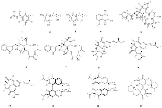
Article Menu Font Type: Arial Georgia Verdana Font Size: Aa Aa Aa Line Spacing: Column Width: Background: Open AccessArticle by Tuxiang Mo 1,2,3, Xuan Zhang 1,2,3, Chenyang Yi 1,2,3, Hua Wu 1,2,3, Xinrui Xia 1,2,3, Xinjie Ge 1,2,3, Hong Xiao 1,2,3, Wanshan Li 1,2,3,* and Guangying Chen 1,2,3,* 1 Key Laboratory of Tropical Medicinal Resource Chemistry of Ministry of Education, Hainan Normal University, Haikou 571158, China 2 Key Laboratory of Tropical Medicinal Plant Chemistry of Hainan Province, School of Chemistry and Chemical Engineering, Hainan Normal University, Haikou 571158, China 3 Key Laboratory of Hainan Provincial International Joint Research Center for Comprehensive Utilization and Efficient Conversion of Tropical Medicinal Resources, Hainan Normal University, Haikou 571158, China * Authors to whom correspondence should be addressed. Submission received: 16 September 2026 / Revised: 29 September 2026 / Accepted: 6 October 2026 / Published: 9 October 2026 Abstract Four new metabolites, including two resorcinol derivatives, (S)-1-(2,4-dihydroxy-3,5-dimethylphenyl)-3-hydroxybutan-1-one (1) and 5-[(8S)-2-hydroxypropyl]-6-methylbenzene-1,3-diol (2), a benzo[c][1,2]dioxine derivative, (S)-3,5,7-trimethyl-3,4-dihydrobenzo[c][1,2]dioxine-6,8-diol (3), and a cytochalasan alkaloid, 7-O-acetylarmochaetoglobin Q (5), along with nine known compounds, were isolated from the marine fungus Chaetomium globosum F0831, obtained from a South China Sea marine sediment. Their structures were accomplished by extensive spectroscopic analysis combined with single-crystal X-ray crystallography and ECD calculations (Electronic Circular Dichroism calculations). The anti-inflammatory activity of the isolated compounds was evaluated. Compound 8 was discovered to potentially reduce inflammation through suppression of the AKT/STAT3 pathway, thereby inhibiting the release of inflammatory mediators (iNOS, COX-2) and inflammatory cytokines (IL-6, TNF-α, and IL-1β). 1. Introduction Marine sediment-derived fungi, an important yet underexplored reservoir for novel bioactive compounds, thrive in extreme environments that promote unique metabolic adaptations [1,2]. The fungus Chaetomium globosum is recognized as a prolific source of structurally diverse secondary metabolites with significant biological activities [3]. The compounds produced by this fungus mainly include cytochalasans, azaphilones, indole alkaloids, and anthraquinones [3]. Among these, cytochalasans and azaphilones represent the most abundant and characteristic components of its metabolic repertoire [4,5,6]. Inflammation functions not only as a protective response to injury and infection but also as a unifying mechanism underlying numerous chronic diseases. Among these, systemic chronic inflammation has been recognized as a central driver across the lifespan, contributing to the onset and progression of cardiovascular disease, cancer, and metabolic disorders [7]. Importantly, the transcription factor STAT3 plays a pivotal role in immune regulation and represents a key molecular link bridging chronic inflammation and tumorigenesis [8,9]. Consequently, therapeutic strategies that target STAT3 and related signaling pathways have emerged as promising approaches for the development of novel anti-inflammatory agents [10,11]. The marine-derived fungus Chaetomium globosum has proven to be a prolific source of structurally diverse polyketides and alkaloids. In the present study, chromatographic investigation of this strain afforded four previously unreported metabolites, encompassing two resorcinol derivatives (1, 2), a rare benzo[c][1,2]dioxine-type endoperoxide (3), and a cytochalasan alkaloid (5), together with nine known congeners (4 and 6–13) (Figure 1). More significantly, compound 8 was found to attenuate the inflammatory response, potentially by inhibiting the AKT/STAT3 signaling pathway, thereby blocking the release of inflammatory mediators (iNOS, COX-2) and inflammatory cytokines (IL-6, TNF-α, and IL-1β). Herein, the detailed isolation, structural elucidation, and bioactivity evaluation of 1–13 have been reported. Figure 1. Structures of compounds 1–13. 2. Results and Discussion 2.1. Chemical Structure Elucidation (S)-1-(2,4-dihydroxy-3,5-dimethylphenyl)-3-hydroxybutan-1-one (1) was isolated as needle-like crystals with the molecular formula of C12H16O4 as determined by HRESIMS peak at m/z 247.0930 [M + Na]+ (calcd for C12H16NaO4+, 247.0941). The IR (Figure S7) spectrum of 1 suggested the presence of hydroxyl (3443 cm−1) and aromatic (1599 cm−1) groups. The 1H and 13C NMR spectra, together with DEPT, showed one carbonyl group, one oxygen-containing methylene group, one methylene group, three methyl groups, and six sp2 hybridized tertiary carbons. Detailed analysis of the 1H and 13C NMR spectra of 1 (Table 1), together with HSQC and HMBC experiments, suggested the presence of a 1,2,3,4-tetrasubstituted butyrophenone. 1H–1H COSY (Figure 2) correlations were observed between H3-10 [δH 1.25 (d, J = 6.3 Hz, 3H)] and H-9 [δH 4.33 (m, 1H)], and between H-9 and the diastereotopic methylene protons H2-8 [δH 2.92 (dd, J = 15.6, 5.0 Hz, 1H) and 3.11 (dd, J = 15.6, 7.5 Hz, 1H)], revealing a 3-hydroxybutanoyl moiety. In the HMBC spectrum, cross-peaks from H2-8 to the ketone carbonyl carbon C-7 (δC 204.6) and from the aromatic proton H-6 [δH 7.45 (s, 1H)] to C-7 established the attachment of the side chain to C-1 of the aromatic ring. The structure of 1 was further confirmed by comprehensive 2D NMR analyses (Figure 2) and single-crystal X-ray diffraction analyses (Figure 3), which finally established the absolute configuration of 1 as 9S. Crystallographic data for compound 1 have been deposited with the Cambridge Crystallographic Data Centre (CCDC 2588332). Thus, we propose that compound 1 is (S)-1-(2,4-dihydroxy-3,5-dimethylphenyl)-3-hydroxybutan-1-one. Figure 2. Key 1H–1H COSY and HMBC correlations of compounds 1–3 and 5. Figure 3. X-ray crystallographic structure of 1. Table 1. 1H (J in Hz, δH in ppm) NMR and 13C (δC in ppm) NMR data of compounds 1 in CD3OD, 2 and 3 in DMSO-d6. Compound 2 was obtained as a colorless solid, with a molecular formula of C10H14O3, as deduced from the HRESIMS at m/z [M-H]− 181.0865 (calcd 181.0870). The 1H and 13C NMR spectra (Table 1) revealed ten carbon resonances, including one aromatic methyl, one aliphatic methyl, one oxygenated methine, two oxygenated tertiary carbons, one aliphatic methylene, and four tertiary carbons. The downfield carbons at δC 155.8 (C-1) and 154.9 (C-3) indicated oxygenated aromatic carbons adjacent to hydroxyl substituents. The singlet protons at δH 6.13 (H-2, δC 100.3) and δH 6.02 (H-4, δC 108.3) confirmed the presence of a 1,3,5,6-tetrasubstituted benzene ring. The methyl group at δH 1.94 (3H, s, δC 11.0, 6-CH3) showed HMBC (Figure 2) correlations to C-1 (δC 155.8), C-5 (δC 139.3) and C-6 (δC 112.7), supporting its attachment at C-6. The 1H–1H COSY (Figure 2) correlations between H2-7/H-8/H3-9 confirmed the connectivity of this side chain, revealing a 2-hydroxypropyl structural fragment. HMBC correlations from H-7 to C-4 and C-5 established the linkage of the side chain to the aromatic ring. The optical rotation of 2 was similar to that reported for 5-[2-hydroxypropane-1-yl]-2,6-dimethylbenzene-1,3-diol [12], confirming the C-8 absolute configuration. Thus, compound 2 was elucidated as 5-[(8S)-2-hydroxypropyl]- 6-methylbenzene-1,3-diol. Compound 3 was obtained as a colorless solid, with a molecular formula of C11H14O4 as established by HRESIMS (m/z [M-H]− 209.0819, calcd for C11H13O4, 209.0819), corresponding to five degrees of unsaturation. Analysis of the 1H and 13C NMR data indicated that compound 3 is a benzene derivative. The methyl group at δH 1.95 (s, 3H, δC 9.7, 2–CH3) showed HMBC correlations (Figure 2) to C-1 (δC 146.1), C-2 (δC 112.2), and C-3 (δC 136.3), supporting its attachment at C-2. The 1H–1H COSY correlations between H2-7/H-8/H3-9 confirmed the connectivity of the side chain, revealing a 2-hydroxypropyl fragment. HMBC correlations from H-7 to C-4 and C-5 established the linkage of the side chain to the aromatic ring at C-5. The methyl protons at δH 1.96 (3H, s, δC 12.9) correlated with C-1 (δC 146.1), C-5 (δC 123.2), and C-6 (δC 111.5), establishing the location of the methyl group at C-6. The 3-OH proton signal at δH 8.23 (1H, s) correlated with C-2 (δC 112.2), C-3 (δC 136.3), and C-4 (δC 139.9), confirming the placement of the hydroxy group at C-3. The remaining unsaturation number, combined with high-resolution mass spectrometry data, suggested that a peroxide ring was likely present in compound 3. The IR (Figure S22) spectrum of compound 3 exhibited a sharp absorption band at 823 cm−1, assigned to the stretching vibration of the peroxide bond (O–O); together with the spectroscopic data of bombaxoin, which shares the same peroxide-containing 3-methyl-3,4-dihydrobenzo[c][1,2]dioxine skeleton [13], this established the planar structure of compound 3. To establish the absolute configuration of compound 3, ECD calculations were performed. The calculated ECD spectrum of (S)-3a was in good agreement with the experimental CD spectrum, allowing the absolute configuration of compound 3 to be assigned as (S). Compound 3 was thus elucidated as (S)-3,5,7-trimethyl-3,4-dihydrobenzo[c][1,2]dioxine-6,8-diol. 7-O-acetylarmochaetoglobin Q (5) was isolated as a brown oily substance with the molecular formula of C34H40N2O7 determined by HRESIMS peak at m/z 587.2770 [M-H]—(calcd for C34H39N2O7, 587.2762). The 1H and 13C NMR data of compound 5 (Table 2) were closely similar to those of chaetoglobosin Q (7) [14], indicating the same cytochalasin skeleton. The remarkable difference between compound 5 and chaetoglobosin Q was that the hydroxyl group at C-7 was replaced by an acetyl group, which was verified by HMBC correlation from H-7 to 7-OCOCH3 (Figure 2). The structure of 5 was finally elucidated by the analysis of the HSQC, COSY, and HMBC experimental data. Table 2. 1H NMR and 13C NMR data of compound 5 (J in Hz, δ in ppm) in CD3OD. The relative stereochemistry of compound 5 was assigned through a combination of NOESY data (Figure 4) and analysis of vicinal coupling constants. The correlations between 11-Me/H-3, H-4/H-5, H-4/H2-10, H-5/H-6, and H-6/H-8 indicated the α-orientations of H-3, 11-Me, and 12-Me, and the β-orientations of H-4, H-5, H-6, H-8, and H2-10 in the perhydro-isoindolone ring. The correlations from H-8 to H-14, from H-14 to H-16, from H-13 and H-17 to 15a, and from H-17 to H-19 and H-22 implied that 16-Me and H-19 were α-configured. The correlations between H-17 and H-19 and the large coupling constants of H-21 and H-22 (J = 16.7 Hz) and H-13 and H-14 (J = 14.7 Hz) confirmed these olefinic protons were trans-configured, and this was consistent with the relative configuration of the analogue chaetoglobosin A (6), which possesses the same macrocyclic ring as that of 5. The absolute configuration of chaetoglobosin A (6) was determined by X-ray diffraction [15]. Biosynthetically, compound 5 is derived from chaetoglobosin A (6) through post-modification reactions. Thus, the absolute configuration of 5 was postulated to be the same as that of 6. Figure 4. Key NOESY correlations of compound 5. To support the above-mentioned conclusion, the absolute configuration of compound 5 was unambiguously determined by comparing its experimental ECD spectrum with those calculated via TDDFT, following the established protocols [16,17]. The calculated ECD spectrum of the (3S, 4R, 5S, 6S, 8R, 9R, 16S, 19R)-isomer matched well with the experimental ECD spectrum of compound 5 (Figure 5). In addition, compounds 5–7 possess the same stereogenic centers, and the CD spectra of compounds 6 and 7 were consistent with that of compound 5, further supporting that compounds 5–7 share the same absolute configuration. Therefore, the stereochemistry of compound 5 was determined to be 3S, 4R, 5S, 6S, 8R, 9R, 16S, and 19R, which is consistent with its biogenetic origin. Figure 5. Experimental and calculated ECD spectra of 3 and 5–7. Compounds 4 and 6–13 were characterized as follows: Xylariol A (4) [18], Chaetoglobosin A (6) [14], Chaetoglobosin Q (7) [19], Chaetomugilin D (8) [20], Chaetoviridin A (9) [21], 5′-epichaetoviridin A (10) [22], Cochliodone A (11) [23], Cochliodone B (12) [23], and Thielavialide E (13) [24]. Their identification was based on comparison of their 1H NMR and 13C NMR spectra with those reported in the literature. 2.2. Biological Activity The inhibitory effects of all isolated compounds (1–13) on NO production were evaluated in LPS-induced RAW 264.7 macrophages using dexamethasone as a positive control. Cell viability was determined by MTT assay to confirm whether the observed inhibition of NO production was attributable to cytotoxicity. The results showed that all compounds exhibited no or weak cytotoxic effects (cell viability > 90%) on RAW 264.7 cells at a concentration of 40 μM. Compounds 8 and 9 demonstrated inhibitory effects on LPS-induced inflammatory responses. Compound 8 exhibited the lowest IC50 value among the isolated compounds (10.99 ± 0.17 μM) in the NO inhibition assay, while compound 9 showed an IC50 value of 28.56 ± 0.39 μM; the positive control dexamethasone showed an IC50 value of 35.44 ± 2.27 μM under the tested conditions. Inflammation plays an important role in the development and prognosis of various diseases. In this study, we evaluated the anti-inflammatory activity of compound 8 in an LPS-induced inflammation model. NO, a critical pro-inflammatory mediator, serves as a signaling molecule in the induction of inflammation. Under pathological conditions, NO is primarily synthesized by the iNOS enzyme and is subsequently oxidized to the cytotoxic peroxynitrite [25]. Both iNOS and COX-2 play pivotal roles in the inflammatory response: iNOS produces NO, while COX-2 facilitates the production of pro-inflammatory prostaglandins [26]. In this study, pretreatment with compound 8 significantly suppressed NO production at non-cytotoxic concentrations (Figure 6). Western blot analysis further revealed that compound 8 downregulated iNOS protein levels in LPS-treated RAW264.7 cells. Additionally, we observed that compound 8 sharply reduced the protein levels of COX-2. In conclusion, compound 8 mitigates LPS-induced inflammation by inhibiting the overproduction of NO through the downregulation of both COX-2 and iNOS protein levels. Figure 6. Pretreatment with compound 8 attenuated LPS-mediated inflammatory outcomes in RAW264.7 cells. (A) Chemical structure of compound 8. (B) Cell culture supernatants were collected and NO concentrations determined by a commercial kit. (C) Cell viability of compound 8 at doses of 5, 10, 20, and 40 μM. (D) Cell lysates were obtained and used to determine COX-2, iNOS, and β-actin concentrations by Western blotting. ## p < 0.01 vs. Con, * p < 0.05, ** p < 0.01 vs. LPS. n ≥ 3. Activated inflammatory cells produce high amounts of ROS, as well as inflammatory cytokines. Excessive ROS is associated with cell damage and apoptosis; ROS can also synergistically act with NO to enhance oxidative stress and damage [27]. In compound 8-pre-treated LPS-treated RAW264.7 cells, we observed a reduced green fluorescence intensity, implying that excessive ROS was effectively scavenged (Figure 7). Cytokines mediate cell–cell communication. Excessive IL-6, TNF-α, and IL-1β promote inflammation, leading to pain, fever, and increased permeability. While these cytokines are protective at normal levels, their overproduction drives inflammatory diseases, making their inhibition crucial for controlling inflammation [28]. We found that pretreatment with compound 8 significantly suppressed pro-inflammatory factor levels, including those of IL-6, TNF-α, and IL-1β, implying that it exhibits good anti-inflammation effects against LPS-treated RAW 264.7 cells. Figure 7. Impact of compound 8 on cytokine secretion and ROS release in LPS-treated RAW264.7 cells. Cell pre-treatment was done for 1 h using different compound 8 concentrations (5, 10 and 20 μM) followed by LPS (1 μg/mL) treatment for 24 h. (A) Supernatants of the cell cultures were obtained and used to determine IL-1β and IL-6 as well as TNF-α concentrations by ELISA. (B) After treating cells with DCFH-DA fluorescent probe, ROS levels were detected by fluorescence microscopy. Red arrows indicate inflammatory cells with ROS accumulation. Scale bar, 50 µm. ns: not significant; ## p < 0.01 vs. Con, ** p < 0.01 vs. LPS. n ≥ 3. Protein kinase B (Akt/PKB), a serine/threonine protein kinase, plays a crucial role in the activation of various signaling pathways, including STAT3. Previous studies have shown that Akt regulates transcriptional activity of STAT3 by inducing its phosphorylation [29]. To explore the effect of compound 8 on Akt signaling, we assessed the phosphorylation levels of Akt in LPS-treated cells. As shown in Figure 8A, pretreatment with compound 8 attenuated LPS-induced inflammatory responses and was accompanied by reduced phosphorylation of AKT and STAT3, suggesting a possible association between its anti-inflammatory effects and the AKT/STAT3 signaling pathway. Figure 8. Pretreatment with compound 8 modulated AKT/STAT3 signaling pathway. RAW264.7 cells were treated with different concentrations (5, 10 and 20 μM) of compound 8 and LPS (1 μg/mL) for 24 h. (A) The effect of compound 8 on the Akt and STAT3 pathways in LPS induced RAW264.7 cells, with total Akt and total STAT3 as sample controls. (B) The NF-κB nuclear localizations of the treated cells were visualized through immunofluorescence analysis with an anti-NF-κB p65 (red) antibody. The nuclei were DAPI-stained (blue). Scale bar: 50 μm. Blue = nuclei, red = NF-κB, arrows = NF-κB nuclear translocation. Data are expressed as three independent experiments mean ± SD. ns: not significant; ## p < 0.01, compared with the control group. * p < 0.05, ** p < 0.01, compared with the LPS group. In addition, NF-κB is one of the most classical pathways regulating the initiation and progression of inflammation. Once activated, the NF-κB p65 subunit is released from the combination with inhibitory subunit IκB and subsequently translocated into the nucleus to engage in the transcription of multiple target genes, ultimately resulting in the release of proinflammatory factors. NF-κB expression correlates with elevated total cellular levels of STAT3 and supports expression and activation of STAT3 [30]. As shown in Figure 8B, confocal immunofluorescence analysis showed that pretreatment with compound 8 reduced the LPS-induced nuclear accumulation of NF-κB p65. 3. Materials and Methods 3.1. General Experimental Procedures Infrared (IR) spectra were acquired using a Nicolet 6700 spectrophotometer (Thermo Fisher Scientific, Waltham, MA, USA). High-resolution electrospray ionization mass spectrometry (HR-ESI-MS) spectra were obtained on a Bruker Daltonics Apex-Ultra 7.0 T instrument (Bruker Corporation, Billerica, MA, USA). Nuclear magnetic resonance (NMR) spectra were measured using a Bruker AV-400 spectrometer (Bruker Switzerland AG, Fällanden, Switzerland). Optical rotations were recorded using a Modular Circular Polarimeter (Anton Paar GmbH, Graz, Austria, MCP 5100). Semi-preparative HPLC was performed on an LabTotal LC-20A (Shimadzu Corporation, Kyoto, Japan) series with a DAD detector using an Agilent Eclipse XDB-C18 (Agilent Technologies, Santa Clara, CA, USA) column (250 × 9.4 mm, 5 µm). 3.2. Fungal Material Chaetomium globosum F0831 (GenBank accession number QB007695) was isolated from a marine sediment sample collected in the northern part of the South China Sea (GPS coordinates 115.1540° E, 20.1404° N) at a water depth of 500 m in July 2020. This fungal strain is currently preserved at the Key Laboratory of Tropical Medicinal Resource Chemistry, Ministry of Education, Hainan Normal University. Fermentation, extraction and isolation. Two hundred Erlenmeyer flasks (1000 mL each), containing sterile solid medium composed of 75 g of rice and 120 mL of water with 3‰ sea salt, were inoculated with the Chaetomium globosum F0831 strain and incubated at 28 °C for 35 days. The rice-based medium underwent exhaustive extraction using EtOAc, followed by vacuum concentration to yield a dry EtOAc extract (230.0 g). The EtOAc extract was subjected to silica gel column chromatography with a gradient elution of petroleum ether and ethyl acetate (100:0 to 0:100), yielding six fractions (Fr.A–Fr. F). Fr. C was further fractionated using reversed-phase column chromatography eluted with MeOH–H2O (30:70 to 100:0), affording four subfractions (Fr. C.1–Fr. C.4). Fr. C.2 was subsequently separated on a Sephadex LH-20 column (MeOH), giving three subfractions (Fr. C.2.1–Fr. C.2.3). Purification of Fr. C.2.1 by semi-preparative HPLC with CH3CN–H2O (36:64) yielded compounds 2 (5.2 mg) and 3 (10.3 mg). Similarly, Fr. C.2.3 was subjected to semi-preparative HPLC (CH3CN–H2O, 65:35) to afford compounds 8 (30.1 mg), 9 (50.0 mg), and 10 (15.0 mg). Fr. C.4 was chromatographed over a Sephadex LH-20 column eluted with MeOH, resulting in three subfractions (Fr. C.4.1–Fr. C.4.3). From Fr. C.4.2, compounds 1 (25.2 mg) and 4 (11.0 mg) were obtained via semi-preparative HPLC (CH3CN–H2O, 35:65). Fr. C.4.3 was purified by semi-preparative HPLC (CH3CN–H2O, 51:49) to yield compounds 11 (10.2 mg), 12 (11.7 mg), and 13 (15.3 mg). Fr. D was fractionated using reversed-phase column chromatography (MeOH–H2O, 20:80 to 100:0), providing five subfractions (Fr. D.1–Fr. D.5). Fr. D.3 was further separated on a Sephadex LH-20 column (MeOH), giving four subfractions (Fr. D.3.1–Fr. D.3.4). Final purification of Fr. D.3.3 by semi-preparative HPLC (CH3CN–H2O, 33:67) afforded compounds 5 (6.1 mg), 6 (15.6 mg), and 7 (12.0 mg). X-Ray Crystallographic Data for Compound 1. The crystals of compound 1 were obtained by slow evaporation from a MeOH solution. The crystallographic data for compound 1 have been deposited in the Cambridge Crystallographic Data Centre (CCDC) with the deposition number CCDC 2588332. Crystal Data for C12H16O4 (M = 224.26 g/mol): orthorhombic, space group C2221, a = 7.0987(3) Å, c = 15.4116(6) Å, V = 15.4116(6) Å3, Z = 8, T = 293 K, μ(Cu Kα) = 0.803 mm−1, Dcalc = 1.2974 g/cm3, 6467 reflections measured (8.42° ≤ 2Θ ≤ 133.78°), 2037 unique (Rint = 0.0812, Rsigma = 0.0547), which were used in all calculations. The final R1 was 0.0606 (I > 2σ(I)), and wR2 was 0.1524 (all data). (S)-1-(2,4-dihydroxy-3,5-dimethylphenyl)-3-hydroxybutan-1-one (1): colorless block-shaped crystals; mp 129.6–130.3 °C; [α]D20" role="presentation"> ] D 20 + 27.8 (c 0.1, MeOH); IR (KBr) νmax 3443, 2833, 1599, 1360 cm−1; HR-ESI-MS m/z 247.0930 [M + Na]+ (calcd for C12H16NaO4+, 247.0941). For 1H and 13C NMR data, see Table 1. 5-[(8S)-2-hydroxypropyl]- 6-methylbenzene-1,3-diol (2): brown oily substance; [α]D20" role="presentation"> ] D 20 −13.5 (c 0.1, MeOH); IR (KBr) νmax 3446, 1056, 1028, 1008 cm−1; HR-ESI-MS m/z 181.0865 [M-H]—(calcd for C10H13O3−, 181.0870). For 1H and 13C NMR data, see Table 1. (S)-3,5,7-trimethyl-3,4-dihydrobenzo[c] [1,2] dioxine-6,8-diol (3): brown oily substance; [α]D20" role="presentation"> ] D 20 −10.5 (c 0.1, MeOH); IR (KBr) νmax 3420, 1652, 1635, 1120, 823 cm−1; HR-ESI-MS m/z 209.0819 [M-H]—(calcd for C11H13O4−, 209.0819). For 1H and 13C NMR data, see Table 1. 7-O-acetylarmochaetoglobin Q (5): brown oily substance; [α]D20" role="presentation"> ] D 20 −135.3 (c 0.1, MeOH); IR (KBr) νmax 3445, 2832, 1609, 1364, 779 cm−1; HR-ESI-MS m/z 587.2770 [M-H]—(calcd for C34H39N2O7, 587.2762). For 1H and 13C NMR data, see Table 2. 3.3. Computational Section Conformational analysis of all compounds was performed using Spartan 14 software with the MMFF94S force field. The conformers were then imported into the Gaussian 16 package. Geometry optimization was carried out at the B3LYP/6-31+G(d) level. The optimized conformers were subjected to electronic circular dichroism (ECD) calculations using time-dependent density functional theory (TDDFT) at the B3LYP/6-311G* level in methanol, with the polarizable continuum model (PCM) employed to account for solvation effects. The calculated ECD curves were generated using SpecDis 1.71. The calculated spectra were Boltzmann-weighted according to the relative populations of the low-energy conformers in MeOH. 3.4. Cell Culture and Treatment The RAW 264.7 cells were purchased from the Chinese Academy of Sciences Cell Bank (Shanghai, China).RAW 264.7 cells were maintained in Dulbecco’s Modified Eagle Medium (DMEM) (C11995500BT, Gibco, Waltham, MA, USA), supplemented with 10% (v/v) fetal bovine serum (FBS) (10099141C, Gibco, Waltham, MA, USA) and 1% penicillin–streptomycin (PS) (15140122, Gibco, Waltham, MA, USA). Cells were incubated in a humidified incubator with 5% CO2 and 95% air at 37 °C. To evaluate the anti-inflammatory effects of compound 8, RAW 264.7 cells were seeded at a density of 2 × 10⁴ cells per well in a 96-well plate. The cells were treated with 1 μg/mL lipopolysaccharide (LPS) in the presence or absence of compound 8 (20 μM) for 12 h. Inflammatory mediator production, including nitric oxide (NO), and pro-inflammatory cytokines (TNF-α, IL-6, IL-1β), was measured to evaluate the inhibitory effects of compound 8 on LPS-stimulated inflammatory responses. 3.5. Determination of NO Production Nitric oxide (NO) production was evaluated by measuring nitrite (NO2−) levels in the culture supernatants using the Griess Reagent method with a commercial Nitric Oxide Assay Kit (Beyotime, S0021, Shanghai, China), according to the manufacturer’s instructions. Briefly, culture supernatants were collected after treatment and centrifuged when necessary to remove cellular debris. A NaNO2 standard curve was prepared using concentrations of 0, 1, 2, 5, 10, 20, 40, 60, and 100 μM in the same culture medium used for the samples. Subsequently, 50 μL of each standard or sample was mixed sequentially with 50 μL of Griess Reagent I and 50 μL of Griess Reagent II in a 96-well plate. After incubation at room temperature, absorbance was measured at 540 nm. Nitrite concentrations were calculated from the NaNO2 standard curve (R2 > 0.99). Cell viability was evaluated in parallel using the CCK-8 assay to assess potential cytotoxic interference. NO production was normalized to the LPS-treated group, which was defined as 100%. Relative NO production was calculated as: relative NO production (%) = (NO2− concentration of sample/NO2− concentration of LPS group) × 100. The NO inhibition rate was calculated as: inhibition rate (%) = [(NO2− concentration of LPS group − NO2− concentration of sample)/(NO2− concentration of LPS group − NO2− concentration of control group)] × 100. All experiments were performed independently at least three times, and the results are expressed as the mean ± SD. 3.6. ELISA Assay To evaluate cytokine concentrations in the cell culture supernatant, RAW264.7 cells were cultured under the indicated experimental conditions. The concentration of TNF-α (RayBiotech, Peachtree Corners, GA, USA, #ELM-TNFα), IL-1β (RayBiotech, Peachtree Corners, GA, USA, #ELM-IL-1b), and IL-6 (RayBiotech, Peachtree Corners, GA, USA, #ELM-IL6) in cell culture supernatant was detected using ELISA kits according to manufacturers’ instructions. Briefly, the assay was based on a double-antibody sandwich method, and the absorbance of samples was measured at 450 nm using a microplate reader. 3.7. Intracellular ROS Detection by Fluorescence Microscopy RAW264.7 cells were pretreated with compound 8 (5, 10, or 20 μM) for 1 h and then stimulated with LPS (1 μg/mL) for 24 h. Intracellular ROS were detected using the DCFH-DA fluorescent probe. Following treatment, cells were incubated with DCFH-DA (diluted 1:1000) at 37 °C for 30 min and subsequently washed with serum-free culture medium. Fluorescence images were acquired using an Olympus FV3000 confocal laser scanning microscope (Olympus, Tokyo, Japan) under identical imaging settings for all groups. Fluorescence intensity was quantified using ImageJ 1.53c. Background fluorescence was subtracted, and the mean fluorescence intensity within the selected regions of interest (ROIs) was calculated and normalized to that of the control group. 3.8. Western Blotting Analysis Cells were lysed in RIPA buffer (Beyotime, Shanghai, China) with added protease inhibitors for 30 min on ice. The resulting homogenates were then centrifuged at 12,000 rpm for 10 min at 4 °C. Total protein concentrations in the supernatants were measured using the BCA assay. Protein samples, adjusted to equal concentrations, were separated on SDS-PAGE gels and subsequently transferred to PVDF membranes (Millipore, Burlington, MA, USA). After a 2-h sealing treatment with 5% BSA, the membrane was further incubated at 4 °C overnight to allow it to bind to the specific antibodies related to the target protein. After washing, the membranes were incubated with the corresponding secondary antibodies for 2 h at room temperature (RT). The immune complexes were detected with an ECL reagent (Millipore, Burlington, MA, USA). Following incubation with horseradish peroxidase (HRP)-conjugated secondary antibodies at room temperature. Immunoblots were analyzed using analytikjena (Analytik Jena, Burlington, MA, USA). 3.9. Immunofluorescence Staining RAW264.7 cells were seeded on coverslips and cultured overnight. After the indicated treatments, the cells were fixed with 4% paraformaldehyde for 15 min, permeabilized with 0.1% Triton X-100 for 10 min, and blocked with 5% bovine serum albumin (BSA) for 1 h at room temperature. The cells were then incubated with an anti-NF-κB p65 primary antibody overnight at 4 °C, followed by incubation with an appropriate fluorescence-conjugated secondary antibody for 1 h at room temperature in the dark. Nuclei were counterstained with DAPI. Fluorescence images were acquired using an Olympus FV3000 confocal laser scanning microscope (Olympus, Tokyo, Japan) under identical imaging settings for all groups. 3.10. Statistical Analysis Data were analyzed using GraphPad Prism 10.4. The figures and/or legends contained statistical tests, n values, replicate experiments, and p values. A two-tailed Student’s t-test was used to assess significance between the two groups, while one-way ANOVA with Tukey’s post hoc test was employed to evaluate significance among multiple groups. Statistical significance was defined as a p value < 0.05. Error bars indicate mean ± SD. A difference at * p < 0.05 was considered significant, and that at ** p < 0.01 was considered highly significant. Ns: not significant. 4. Conclusions In our continuing search for bioactive secondary metabolites from marine-derived fungi, a chemical investigation of Chaetomium globosum obtained from marine sediments led to the isolation of four undescribed compounds (1–3 and 5), representing diverse structural classes including two resorcinol derivatives, a rare benzo[c][1,2]dioxine endoperoxide, and a cytochalasan alkaloid, together with nine known congeners. Bioassay results demonstrated that compound 8 was found to attenuate the inflammatory response, potentially by inhibiting the AKT/STAT3 signaling pathway, thereby suppressing the expression of inflammatory mediators (iNOS and COX-2) and pro-inflammatory cytokines (IL-6, TNF-α, and IL-1β). SAR analysis of the chaetomugilin class indicates that the C-1 lactone ring and the C-8 hydroxy group are favorable for anti-inflammatory activity. Chaetomugilins E and F, which retain both features, inhibited NO production with IC50 values of 5.8 and 1.9 μM, respectively [31]. Similarly, chaetomugilin I and 11-epi-chaetomugilin I suppressed TNF-α-induced NF-κB activity (IC50 = 0.9 μM) and NO release (IC50 = 0.3 and 0.8 μM, respectively) [32]. In contrast, chaephilones C and D, lacking these features, showed only moderate NO inhibition (IC50 = 15.12 ± 1.48 and 20.65 ± 0.95 μM) [33]. In the present study, compound 8 (IC50 = 10.99 ± 0.17 μM) also exhibited greater activity than compound 9 (IC50 = 28.56 ± 0.39 μM), consistent with the favorable roles of the lactone ring and C-8 hydroxy group. Among the compounds isolated in this work, compound 8 was the most potent NO inhibitor, and its C-1 lactone ring and C-8 hydroxy group may provide a structural basis for further optimization of azaphilone-based anti-inflammatory leads. These findings also expand the chemical diversity of C. globosum. Supplementary Materials The following supporting information can be downloaded at https://www.mdpi.com/article/10.3390/md24100355/s1. Figure S1−S8: 1H and 13C NMR, DEPT, HSQC, HMBC, 1H−1H COSY, IR, and (−)-HR-ESI-MS spectra of compound 1; Figure S9−S16: 1D and 2D NMR, IR, and HR-ESI-MS spectra of compound 2; Figure S17−S23: 1D and 2D NMR, IR, and (-)-HR-ESI-MS spectra of compound 3; Figure S24−S31: 1D and 2D NMR, NOESY, IR, and (+)-HR-ESI-MS spectra of compound 5; Figure S32−S33: Original, uncropped Western blot images corresponding to Figure 6D and Figure 8A; Table S1−S6: Crystallographic data and structure refinement, fractional atomic coordinates, bond lengths/angles, and hydrogen atom coordinates for compound 1. Author Contributions T.M., W.L. and G.C. designed experiments, collected fungal samples, elucidated chemical structures, and wrote the article. Additionally, T.M., X.Z., C.Y., H.W., X.X., H.X. and X.G. isolated compounds and evaluated their activities. All authors have read and agreed to the published version of the manuscript. Funding This work was supported by the National Natural Science Foundation of China (Nos. 22477021 and 22267006), the Hainan Provincial Natural Science Foundation of China (222QN303), the Key Science and Technology Program of Hainan Province (No. ZDKJ202008), the Innovation Platform for Academicians of Hainan Province, the Central Government Guides Local Science and Technology Development (ZY2022HN08), Scientific Research Project of Hainan Innovation Platform for Flexible Talent Introduction (No: RXPTXM202620), and Hainan Provincial Graduate Student Innovative Scientific Research Project (No. Qhyb2023-139). Data Availability Statement The raw data supporting the conclusions of this article will be made available by the authors on request. Conflicts of Interest The authors declare no conflicts of interest. References Li, Y.; Zhang, C.; Zhong, M.; Hu, S.; Cui, Y.; Fang, J.; Yu, X. Revealing the Metabolic Potential and Environmental Adaptation of Nematophagous Fungus, Purpureocillium lilacinum, Derived from Hadal Sediment. Front. Microbiol. 2024, 15, 1474180. [Google Scholar] [CrossRef] [Scilit] Pan, C.; Hassan, S.S.U.; Muhammad, I.; Jin, H. Marine Fungi as a Goldmine for Novel Antibiotics: A 2024 Perspective. Front. Mar. Sci. 2024, 11, 1538136. [Google Scholar] [CrossRef] [Scilit] Chen, Q.; Zhao, P.; Wang, M.; Li, L.; Qi, J.; Zhang, L.; Xia, X. Comprehensive Review—Chemistry and Bioactivities of Secondary Metabolites from Chaetomium globosum. Chem. Biodivers. 2025, 22, e03201. [Google Scholar] [CrossRef] [Scilit] Wang, W.; Gong, J.; Liu, X.; Dai, C.; Wang, Y.; Li, X.N.; Wang, J.; Luo, Z.; Zhou, Y.; Xue, Y.; et al. Cytochalasans Produced by the Coculture of Aspergillus flavipes and Chaetomium globosum. J. Nat. Prod. 2018, 81, 1578–1587. [Google Scholar] [CrossRef] [Scilit] Li, T.; Wang, Y.; Li, L.; Tang, M.; Meng, Q.; Zhang, C.; Hua, E.; Pei, Y.; Sun, Y. New Cytotoxic Cytochalasans from a Plant-Associated Fungus Chaetomium globosum kz-19. Mar. Drugs 2021, 19, 438. [Google Scholar] [CrossRef] [Scilit] Wang, W.; Yang, J.; Liao, Y.Y.; Cheng, G.; Chen, J.; Cheng, X.D.; Qin, J.J.; Shao, Z. Cytotoxic Nitrogenated Azaphilones from the Deep-Sea-Derived Fungus Chaetomium globosum MP4-S01-7. J. Nat. Prod. 2020, 83, 1157–1166. [Google Scholar] [CrossRef] [Scilit] Furman, D.; Campisi, J.; Verdin, E.; Carrera-Bastos, P.; Targ, S.; Franceschi, C.; Ferrucci, L.; Gilroy, D.W.; Fasano, A.; Miller, G.W.; et al. Chronic Inflammation in the Etiology of Disease across the Life Span. Nat. Med. 2019, 25, 1822–1832. [Google Scholar] [CrossRef] [Scilit] Zhang, H.X.; Xu, Z.S.; Lin, H.; Li, M.; Xia, T.; Cui, K.; Wang, S.Y.; Li, Y.; Shu, H.B.; Wang, Y.Y. TRIM27 Mediates STAT3 Activation at Retromer-Positive Structures to Promote Colitis and Colitis-Associated Carcinogenesis. Nat. Commun. 2018, 9, 3441. [Google Scholar] [CrossRef] [Scilit] Akkız, H.; Şimşek, H.; Balcı, D.; Ülger, Y.; Onan, E.; Akçaer, N.; Delik, A. Inflammation and Cancer: Molecular Mechanisms and Clinical Consequences. Front. Oncol. 2025, 15, 1564572. [Google Scholar] [CrossRef] [Scilit] Amin, N.H.; Hamed, M.I.A.; Abdel-Fattah, M.M.; Abusabaa, A.H.A.; El-Saadi, M.T. Design, Synthesis and Mechanistic Study of Novel Diarylpyrazole Derivatives as Anti-Inflammatory Agents with Reduced Cardiovascular Side Effects. Bioorg. Chem. 2021, 116, 105394. [Google Scholar] [CrossRef] [Scilit] Nilsson, J.; Lichtman, A.; Tedgui, A. Atheroprotective Immunity and Cardiovascular Disease: Therapeutic Opportunities and Challenges. J. Intern. Med. 2015, 278, 507–519. [Google Scholar] [CrossRef] [Scilit] Guo, C.; Lin, X.P.; Liao, S.R.; Yang, B.; Zhou, X.F.; Yang, X.W.; Tian, X.P.; Wang, J.F.; Liu, Y.H. Two New Aromatic Polyketides from a Deep-Sea Fungus Penicillium sp. SCSIO 06720. Nat. Prod. Res. 2020, 34, 1197–1205. [Google Scholar] [CrossRef] [Scilit] Sichaem, J.; Siripong, P.; Khumkratok, S.; Tip-Pyang, S. Chemical Constituents from the Roots of Bombax anceps. J. Chil. Chem. Soc. 2010, 55, 325–327. [Google Scholar] [CrossRef] [Scilit] Jiao, W.; Feng, Y.; Blunt, J.W.; Cole, A.L.; Munro, M.H. Chaetoglobosins Q, R, and T, Three Further New Metabolites from Chaetomium globosum. J. Nat. Prod. 2004, 67, 1722–1725. [Google Scholar] [CrossRef] [Scilit] Silverton, J.V.; Kabuto, C.; Akiyama, T. The Structure of Chaetoglobosin A: A Novel Use of Quartet Invariants. Acta Crystallogr. Sect. B 1978, 34, 588–593. [Google Scholar] [CrossRef] [Scilit] Pescitelli, G.; Bruhn, T. Good Computational Practice in the Assignment of Absolute Configurations by TDDFT Calculations of ECD Spectra. Chirality 2016, 28, 466–474. [Google Scholar] [CrossRef] [Scilit] Nugroho, A.E.; Morita, H. Circular dichroism calculation for natural products. J. Nat. Med. 2014, 68, 1–10. [Google Scholar] [CrossRef] [Scilit] Gu, W.; Ding, H. Two New Tetralone Derivatives from the Culture of Xylaria hypoxylon AT-028. Chin. Chem. Lett. 2008, 19, 1323–1326. [Google Scholar] [CrossRef] [Scilit] Li, H.F.; Jin, T.; Du, J.X.; Luo, X.W.; Zhang, G.; Deng, W.Q.; He, J.; Feng, T. Cytochalasans from the Fungus Chaetomium globosum with Cytotoxic Activities against HepG2 Cell Line. Phytochemistry 2026, 241, 114651. [Google Scholar] [CrossRef] [Scilit] Qin, J.C.; Zhang, Y.M.; Gao, J.M.; Bai, M.S.; Yang, S.X.; Laatsch, H.; Zhang, A.L. Bioactive Metabolites Produced by Chaetomium globosum, an Endophytic Fungus Isolated from Ginkgo biloba. Bioorg. Med. Chem. Lett. 2009, 19, 1572–1574. [Google Scholar] [CrossRef] [Scilit] McMullin, D.R.; Sumarah, M.W.; Blackwell, B.A.; Miller, J.D. New Azaphilones from Chaetomium globosum Isolated from the Built Environment. Tetrahedron Lett. 2013, 54, 568–572. [Google Scholar] [CrossRef] [Scilit] Borges, W.S.; Mancilla, G.; Guimarães, D.O.; Durán-Patrón, R.; Collado, I.G.; Pupo, M.T. Azaphilones from the Endophyte Chaetomium globosum. J. Nat. Prod. 2011, 74, 1182–1187. [Google Scholar] [CrossRef] [Scilit] Phonkerd, N.; Kanokmedhakul, S.; Kanokmedhakul, K.; Soytong, K.; Prabpai, S.; Kongsearee, P. Bis-spiro-azaphilones and Azaphilones from the Fungi Chaetomium cochliodes VTh01 and C. cochliodes CTh05. Tetrahedron 2008, 64, 9636–9645. [Google Scholar] [CrossRef] [Scilit] Wijeratne, E.M.; Espinosa-Artiles, P.; Gruener, R.; Gunatilaka, A.A. Thielavialides A–E, Nor-spiro-azaphilones, and a Bis-spiro-azaphilone from Thielavia sp. PA0001, an Endophytic Fungus Isolated from Aeroponically Grown Physalis alkekengi. J. Nat. Prod. 2014, 77, 1467–1472. [Google Scholar] [CrossRef] [Scilit] Kim, M.E.; Lee, J.S. Advances in the Regulation of Inflammatory Mediators in Nitric Oxide Synthase: Implications for Disease Modulation and Therapeutic Approaches. Int. J. Mol. Sci. 2025, 26, 1204. [Google Scholar] [CrossRef] [Scilit] Mollace, V.; Muscoli, C.; Masini, E.; Cuzzocrea, S.; Salvemini, D. Modulation of Prostaglandin Biosynthesis by Nitric Oxide and Nitric Oxide Donors. Pharmacol. Rev. 2005, 57, 217–252. [Google Scholar] [CrossRef] [Scilit] Mittal, M.; Siddiqui, M.R.; Tran, K.; Reddy, S.P.; Malik, A.B. Reactive Oxygen Species in Inflammation and Tissue Injury. Antioxid. Redox Signal. 2014, 20, 1126–1167. [Google Scholar] [CrossRef] [Scilit] Tanaka, T.; Narazaki, M.; Kishimoto, T. IL-6 in Inflammation, Immunity, and Disease. Cold Spring Harb. Perspect. Biol. 2014, 6, a016295. [Google Scholar] [CrossRef] [Scilit] Sun, Z.; Jiang, Q.; Gao, B.; Zhang, X.; Bu, L.; Wang, L.; Lin, Y.; Xie, W.; Li, J.; Guo, J. AKT Blocks SIK1-Mediated Repression of STAT3 to Promote Breast Tumorigenesis. Cancer Res. 2023, 83, 1264–1279. [Google Scholar] [CrossRef] [Scilit] Fan, Y.; Mao, R.; Yang, J. NF-κB and STAT3 Signaling Pathways Collaboratively Link Inflammation to Cancer. Protein Cell 2013, 4, 176–185. [Google Scholar] [CrossRef] [Scilit] Youn, U.J.; Sripisut, T.; Park, E.J.; Kondratyuk, T.P.; Fatima, N.; Simmons, C.J.; Wall, M.M.; Sun, D.; Pezzuto, J.M.; Chang, L.C. Determination of the Absolute Configuration of Chaetoviridins and Other Bioactive Azaphilones from the Endophytic Fungus Chaetomium globosum. Bioorg. Med. Chem. Lett. 2015, 25, 4719–4723. [Google Scholar] [CrossRef] [Scilit] Omar, A.M.; Mohamed, G.A.; Ibrahim, S.R.M. Chaetomugilins and Chaetoviridins—Promising Natural Metabolites: Structures, Separation, Characterization, Biosynthesis, Bioactivities, Molecular Docking, and Molecular Dynamics. J. Fungi 2022, 8, 127. [Google Scholar] [CrossRef] [Scilit] Gao, W.; Chai, C.; Li, X.-N.; Sun, W.; Li, F.; Chen, C.; Wang, J.; Zhu, H.; Wang, Y.; Hu, Z.; et al. Two Anti-inflammatory Chlorinated Azaphilones from Chaetomium globosum TW1-1 Cultured with 1-Methyl-l-tryptophan and Structure Revision of Chaephilone C. Tetrahedron Lett. 2020, 61, 151516. [Google Scholar] [CrossRef] [Scilit] Figure 1. Structures of compounds 1–13. Figure 2. Key 1H–1H COSY and HMBC correlations of compounds 1–3 and 5. Figure 3. X-ray crystallographic structure of 1. Figure 4. Key NOESY correlations of compound 5. Figure 5. Experimental and calculated ECD spectra of 3 and 5–7. Figure 6. Pretreatment with compound 8 attenuated LPS-mediated inflammatory outcomes in RAW264.7 cells. (A) Chemical structure of compound 8. (B) Cell culture supernatants were collected and NO concentrations determined by a commercial kit. (C) Cell viability of compound 8 at doses of 5, 10, 20, and 40 μM. (D) Cell lysates were obtained and used to determine COX-2, iNOS, and β-actin concentrations by Western blotting. ## p < 0.01 vs. Con, * p < 0.05, ** p < 0.01 vs. LPS. n ≥ 3. Figure 7. Impact of compound 8 on cytokine secretion and ROS release in LPS-treated RAW264.7 cells. Cell pre-treatment was done for 1 h using different compound 8 concentrations (5, 10 and 20 μM) followed by LPS (1 μg/mL) treatment for 24 h. (A) Supernatants of the cell cultures were obtained and used to determine IL-1β and IL-6 as well as TNF-α concentrations by ELISA. (B) After treating cells with DCFH-DA fluorescent probe, ROS levels were detected by fluorescence microscopy. Red arrows indicate inflammatory cells with ROS accumulation. Scale bar, 50 µm. ns: not significant; ## p < 0.01 vs. Con, ** p < 0.01 vs. LPS. n ≥ 3. Figure 8. Pretreatment with compound 8 modulated AKT/STAT3 signaling pathway. RAW264.7 cells were treated with different concentrations (5, 10 and 20 μM) of compound 8 and LPS (1 μg/mL) for 24 h. (A) The effect of compound 8 on the Akt and STAT3 pathways in LPS induced RAW264.7 cells, with total Akt and total STAT3 as sample controls. (B) The NF-κB nuclear localizations of the treated cells were visualized through immunofluorescence analysis with an anti-NF-κB p65 (red) antibody. The nuclei were DAPI-stained (blue). Scale bar: 50 μm. Blue = nuclei, red = NF-κB, arrows = NF-κB nuclear translocation. Data are expressed as three independent experiments mean ± SD. ns: not significant; ## p < 0.01, compared with the control group. * p < 0.05, ** p < 0.01, compared with the LPS group. Table 1. 1H (J in Hz, δH in ppm) NMR and 13C (δC in ppm) NMR data of compounds 1 in CD3OD, 2 and 3 in DMSO-d6. 1 2 3 Position δH (J in Hz) δC (ppm) δH (J in Hz) δC (ppm) δH (J in Hz) δC (ppm) 1 111.9 155.8 146.1 2 162.6 6.13 (s, 1H) 100.3 112.2 3 113.8 154.9 136.3 4 162.2 6.02 (s, 1H) 108.3 139.9 5 117.2 139.3 123.2 6 7.45 (s, 1H) 130.7 112.7 111.5 7 204.6 2.34 (m, 1H), 2.65 (m, 1H) 43.5 2.64 (m, 1H), 3.15 (m, 1H) 36.9 8 2.92 (dd, 15.6, 5.0, 1H)3.11 (dd, 15.6, 7.5, 1H) 47.6 3.73 (m, 1H) 66.7 4.79 (m, 1H) 78.6 9 4.33 (m, 1H) 65.7 1.00 (d, 6.1, 3H) 23.1 1.37 (d, 6.3, 3H) 21.7 10 1.25 (d, 6.3, 3H) 23.4 11 2.05 (s, 3H) 8.0 12 2.16 (s, 3H) 16.4 2-CH3 1.95 (s, 3H) 9.7 6-CH3 1.94 (s, 3H) 11.0 1.96 (s, 3H) 12.9 1-OH 7.40 (s, 1H) 3-OH 8.23 (s, 1H) Table 2. 1H NMR and 13C NMR data of compound 5 (J in Hz, δ in ppm) in CD3OD. 5 Position δH (J in Hz) δC (ppm) 1 174.9 2 3 3.77 (q, 4.9, 1H) 55.5 4 3.11 (t, 4.8, 1H) 43.3 5 2.02 (m, 1H) 41.7 6 73.6 7 4.84 (d, 11.4, 1H) 75.0 8 2.56 (t, 11.4, 1H) 47.1 9 64.0 10 2.76 (dd, 14.6, 6.0, 1H)2.89 (dd, 14.6, 6.0, 1H) 33.2 11 1.12 (d, 7.2, 3H) 13.7 12 1.17 (s, 3H) 24.7 13 5.80 (dd, 14.6, 11.1, 1H) 128.8 14 5.16 (ddd, 14.6, 11.1, 3.6, 1H) 135.4 15 1.92 (m, 1H)2.23 (m, 1H) 43.1 16 2.49 (m, 1H) 33.3 17 5.58 (d, 9.3, 1H) 141.6 18 133.9 19 5.13 (s, 1H) 82.6 20 201.8 21 6.38 (d, 16.7, 1H) 133.4 22 7.84 (d, 16.7, 1H) 137.7 23 198.7 24 (16-Me) 0.98 (d, 6.8, 1H) 21.2 25 (18-Me) 1.35 (s, 3H) 10.9 7-OCOCH3 172.4 7-OCOCH3 1.98 (s, 3H) 20.8 1′-NH 1′a 128.8 2′ 7.03 (s, 1H) 122.4 3′a 138.0 3′ 110.6 4′ 7.48 (d, 7.8, 1H) 119.3 5′ 7.07 (m, 1H) 125.0 6′ 6.99 (m, 1H) 119.9 7′ 7.30 (d, 8.0, 1H) 112.4 Disclaimer/Publisher’s Note: The statements, opinions and data contained in all publications are solely those of the individual author(s) and contributor(s) and not of MDPI and/or the editor(s). MDPI and/or the editor(s) disclaim responsibility for any injury to people or property resulting from any ideas, methods, instructions or products referred to in the content. © 2026 by the authors. Licensee MDPI, Basel, Switzerland. This article is an open access article distributed under the terms and conditions of the Creative Commons Attribution (CC BY) license. Share and Cite MDPI and ACS Style Mo, T.; Zhang, X.; Yi, C.; Wu, H.; Xia, X.; Ge, X.; Xiao, H.; Li, W.; Chen, G. Secondary Metabolites from a Marine-Derived Fungus Chaetomium globosum F0831 and Their Anti-Inflammatory Effects. Mar. Drugs 2026, 24, 355. https://doi.org/10.3390/md24100355 AMA Style Mo T, Zhang X, Yi C, Wu H, Xia X, Ge X, Xiao H, Li W, Chen G. Secondary Metabolites from a Marine-Derived Fungus Chaetomium globosum F0831 and Their Anti-Inflammatory Effects. Marine Drugs. 2026; 24(10):355. https://doi.org/10.3390/md24100355 Chicago/Turabian Style Mo, Tuxiang, Xuan Zhang, Chenyang Yi, Hua Wu, Xinrui Xia, Xinjie Ge, Hong Xiao, Wanshan Li, and Guangying Chen. 2026. "Secondary Metabolites from a Marine-Derived Fungus Chaetomium globosum F0831 and Their Anti-Inflammatory Effects" Marine Drugs 24, no. 10: 355. https://doi.org/10.3390/md24100355 APA Style Mo, T., Zhang, X., Yi, C., Wu, H., Xia, X., Ge, X., Xiao, H., Li, W., & Chen, G. (2026). Secondary Metabolites from a Marine-Derived Fungus Chaetomium globosum F0831 and Their Anti-Inflammatory Effects. Marine Drugs, 24(10), 355. https://doi.org/10.3390/md24100355 Note that from the first issue of 2016, this journal uses article numbers instead of page numbers. See further details here. Article Metrics Citations No citations were found for this article, but you may check on Google Scholar Article Access Statistics For more information on the journal statistics, click here. Multiple requests from the same IP address are counted as one view.
曲贝替定(Yondelis,trabectedin,ET-743)的抗癌作用机制艾瑞布林的抗癌作用机制----来源:药渡头条版权声明:我们分享文章、图片来自网络,版权归原作者及原出处所有。此文出于学习分享、传播更多资讯之目的,无商业用途。若涉及作品内容、版权或其他问题,敬请原作者联系我们(邮箱:xianghaiwenyao@126.com),立即处理。
1. Introduction Cancer remains one of the leading causes of death worldwide and continues to pose a major public health challenge [1]. In 2026, approximately 2.11 million new cancer cases and 626,140 cancer-related deaths are projected to occur in the United States alone [1]. Furthermore, the World Health Organization predicts that global cancer incidence will increase by more than 70% over the next two decades due to population growth, aging, and lifestyle-related risk factors [2]. Cancer is a multifactorial disease driven by both genetic alterations and environmental factors that disrupt normal cellular regulation [3]. The effectiveness of chemotherapy and radiotherapy is limited by severe side effects, poor selectivity toward cancer cells, and the emergence of multidrug resistance [4], [5]. Considerable efforts have been directed toward developing therapies that target key cancer-related mechanisms [6]. Precision oncology has advanced cancer treatment through the identification of molecular targets such as kinases, growth factor receptors, checkpoint proteins, and signaling pathways that drive tumor development and progression [7]. Cancer treatment has increasingly shifted toward molecular targets that control cell survival, proliferation, and apoptosis. Several key proteins have been studied as targets for anticancer drug development, viz B-cell lymphoma 2 (BCL2), nuclear factor kappa-light-chain-enhancer of activated B cells (NFkB), mechanistic target of rapamycin (mTOR), and Cyclin-Dependent Kinase 4 (CDK4). BCL2 is one of the most important anti-apoptotic proteins in cancer. Its overexpression assists cancer cells in avoiding programmed cell death and contributes to treatment resistance [8]. Therefore, inhibition of BCL2 is an effective strategy to restore apoptosis in tumor cells [9]. NFkB is another major therapeutic target [10], since persistent activation of NFkB promotes tumor growth and supports cancer progression [11]. Likewise, abnormal activation of mTOR is frequently observed in cancer and is associated with tumor development, metastasis, and drug resistance [12], [13]. Meanwhile, increased CDK4 activity promotes uncontrolled cell division [14], [15]. In contrast, caspases such as caspase 3 (CASP3), CASP8, and CASP9 function as key mediators of apoptosis [16]. Activation of these caspases promotes cancer cell death, whereas reduced caspase activity allows tumor cells to survive and evade apoptosis [17]. Therefore, inhibiting oncogenic targets such as BCL2, NFkB, mTOR, and CDK4 while avoiding unintended inhibition of apoptosis-related caspases may provide a useful target profile for compound prioritization. On the other hand, seaweeds are well-known marine organisms that produce a wide range of bioactive compounds, many of which have shown promising anticancer properties [18], [19]. Numerous studies have explored seaweed species from different phyla to identify novel metabolites with potential applications in cancer treatment [20], [21], [22]. However, the discovery of new anticancer compounds remains a time-consuming process that requires extensive extraction, isolation, and biological evaluation [23], [24], [25]. These conventional approaches often involve substantial laboratory resources and chemical consumption [26], [27], [28]. Alternative strategies have been developed to accelerate compound screening and improve the efficiency of drug discovery. One such approach is the application of machine learning (ML). By learning patterns from known anticancer compounds, ML models can predict the biological activity of new compounds before experimental testing [29]. Although laboratory validation is still necessary, this approach assists in prioritizing the promising candidates and reduces the number of compounds requiring further investigation [30]. In addition, ML enables the assessment of compound activity against multiple cancer-related targets, providing insights into target selectivity and potential mechanisms of action [31], [32]. Therefore, this study aimed to computationally prioritize marine macroalgal metabolites with favorable predicted profiles across oncogenic targets and apoptosis-related caspases, and to identify the seaweed species reported to contain these prioritized metabolites. The findings are expected to support the prioritization of seaweed species and metabolites for future experimental studies. 2. Materials and methods 2.1. Data curation This study utilized two approaches to collect the data. The first approach involved extracting compound information from journal articles identified through the Scopus and Web of Science databases and published within the last 5 years. The search keywords and literature-screening procedure are provided in the Supplementary Material. The second approach was to manually curate the compound list from the data repository that had been identified from seaweed https://np-mrd.org/natural_products/ [33]. The keyword used in the search was obtained by inputting the seaweed genus into the web repository, and the compound's name was retrieved. The seaweed genus used in the search was derived from the algae database (algaebase.org) [34]. 2.2. Data filtering and classification During the compound listing, this study excluded the primary metabolites and macromolecules such as polysaccharides, proteins, and lipids. This study focused on small molecules and other secondary metabolites (Table S1). From the journal references [26], [35], [36], [37], [38], [39], [40], [41], [42], [43], [44], [45], [46], [47], [48], [49], [50], [51], [52], [53], [54], [55], [56], [57], [58], [59], [60], [61], [62], [63], [64], [65], [66], [67], [68], [69], [70], [71], [72], [73], [74], [75], [76], [77], [78], [79], [80], [81], [82], [83], [84], [85], [86], [87], [88], [89], [90], [91], [92], [93], [94], [95], [96], [97], [98], [99], [100], [101], [102], [103], [104], [105], [106], [107], [108], [109], [110], [111], [112], [113], [114], [115], [116], [117], [118], [119], [120], [121], [122], [123], [124], this study grouped the collected compounds into four categories, i.e. 1) compounds that were isolated and characterized as a single pure compound with anticancer properties (Table S2, 64 compounds); 2) compounds that were isolated and characterized as a single pure compound without any anticancer assay (Table S3, 208 compounds); 3) compounds identified in the seaweed extract with anticancer properties (Table S4, 151 compounds); and 4) compounds identified in the seaweed extract without any anticancer assay (Table S5, 307 compounds). Thus, this study retrieved 730 compounds from journal references. These compounds were collected from different seaweed species across phyla. Meanwhile, web-repository exploration was carried out to enrich the compound dataset from seaweed. From the https://np-mrd.org/natural_products/, this study retrieved 2446 compounds from 132 red seaweed species, 1686 compounds from 100 brown seaweed species, and 381 compounds from 32 green seaweed species (Table S6). Then, the compound-species duplications retrieved from the journal references and the web repository were removed to avoid redundancy in further analysis. Following duplicate checking, structure standardization, and compound–species mapping, 5203 valid occurrence records were retained for the final screening dataset. These comprised 2785 records from Rhodophyta, 1965 from Ochrophyta, and 453 from Chlorophyta (Fig. 1). At the structure level, these records represented 4083 unique standardized structures. The collected compounds were classified based on the Natural Products Classifier according to https://npclassifier.ucsd.edu/ web server into their respective pathway, superclass, and class [125]. Then, the natural products (np) class richness was calculated as Eq. (1), following the species richness measurement by Domingo-Fernández et al. [126].(1) R j = ∑ c = 1 K I n jc > 0 where, R j is the natural products (np) class richness of species j , K is the total number of np classes, n jc is the number of compounds belonging to class c in species j , and I ∙ is an indicator function that equals 1 when n jc > 0 ad 0 otherwise. Missing or unclassified np-class assignments were excluded. Download: Download high-res image (1MB) Download: Download full-size image Fig. 1. The general schematic protocol from data curation to model development in this study. 2.3. Protein target selection Seven proteins were selected as molecular targets, as listed in Table 1. These proteins were selected because of their different roles in cancer-related survival and apoptosis. BCL2, NFkB, mTOR, and CDK4 were treated as oncogenic targets, whereas CASP3, CASP8, and CASP9 were selected as apoptosis-related caspases. Then, the Chemical Database of the European Molecular Biology Laboratory (ChEMBL) database was explored to collect the experimental activity (in terms of IC50) of known compounds against those seven selected protein targets [127]. Table 1. The selected protein target for the collected compounds. Type Protein Role Ref Caspase family Caspase 3 (CASP3) Tumor suppressor in apoptosis [128] Facilitate tumor progression [129] Caspase 8 (CASP8) Tumor suppressor in apoptosis [130] Promoting cancer survival, motility, and treatment resistance in some cancer type [131] Caspase 9 (CASP9) Initiator in the intrinsic (mitochondrial) pathway of apoptosis [132] Oncogenic B-cell lymphoma 2 (BCL2) Regulate apoptosis, prolonging cell survival, and contributing to chemoresistance [133] Nuclear factor kappa B (NFkB) Anti-apoptotic, stimulates cancer growth and survival [134] Mammalian target of rapamycin (mTOR) Regulates cell growth, division, and metabolism [135] Cyclin-dependent kinase 4 (CDK4) Controls cell division [15] 2.4. Training-set leakage and chemical-space overlap assessment A structural overlap analysis was performed between the seaweed screening library and the final model-development dataset of each target. This analysis was conducted independently for BCL2, CASP3, CASP8, CASP9, CDK4, mTOR, and NFkB. The seaweed library and training compounds were processed using the same molecular standardization procedure. Duplicate seaweed records were collapsed by standardized structure for this analysis, resulting in 4083 unique seaweed structures. The final target-specific training datasets contained 350 compounds for BCL2, 1751 for CASP3, 252 for CASP8, 25 for CASP9, 226 for CDK4, 4285 for mTOR, and 294 for NFkB. Exact overlap was identified by matching the standardized structure keys between the seaweed library and the corresponding training dataset. Structural similarity was then evaluated using a 2048-bit Extended-Connectivity Fingerprint, up to a maximum diameter of 4 bonds (ECFP4) generated with the Morgan algorithm at radius 2, followed by pairwise Tanimoto similarity calculation as Eq. (2) [136]. For each seaweed structure, the maximum similarity to any compound in the corresponding training set was recorded. Exact matches were separated before analysis of non-identical analogues. For descriptive analysis, non-exact structures with a Tanimoto similarity ≥0.80 were classified as highly similar analogues [137].(2) T A B = c a + b − c where, T A B is the Tanimoto similarity between compounds A and B , a is the number of fingerprint bits present in compound A , b is the number of fingerprint bits present in compound B , and c is the number of fingerprint bits shared by both compounds. Scaffold-level overlap was examined using Bemis–Murcko molecular approach as Eq. (3) [138]. A seaweed structure was recorded as having scaffold overlap when its molecular framework was also present in the corresponding target-specific training dataset. Exact structure matches, maximum non-exact Tanimoto similarity, and shared-scaffold occurrence were summarized separately for each target. The compound-level results were retained to allow individual screening compounds with direct training-set overlap to be identified and flagged.(3) P scaffold = N shared scaffold N seaweed × 100 where, P scaffold is the percentage of novel seaweed structures sharing a training-set scaffold, N shared scaffold is the number of novel seaweed structures with a scaffold also present in the corresponding target training set, and N seaweed is the number of seaweed structures after exact training-set overlaps were excluded. 2.5. Machine-learning model development and molecular representation The curated target-specific datasets contained 350 compounds for BCL2, 1751 for CASP3, 252 for CASP8, 25 for CASP9, 226 for CDK4, 4285 for mTOR, and 294 for NFkB. Median pIC50 values after structure-level curation were used as the regression response. Standardized molecular structures were converted to Morgan circular fingerprints with radius 2 and 2048 bits [136]. Morgan/extended-connectivity fingerprints provide a compact representation of local molecular environments and are widely used for structure–activity modelling [139], [140]. All IC50 values were standardized to nM and transformed to pIC50 before model training using pIC50 = 9 − log10(IC50 [nM]), equivalent to −log10(IC50 [M]). The logarithmic transformation was used to reduce the wide numerical range of IC50 values and provide a more suitable continuous response variable for regression modelling [140]. Four regression algorithms were evaluated for every target, viz. Random Forest (RF), Extra Trees Regressor (ETR), Extreme Gradient Boosting (XGB), and Support Vector Regression (SVR). RF and ETR are tree-ensemble approaches [141], [142], XGB is a gradient-boosted tree method [143], and SVR performs nonlinear regression through kernel-based function estimation [144], [145]. The formula for each model was presented in Table 2. Table 2. The formula for each model. Model formula Symbol meaning ref SVR Prediction function: f x = ω T ∅ x + b (4)Optimation function: 1 2 ω 2 + C ∑ i = 1 n ξ i + ξ i ∗ (5) x : Input feature vector of compound [144] ∅ x : Transformation of x into the kernel feature space ω : Weight vector defining the regression function in feature space ω T : Transpose of the weight vector w b : Bias/intercept ω : Euclidean norm of the weight factor C : Regularization parameter ξ i : Positive error variable for one side of the ε -tube ξ i ∗ : Error variable for the opposite side of the ε -tube RF ŷ = 1 N ∑ i = 1 N T i x (6) ŷ : Final predicted pIC50 [146], [147] ETR N : Number of trees in the forest T i x : Prediction from tree i XGB Ensemble prediction: ŷ = ∑ k = 1 K f k x (7)Sequential update: y ̂ i t = y ̂ i t − 1 + η f t x i (8)Regularize objectives: Obj = ∑ i = 1 n l y i ŷ i + ∑ k = 1 K Ω f k (9)Tree regularization: Ω f = γT + 1 2 λ ∑ j = 1 T w j 2 (10) K : Total number of trees [148] f k x : Prediction from tree k f t The tth regression tree xi : The molecular feature vector of compound i γ : Penalty for adding a terminal leaf to the tree η : learning rate Obj : Optimization objective l y i ŷ i : Prediction loss Ω f k : Regularization penalty y i : Experimental pIC50 ŷ i : Predicted pIC50 T : Number of terminal leaves in the tree λ : L2 regularization coefficient applied to the leaf weights w j 2 : Prediction score The following hyperparameter space was used. SVR used a radial basis function kernel with C values of 1, 10, and 100, epsilon values of 0.05 and 0.10, and gamma = scale as Eqs. (4) and (5). RF and ETR consisted of 500 trees, with max_features evaluated at sqrt and 0.5 and min_samples_leaf at 1 and 2 as Eq. (6) [147], [149], [150]. XGB used 500 estimators, subsample = 0.8, colsample_bytree = 0.8, and evaluated maximum depths of 4 and 6 and learning rates of 0.03 and 0.05 as Eqs. (7)–(10). All ML models were executed using high-performance computing (HPC) Mahameru at the National Research and Innovation Agency of Indonesia (BRIN). The ML script was provided in the supplementary material. 2.6. Uniform true nested cross-validation The same nested cross-validation procedure was applied to each of the seven targets by adopting the protocol of previous studies [151], [152]. The outer loop consisted of fivefold cross-validation repeated three times, producing 15 held-out outer evaluations for each target. Within each outer training partition, fourfold cross-validation with shuffling was used as the inner loop. RF, ETR, XGB, and SVR were optimized separately using only the compounds in the outer training partition. For each algorithm, the hyperparameter combination with the highest mean inner-cross-validation predictive squared correlation (Q2) was selected [141], [143], [147], [148], [150]. When two configurations showed similar Q2 values, the lower inner-cross-validation root mean squared error (RMSE) was used as the secondary selection criterion. The tested hyperparameter ranges and selected settings for each algorithm are provided in Table S7 and S8 (Supplementary). For each outer iteration, the best-performing algorithm and hyperparameter combination identified in the inner loop were refitted using the complete outer training partition. The resulting model was then evaluated only on the corresponding held-out outer test fold. Thus, compounds in the outer test fold were not used for algorithm selection or hyperparameter optimization. The predictive performance for each held-out outer fold was tested using predictive squared correlation (Q2) as Eq. (11) [153], root mean squared error (RMSE) as Eq. (12) [154], mean absolute error (MAE) as Eq. (13) [155], and Lin's concordance correlation coefficient (CCC) as Eq. (14) [156]. The mean (Eq. 15) and standard deviation (Eq. 16) for each of the 15 outside evaluations were used to summarize performance. Repeated outer forecasts for each compound were averaged to generate aggregated out-of-fold predictions and pooled out-of-fold Q2, RMSE, MAE, and CCC values. Following the completion of nested performance estimation, model selection was performed on the whole curated dataset for each target using fivefold internal cross-validation.(11) Q 2 = 1 − ∑ i = 1 n y i − ŷ i 2 ∑ i = 1 n y i − y ¯ 2 (12) RMSE = 1 n ∑ y i − ŷ i 2 (13) MAE = 1 n ∑ y i − ŷ i (14) CCC = 2 cov y y ̂ σ y 2 + σ y ̂ 2 + μ y − μ y ̂ 2 (15) M ¯ = 1 K ∑ k = 1 K M k (16) SD M = ∑ k = 1 K M k − M ¯ 2 K − 1 where, y i is observed pIC50 of compound i, ŷ i is predicted pIC50 of compound i, y ¯ is mean pIC50 of observed pIC50 values, n is number of compounds in the evaluated set, μ y is mean of the observed values, μ y ̂ is mean of the predicted values, σ y 2 is variance of the observed values, μ y ̂ is variance of the predicted values, cov y y ̂ is covariance between observed and predicted values, M ¯ is the mean across the 15 outer evaluations, M k is the metric obtained in outer evaluation k, SD M is standard deviation, and K = 15. 2.7. Applicability-domain analysis An applicability-domain (AD) assessment was performed for each target-specific machine-learning model after model development and validation and before the interpretation and multi-target prioritization of the seaweed metabolites following previous studies [157], [158]. Molecular frameworks were standardized with RDKit and represented as ECFP4 fingerprints generated using the Morgan algorithm with radius 2 and 2048 bits without chirality. Structural similarity was calculated using the Tanimoto coefficient as Eq. (2). For each target, the structural AD threshold was determined independently from the empirical distribution of these leave-one-out (LOO) nearest-neighbour similarities as Eq. (17) [159].(17) s i , t LOO = max j = 1 , … , n t j ≠ i T F i F j where, s i , t LOO is the highest similarity of the training compound i to another compound in the same target-specific training set, n t is the training set for target t , F i and F j are the ECFP4 fingerprints of compounds i and j , respectively, and T F i F j is their Tanimoto similarity. The fifth percentile (Q05) of this distribution was used as the primary criterion as Eq. (18).(18) θ t = Q 05 s 1 , t LOO s 2 , t LOO … s n t , t LOO where, θ t is the structural AD threshold for target t , Q 05 denotes the fifth percentile, and n is the number of compounds in the target-specific training set. This approach allowed the AD boundary to follow the structural diversity of each training dataset instead of applying a single Tanimoto cutoff to all seven models. For each seaweed compound x , its maximum similarity to the training set of target t was calculated as Eq. (19) [159], [160].(19) s t x = max j = 1 , … , n t T F x F j where s t x is the maximum similarity of seaweed compound x to the target-specific training set, F x is its ECFP4 fingerprint, and F j represents the fingerprint of training compound j . The compounds were then assigned to the structural AD based on the highest resemblance to the associated target-specific training set as Eq. (20).(20) AD struct , t x = 1 , s t x ≥ θ t 0 , s t x < θ t where, AD struct , t x = 1 means compound x is inside the structural AD for target t , AD struct , t x = 0 means compound x is outside the structural AD, s t x is maximum similarity of compound x to the target-specific training set, θ t is target-specific AD threshold. By repeating the classification using the first, fifth, and tenth percentiles (Q01, Q05, and Q10) of the training leave-one-out similarity distribution, the robustness of this definition was investigated. The similarity categories of 0.60 ≤ Tanimoto < 0.80 and Tanimoto ≥ 0.80 were not employed as universal AD criteria; rather, they were primarily utilized for descriptive chemical-space summaries. A response-domain criterion was also evaluated for each target. A prediction was considered within the response domain when its predicted pIC50 value remained within the experimental pIC50 range represented by the corresponding training dataset as Eq. (21) [161].(21) y t , min ≤ y ̂ t x ≤ y t , max where y ̂ t x is the predicted pIC50 of compound x for target t , and y t , min and y t , max are the minimum and maximum experimental pIC50 values in the target-specific training set, respectively. A novel compound was considered within the combined AD when it satisfied both the structural and response-domain criteria. Exact standardized-structure matches between the seaweed library and the training data were identified separately and were flagged as training-set overlaps rather than novel external predictions. 2.8. Multi-objective prioritization and uncertainty analysis Following the research of Fromer et al. and Angelo et al. [162], [163], a multi-objective Pareto approach was used because the seven target-specific models differed in training-set size, chemical space, predictive performance, error, and response distribution. All experimental IC50 values had been standardized to molar concentration and transformed to pIC50 before model development. Experimental IC50 values were converted to pIC50 using p IC 50 = − log 10 IC 50 in M ; equivalently, p IC 50 = 9 − log 10 IC 50 in nM . All seven models therefore generated predictions on the same logarithmic molar pIC50 scale. Pairwise pIC50 differences were retained as the primary screening contrasts because they preserve both the direction and magnitude of the predicted potency difference. However, the target-specific models were trained independently using datasets with different sizes, chemical spaces, prediction errors, and response distributions. Thus, ΔpIC50 was interpreted as a model-based screening contrast rather than an experimentally calibrated cross-target selectivity value. Target-specific prediction error was considered through uncertainty propagation. Sensitivity to differences in prediction distribution was examined separately using percentile normalization. BCL2, CDK4, mTOR, and NFkB were the four oncogenic targets that were considered as inhibition targets, with larger anticipated pIC50 being advantageous. CASP3, CASP8, and CASP9 were addressed differently since significant suppression of these apoptosis-related proteins was undesirable in the proposed priority scheme. In this study, “caspase sparing” refers only to lower predicted inhibitory potency toward CASP3, CASP8, and CASP9. It does not indicate predicted or experimentally demonstrated caspase activation or apoptosis induction. 2.8.1. Monte Carlo propagation of target-level prediction error Target-level prediction error was propagated using the mean outer-fold RMSE obtained from nested cross-validation for each target. The RMSE represents the overall prediction error of a target-specific model and was not treated as a compound-specific uncertainty estimate. For the Monte Carlo sensitivity analysis, this target-level RMSE was used as the standard deviation of a Gaussian perturbation around each external seaweed prediction [164]. Thus, compounds evaluated by the same target model shared the same error scale, whereas targets with larger cross-validated RMSE values generated broader perturbations. The Gaussian distribution was used as an error-model assumption for sensitivity analysis and was not intended to represent an experimentally calibrated predictive distribution. The target-specific RMSE was not used as an explicit target weight. Instead, models with larger cross-validated prediction error generated broader perturbations during Monte Carlo sampling. Consequently, predictions from a less certain model had a lower probability of producing a stable pairwise contrast. Therefore, it provided less robust support during prioritization [165], [166]. The Monte Carlo perturbation was calculated as Eq. (22) and Eq. (23).(22) y ~ i , t k = y ̂ i , t + ε i , t k (23) ε i , t k = N 0 RMSE t 2 where, y ~ i , t k is the perturbed prediction for compound i , protein target t , at Monte Carlo iteration k ; y ̂ i , t is ML-predicted pIC50 for compound i against target t ; ε i , t k is random perturbation added to the original prediction; N is a Gaussian distribution. For every iteration, the Pareto Front 1 of the four oncogenic-target predictions was recalculated. Pareto Front-1 stability was formulated as Eq. (24).(24) P F 1 , i = 1 K ∑ k = 1 K I i ∈ F 1 k where, P F 1 , i is frequency of compound i belongs to Pareto Front 1, K is total number of Monte Caro iterations, i ∈ F 1 k is compound i belongs to Pareto Front 1 in iteration k. 2.8.2. Oncogenic–caspase pair stability Twelve pairwise contrasts were defined from the four oncogenic targets and three caspase targets. Differences in predicted pIC50 values were used as model-based contrasts between an oncogenic target and a caspase target for the same compound. Because these predictions were generated by independently trained models, ΔpIC50 was not interpreted as an experimentally calibrated measure of target selectivity. It has been used to evaluate target selectivity in multi-target bioactivity modelling [167]. The predicted contrast was calculated as Eq. (25).(25) ∆ p IC 50 o c = p IC 50 o − p IC 50 c where c stands for a caspase target and o for an oncogenic target. During each Monte Carlo iteration, pair stability under the assumed RMSE-based Gaussian error model was estimated as Eq. (26). Probabilistic comparison of uncertain objective values provides a way to estimate whether an apparent advantage remains after prediction uncertainty is considered [168].(26) P i , o , c = 1 K ∑ k = 1 K I p ~ IC 50 i , o k − p ~ IC 50 i , c k > 0 where, P i , o , c is stability probability for compound i for oncogenic target o versus caspase target c, K is the total number of Monte Carlo iterations, p ~ IC 50 i , o k is the perturbed predicted pIC50 of compound i for oncogenic target o at iteration k, and p ~ IC 50 i , c k is the perturbed predicted pIC50 of compound i for caspase c at iteration k. A target pair was classified as robust when P i , o , c ≥ 0.95 . Here, P i , o , c represents the fraction of Monte Carlo perturbations in which the predicted oncogenic–caspase contrast remained positive. It is a model-based stability measure conditional on the assumed Gaussian perturbation and target-level RMSE, rather than an experimentally calibrated probability of biological selectivity. For each compound, the number of robust positive target pairs was calculated as Eq. (27).(27) N robust , i = ∑ o , c I P i , o , c ≥ 0.95 The minimum pairwise stability was also calculated as P min , i = min P i , o , c . The final robust priority ranking was obtained by Pareto non-dominated sorting while maximizing P F 1 , i , N robust , i , P min , i . Compounds assigned to Robust Priority Front 1 were retained as the final computational priority set. These two criteria have different roles in relation to an uncertain target. A target with highly uncertain predictions may contribute few or no pairs to N robust , while low pair-stability values can still reduce P min . A highly uncertain target acts mainly as a conservative constraint rather than providing strong positive evidence for prioritization. 2.8.3. Target-distribution sensitivity analysis Since the seven target models were trained independently and their predicted pIC50 distributions had different locations and dispersions, a separate sensitivity analysis was performed [169]. Predictions were independently converted to empirical percentile scores for each target as Eq. (28).(28) P i t = rank i t − 0.5 N where rank i t is the rank of compound i among all compounds for target t and N = 5203. This transformation projects each target prediction into a common interval that preserves the within-target order of compounds. Twelve oncogenic-caspase contrasts were then calculated based on percentiles as Eq. (29).(29) ∆ P o c = P o − P c and repeat the multi-objective prioritization as an additional sensitivity analysis. The agreement of the primary and percentile-normalized rankings was quantified using Spearman rank correlation. The membership of the Robust Front-1 was also compared in terms of the number of compounds in common and the Jaccard similarity coefficient as Eq. (30) [170].(30) J A B = A ∩ B A ∪ B where A and B represent the priority sets obtained from the primary and sensitivity analyses, respectively. Overlap among several top-ranked fractions was also examined. This analysis was used as a sensitivity analysis rather than as a replacement for the primary pIC50 analysis. Percentile normalization reduces the influence of differences in target-specific prediction distributions. However, it does not retain the magnitude of predicted potency differences. 2.9. Chemical-class enrichment analysis Chemical-class enrichment was investigated to see if certain chemical classes were more common among prioritized compounds than in the entire dataset. The analysis was performed separately for chemical class and chemical superclass. Three groups of prioritized compounds were evaluated, i.e., the top 5%, the top 10%, and the final Robust Priority Front 1 [171]. The complete set of 5203 compounds was used as the background. The enrichment factor ( EF class ) was calculated as Eq. (31).(31) EF class = a / a + b a + c / a + b + c + d The numerator represents the frequency of the tested chemical class in the prioritized group. The denominator represents its frequency in the complete dataset. Where, a is number of compounds from the tested chemical class in the prioritized subset, b is number of compounds from all other chemical classes in the prioritized subset, c is the number of compounds from the tested chemical class outside the prioritized subset, and d is the number of compounds from all other chemical classes outside the prioritized. An ( EF > 1) indicates enrichment. An ( EF = 1) indicates the same frequency as the background. An ( EF < 1) indicates depletion. The odds ratio ( OR ) was calculated as Eq. (32). An OR > 1 indicates that the tested chemical class occurred more frequently among prioritized compounds relative to the background, whereas OR < 1 indicates under-representation [172].(32) OR = ad bc Statistical significance was tested using a one-sided Fisher's exact test as Eq. (33). For the enrichment test, the probabilities of the observed table and all tables showing stronger enrichment were summed to obtain the one-sided ( P ) value. Fisher's exact test was selected because some chemical classes contained small numbers of compounds [173].(33) P enrich = ∑ k = a min a + b a + c a + b a c + d c a + b + c + d a + c Because several chemical classes were tested, multiple comparisons were controlled using the Benjamini-Hochberg false discovery rate method, Eq. (34). The ( P ) values were ordered from the smallest to the largest [172], [173], [174].(34) p i ≤ i m α where ( i ) is the rank of the ( p ) value, ( m ) is the total number of tests, and α = 0.05. To support reproducibility, fixed random seeds were used for stochastic procedures, including data splitting, cross-validation, model fitting, hyperparameter optimization, and random sampling, where applicable. The data sets used for model development, train/test split information, selected model parameters and hyperparameters, and scripts used to generate the main results are provided in the Supplementary material. 2.10. Molecular docking and retrospective active–decoy benchmarking The docking analysis was performed after completion of the multi-objective prioritization. The final Robust Priority Front 1 contained 16 occurrence records corresponding to 12 unique standardized structures. Because molecular docking was performed at the structure level, repeated occurrence records sharing the same standardized structure were represented once in the docking set. The 12 unique structures were each docked against CASP3 (PDB ID: 2XYG) [175], CASP8 (PDB ID: 1QTN) [176], CASP9 (PDB ID: 1JXQ) [177], BCL2 (PDB ID: 6O0K) [178], CDK4 (PDB ID: 7SJ3) [179], mTOR (PDB ID: 4JT6) [180], [181], and NFkB (PDB ID: 1IKN) [182]. Thus, the final candidate docking analysis contained 84 structure–target combinations (12 structures × 7 targets). All docking and validation were executed using high-performance computing (HPC) Mahameru at the National Research and Innovation Agency of Indonesia (BRIN). The protein crystallization model was obtained from the Research Collaboratory for Structural Bioinformatics (RCSB) database (https://www.rcsb.org/) [183]. AutoDock Vina was used for molecular docking, and more negative Vina scores (kcal/mol) were interpreted as more favorable docking scores within the same protein target [184]. Docking scores were interpreted separately for each protein target. Because Vina scores from different receptor systems are not assumed to be directly comparable on an absolute scale, cross-target mean scores were not used for compound ranking or as estimates of overall binding affinity. Three-dimensional ligand structures were generated from standardized Simplified Molecular Input Line Entry System (SMILES). Hydrogen atoms were added and initial conformations were generated using the Experimental Torsion Knowledge Distance Geometry (ETKDG) approach implemented in RDKit [185]. Geometry optimization was performed using Merck Molecular Force Field (MMFF) when the required parameters were available and Universal Force Field (UFF) otherwise. The structures were subsequently converted to the Protein Data Bank + Partial Charge + Atom Type (PDBQT) format for AutoDock Vina calculations. The same receptor preparation, docking box, and Vina parameters were used for prioritized metabolites and reference compounds to allow direct target-wise comparison (Table S10, supplementary). Native-ligand redocking was retained as a test of pose reproduction (RMSD < 2) [186]. However, redocking Root-Mean-Square Deviation (RMSD) was not considered evidence of virtual-screening enrichment. Therefore, a separate active–decoy benchmark was performed for each target [187]. Twenty known active compounds and 100 decoys were used for each protein. Database of Useful Decoys - Enhanced (DUD-E) reference decoys were used where suitable target-specific sets were available [188], while LIDeB's Useful Decoys (LUDe)-generated decoys were used for CDK4 and NFkB [189]. These approaches provide property-matched decoys for retrospective evaluation of virtual-screening methods. All reference molecules were processed and docked using the same workflow as the seaweed compounds. Docking discrimination was evaluated using the Receiver Operating Characteristic–Area Under the Curve (ROC-AUC) and enrichment factors (EF) at the top 1%, 5%, and 10% of each target-specific ranked list. ROC-AUC describes the ability of a ranking method to separate known actives from decoys over the complete ranked set, while the enrichment factor measures the concentration of actives near the top of the ranked list [190]. The true-positive and false-positive rates were defined as Eqs. (35)–(37).(35) ROC − AUC = ∫ 0 1 TPR FPR d FPR (36) TPR = TP TP + FN (37) FPR = FP FP + TN where TP and FN are active compounds classified above and below a given ranking threshold, respectively, and FP and TN are the corresponding decoys. For the top fraction (x) of the ranked library, the enrichment factor ( EF x % ) was calculated as Eq. (38).(38) EF x % = a x N n x A where ax is the number of known actives found within the top x fraction, nx is the number of compounds in that fraction, A is the total number of actives, and N is the total number of benchmark compounds. The retrospective active–decoy analysis was used to characterize docking performance separately for each target. Because discrimination differed among targets, docking scores were interpreted according to the corresponding target-specific benchmarking performance. Docking was used as complementary structural support and not as validation of the ML-predicted activities. 3. Result 3.1. Compounds' diversity The distribution of natural products was uneven across the investigated seaweed species and phyla (Fig. 2a). Based on the compounds collected from literature and databases, red seaweeds (Rhodophyta) contributed the largest number of compounds in the curated dataset. In comparison, brown seaweeds (Ochrophyta) and green seaweeds (Chlorophyta) contributed fewer compounds. The large number of compounds reported from red seaweeds reflects both their rich secondary metabolism and the extensive investigation of this group in marine natural-product research. Download: Download high-res image (5MB) Download: Download full-size image Fig. 2. The compound diversity across seaweed species and phylum: (a) The diversity of compound superclass and class based on the seaweed species retrieved in this study, (b) the top 30 species with the richest compounds' class, and (c) the top 30 compound class distribution across three seaweed phyla. The NP class richness analysis showed considerable variation among species (Fig. 2b). Class richness was used as an indicator of chemical diversity by counting the number of distinct natural product classes reported for each species. Laurencia dendroidea exhibited the highest richness score, followed by Dictyota dichotoma and Laurencia obtusa. Species belonging to the genus Laurencia dominated the top-ranked species, with several representatives appearing among the 30 most chemically diverse seaweeds. Other red seaweed genera, including Gracilaria and Plocamium, were also frequently represented. Among brown seaweeds, Dictyota, Undaria, Saccharina, Fucus, and Sargassum appeared within the top 30 species. This trend indicated substantial chemical diversity in selected members of Ochrophyta. Green seaweeds were less represented, although species from the genera Ulva and Halimeda were also present among the most compound-rich taxa. The distribution of natural product classes revealed clear chemotaxonomic differences between seaweed phyla (Fig. 2c). Red seaweeds were strongly enriched in sesquiterpene-derived metabolites, particularly chamigrane sesquiterpenes, laurene sesquiterpenes, and bisabolene sesquiterpenes. These classes were abundant in Rhodophyta, although they occurred in limited numbers in brown seaweeds. In contrast, brown seaweeds were characterized by prenyl quinone meroterpenoids, dolabellane diterpenoids, and tetraketide meroterpenoids, which were much less common in red seaweeds. The heatmap also showed that several classes were almost exclusively associated with one phylum, suggesting distinct biosynthetic capabilities among macroalgal lineages. Green seaweeds generally contained fewer compounds and fewer dominant classes, although some metabolite groups were shared with both Rhodophyta and Ochrophyta. 3.2. Target-specific training datasets and activity distributions The final training datasets differed considerably in size among the seven targets. The largest dataset was obtained for mTOR, with 4285 compounds, followed by CASP3 with 1751 compounds. The remaining datasets contained 350 compounds for BCL2, 294 for NFkB, 252 for CASP8, 226 for CDK4, and only 25 for CASP9. Thus, the amount of experimental activity data available for model development was strongly target-dependent. The experimental pIC50 distributions also differed among the targets (Fig. 3). The pIC50 ranges were 2.37–9.09 for BCL2, 3.05–10.00 for CASP3, 4.00–9.92 for CASP8, 4.03–8.70 for CASP9, 3.63–9.15 for CDK4, 2.00–10.15 for mTOR, and 4.01–12.00 for NFkB. Median experimental pIC50 values also varied among the datasets, with the lowest median observed for CASP3 (5.29) and the highest for CDK4 (7.49). Download: Download high-res image (592KB) Download: Download full-size image Fig. 3. Final sizes and experimental pIC50 distributions of the seven target-specific ChEMBL datasets after data audit and curation. The dashed vertical lines indicate the median experimental pIC50 for each target. 3.3. Training-set overlap and chemical-space comparison The seaweed library contained 4083 unique standardized structures. These structures were compared with the final target-specific model-development datasets to examine possible structural overlap (Table 3). Only one exact structural match was identified. Caffeine occurred in both the seaweed library and the mTOR model-development dataset and was therefore treated as a non-novel prediction for this target. No exact match was found for the other six targets (Table S9, supplementary). Among the remaining non-exact structures, no seaweed compound showed a Tanimoto similarity ≥0.80 to a compound in any of the model-development datasets. The highest non-exact similarity ranged from 0.279 for CDK4 to 0.778 for NFkB. Median maximum similarity was also low, ranging from 0.116 for CASP9 to 0.197 for NFkB. Some scaffold-level overlap was nevertheless observed. A shared Bemis–Murcko scaffold was found for 74 BCL2 compounds (1.81%), 293 CASP3 compounds (7.18%), 254 CASP8 compounds (6.22%), 253 CASP9 compounds (6.20%), and 259 NFkB compounds (6.34%). No non-exact shared scaffold was detected for CDK4 or mTOR. Thus, direct structural overlap with the model-development compounds was limited, although several targets showed broader scaffold-level similarity. Table 3. Structural similarity and overlap between the seaweed screening library and the final target-specific model-development datasets. Target Final model-development compounds (n) Exact seaweed–training matches (n) Maximum non-exact Tanimoto Non-exact analogues with Tanimoto ≥ 0.80, n (%) Median maximum Tanimoto Non-exact shared Murcko scaffolds, n (%) BCL2 350 0 0.517 0 (0.00) 0.147 74 (1.81) CASP3 1751 0 0.677 0 (0.00) 0.172 293 (7.18) CASP8 252 0 0.375 0 (0.00) 0.143 254 (6.22) CASP9 25 0 0.309 0 (0.00) 0.116 253 (6.20) CDK4 226 0 0.279 0 (0.00) 0.126 0 (0.00) mTOR 4285 1 0.514 0 (0.00) 0.158 0 (0.00) NFkB 294 0 0.778 0 (0.00) 0.197 259 (6.34) 3.4. Model comparison and selection under nested cross-validation Four regression algorithms were evaluated for each target using the same nested cross-validation framework. Model and hyperparameter selection were performed within the inner cross-validation loop, while the outer folds were used to estimate predictive performance. The final algorithm differed among targets. SVR was selected for BCL2, CASP9, CDK4, and mTOR, ETR was selected for CASP3 and NFkB, and RF was selected for CASP8 (Table 4). Predictive performance varied among the seven targets. CASP3 showed the highest mean outer-fold Q2 at 0.816 ± 0.025, followed by mTOR at 0.788 ± 0.014. BCL2 and CDK4 gave Q2 values of 0.682 ± 0.074 and 0.663 ± 0.118, respectively. NFkB showed a Q2 of 0.627 ± 0.104, while CASP8 gave 0.570 ± 0.103. CASP9 remained the weakest model, with a mean outer-fold Q2 of 0.292 ± 0.447, RMSE of 0.802, and CCC of 0.573. Its large variation among outer folds was consistent with the very small training set of only 25 compounds. Consequently, CASP9 predictions were retained in the seven-target analysis yet were interpreted as the least certain component of the predicted profile. Their larger prediction error was propagated into the subsequent Monte Carlo pair-stability analysis. Table 4. Predictive performance of the final target-specific models under nested cross-validation. Target N Final model Outer Q2, mean ± SD RMSE MAE CCC Pooled OOF Q2 BCL2 350 SVR 0.682 ± 0.074 0.614 0.443 0.81 0.695 CASP3 1751 ETR 0.816 ± 0.025 0.566 0.398 0.897 0.824 CASP8 252 RF 0.570 ± 0.103 0.625 0.442 0.723 0.618 CASP9 25 SVR 0.292 ± 0.447 0.802 0.69 0.573 0.528 CDK4 226 SVR 0.663 ± 0.118 0.771 0.559 0.805 0.704 mTOR 4285 SVR 0.788 ± 0.014 0.552 0.403 0.882 0.798 NFkB 294 ETR 0.627 ± 0.104 0.703 0.512 0.762 0.658 3.5. Applicability domain of seaweed metabolites The structural applicability domain differed strongly among the seven target-specific models (Fig. 4, Table 5). The analysis was performed for 4083 unique standardized seaweed structures, with exact target-specific training-set matches excluded from the novel-compound coverage calculation. The primary structural AD was defined using the fifth percentile (Q05) of the leave-one-out nearest-neighbour Tanimoto similarity distribution of each training set. The Q05 threshold ranged from 0.207 for CASP9 to 0.658 for mTOR. NFkB showed the largest coverage of the seaweed chemical space, with 526 structures (12.88%) inside the Q05 structural AD. Coverage was much lower for CASP9 (102 structures (2.50%)), CASP3 (84 (2.06%)), and BCL2 (65 (1.59%)). No novel seaweed structure met the Q05 threshold for CASP8, CDK4, or mTOR. Coverage changed when Q01 or Q10 was used, confirming that the AD classification was sensitive to the selected boundary. No novel seaweed structure was inside the Q05 structural AD of all seven targets. Thus, most large-scale predictions represented extrapolation relative to the corresponding target-specific training chemical space and were interpreted with greater caution. Download: Download high-res image (375KB) Download: Download full-size image Fig. 4. Applicability-domain assessment of the seven target-specific machine-learning models: (a) Percentage of seaweed structures inside the primary structural applicability domain defined at the Q05 threshold, (b) Target-specific Tanimoto similarity thresholds derived from the Q01, Q05, and Q10 percentiles of leave-one-out nearest-neighbour similarities in each training set, (c) Sensitivity of structural applicability-domain coverage to the selected threshold. Table 5. Target-specific structural applicability-domain coverage of the seaweed screening library. Target Q05 Tanimoto threshold Novel structures evaluated, n Inside structural AD, n (%) Outside structural AD, n (%) Exact training matches, n BCL2 0.2765 4083 65 (1.59%) 4018 (98.41%) 0 CASP3 0.3226 4083 84 (2.06%) 3999 (97.94%) 0 CASP8 0.5801 4083 0 (0.00%) 4083 (100.00%) 0 CASP9 0.2075 4083 102 (2.50%) 3981 (97.50%) 0 CDK4 0.3764 4083 0 (0.00%) 4083 (100.00%) 0 mTOR 0.6575 4082 0 (0.00%) 4082 (100.00%) 1 NFkB 0.2753 4083 526 (12.88%) 3557 (87.12%) 0 3.6. Robust multi-target prioritization under prediction uncertainty The target-distribution sensitivity analysis showed a Spearman correlation of ρ = 0.601 between the primary pIC50-based and percentile-normalized rankings across the 5203 records. Twelve of the 16 primary Robust Priority Front-1 records (75.0%) were also included in the percentile-normalized Front 1. This result indicates that most final candidates remained supported after removal of target-specific prediction-scale differences, although the overall ranking was moderately sensitive to normalization. CDK4 was the strongest predicted oncogenic target for 5169 compounds (99.35%). CASP3 had the lowest predicted inhibitory potency among the three caspases for 5182 compounds (99.60%) and was described as the most-spared caspase in the computational screening. Here, sparing refers to weaker predicted inhibition and not to caspase activation. The largest median oncogenic–caspase contrasts involved CDK4-CASP3 and CDK4-CASP8, followed by mTOR-CASP3. Propagation of target-level model error changed the stability of several contrasts. Under the RMSE-based Gaussian perturbation model, CDK4-CASP3 remained the most stable oncogenic–caspase comparison, while CDK4-CASP8 also provided substantial robust-pair support. In contrast, none of the four oncogenic-CASP9 pairs reached the predefined pair-stability threshold of ≥0.95. Thus, CASP9 did not contribute a robust positive pair to the criterion for any compound. Its influence on the final ranking was mainly through the lower pair-stability values and potentially the minimum pair-stability criterion. CASP9 acted as an uncertainty-related constraint rather than as positive evidence supporting final candidate selection. The final ranking was based on the joint trade-off among Monte Carlo oncogenic Pareto Front-1 stability, the number of robust positive oncogenic–caspase pairs, and minimum pair stability (Fig. 5). Sixteen occurrence records, corresponding to 12 unique standardized structures, formed the final Robust Priority Front 1, including 5-OHKF (SW5194), hydroxypheophytin a (SW0481), pheophytin a (SW0482), aplysiatoxin (SW2423), 6-hydroxystigmasta-4,22-dien-3-one (SW0494), pheophorbide A (SW0476), pheophorbide B (SW0325), and clerosterol 3-glucoside (SW4827) (Fig. 6, Fig. 7). Since some compounds have International Union of Pure and Applied Chemistry (IUPAC) names, and those are long names, we put the complete list of Robust Priority Front 1 into Table S11 (Supplementary). Download: Download high-res image (557KB) Download: Download full-size image Fig. 5. Monte Carlo Pareto Front-1 stability frequency plotted against the minimum oncogenic–caspase pair stability for 5203 compounds. Download: Download high-res image (464KB) Download: Download full-size image Fig. 6. Predicted pIC50 profiles of the 16 Robust Priority Front-1 occurrence records across the seven targets. Download: Download high-res image (540KB) Download: Download full-size image Fig. 7. Pairwise Monte Carlo stability of the 16 Robust Priority Front-1 occurrence records. Stability values represent the fraction of RMSE-based Gaussian perturbations in which each of the 12 oncogenic–caspase contrasts remained positive. These values are model-based stability measures and not experimentally calibrated probabilities. 3.7. Chemical-class enrichment across compound-priority thresholds Chemical-class enrichment was evaluated at the Top 5%, Top 10%, and final Robust Priority Front-1 thresholds (Fig. 8). Here, enrichment refers to statistical overrepresentation of a chemical class among computationally prioritized compounds relative to its background frequency in the complete dataset. A high EF does not indicate greater biological potency or stronger in vitro or in vivo anticancer activity of compounds belonging to that class. At the Top 5% threshold, several chemical classes remained significantly enriched after FDR correction. Related enrichment patterns were also observed in the Top 10% set, although the magnitude and statistical support varied among classes and priority thresholds. Download: Download high-res image (406KB) Download: Download full-size image Fig. 8. Chemical-class enrichment across the compound-priority thresholds: (a) top 5%, (b) top 10%, and (c) robust priority Front-1. Red bars indicate classes significant after Benjamini–Hochberg correction (FDR < 0.05), whereas blue bars indicate non-significant enrichment. The dashed line indicates EF = 1, *FDR < 0.05. only classes with ≥10 compounds in the complete dataset are shown. (For interpretation of the references to color in this figure legend, the reader is referred to the web version of this article.) The enrichment pattern changed in the final 16-record Robust Priority Front 1. Four occurrence records were classified as stigmastane steroids, while two records were classified as prenyl quinone meroterpenoids. Aminosugars, cyclic peptides/depsipeptides/lipopeptides, aplysiatoxins, and carboline alkaloids were each represented by one Front-1 record. Several of these classes showed high enrichment factors because their background frequencies in the complete dataset were low. However, high EF values based on only one or a few Front-1 records should be interpreted cautiously, particularly when statistical significance was not retained after multiple-testing correction. 3.8. Molecular docking investigation The docking protocol was evaluated using 20 known actives and 100 decoys for each target (Table 6). Its ability to discriminate actives from decoys differed strongly among targets. CDK4 showed the highest ROC-AUC of 0.970, followed by mTOR at 0.899. BCL2 showed moderate discrimination (AUC = 0.704), whereas CASP3, NFkB, CASP9, and CASP8 showed substantially weaker discrimination, with AUC values of 0.572, 0.536, 0.532, and 0.438, respectively. Early enrichment showed a similar pattern, with EF5 = 6.0 for CDK4 and mTOR but little or no enrichment for most other targets. Docking reliability was strongly target-dependent. Results for CDK4 and mTOR had greater support from the retrospective benchmarking, whereas results for BCL2 should be interpreted more moderately and those for CASP3, NFkB, CASP9, and CASP8 with greater caution. Docking was used only as complementary structural evidence and not as confirmation or independent validation of the ML predictions. Table 6. Retrospective active–decoy validation of the target-specific molecular docking protocols. Target PDB ID Actives Decoys ROC-AUC (95% CI) EF1 EF5 EF10 Median active score Median decoy score BCL2 6O0K 20 100 0.704 (0.604–0.797) 0 1 1 −9.204 −8.475 CDK4 7SJ3 20 100 0.970 (0.930–0.997) 6 6 6 −11.538 −9.481 mTOR 4JT6 20 100 0.899 (0.811–0.967) 6 6 5 −10.682 −8.564 NFkB 1IKN 20 100 0.536 (0.408–0.657) 0 0 1 −8.707 −8.402 CASP3 2XYG 20 100 0.572 (0.435–0.709) 0 1 2 −7.437 −7.258 CASP8 1QTN 20 100 0.438 (0.324–0.554) 0 0 0 −6.928 −7.016 CASP9 1JXQ 20 100 0.532 (0.401–0.663) 0 0 1 −6.877 −6.766 The final Robust Priority Front 1 contained 16 occurrence records corresponding to 12 unique standardized structures. Repeated occurrence records sharing the same standardized structure were represented once in the docking analysis. These 12 structures were docked against all seven targets, giving 84 structure–target combinations (Fig. 9). Vina scores ranged from −10.637 to −4.223 kcal/mol. The most negative scores were observed for SW0500 against mTOR (−10.637 kcal/mol) and for SW0494 against CDK4 (−10.507 kcal/mol). SW0500 also showed a low score against NFkB (−9.247 kcal/mol), while SW0494 gave the lowest score for BCL2 (−8.788 kcal/mol) and CASP8 (−8.004 kcal/mol). Within the other caspases, the lowest scores were obtained for SW0500 against CASP3 (−7.767 kcal/mol) and SW4832 against CASP9 (−7.818 kcal/mol). Download: Download high-res image (769KB) Download: Download full-size image Fig. 9. Molecular docking-score profiles of the final Robust Priority Front-1 structures across seven targets. Agreement between the ML predictions and docking rankings was target-dependent and generally limited (Fig. 10). Spearman correlations ranged from −0.816 for CASP3 to 0.116 for mTOR. Negative correlations were observed for CASP3 (ρ = −0.816), CASP8 (ρ = −0.599), BCL2 (ρ = −0.508), CASP9 (ρ = −0.403), and CDK4 (ρ = −0.144), while weak positive correlations were obtained for mTOR (ρ = 0.116) and NFkB (ρ = 0.053). The inverse CASP3 correlation was statistically significant and remained significant after Benjamini–Hochberg correction (p = 0.0012; FDR = 0.0084). CASP8 showed a nominal association (p = 0.0396), yet it did not remain significant after multiple-testing correction (FDR = 0.139). The remaining target-wise correlations were not statistically significant after correction. The significant inverse CASP3 association indicates disagreement between the ML and docking rankings rather than independent validation of the ML predictions. Docking was retained as a complementary structural assessment and was not used to rerank or confirm the ML predictions. Download: Download high-res image (344KB) Download: Download full-size image Fig. 10. Target-wise ML–docking rank concordance for the 12 final unique structures. Points show Spearman ρ between predicted pIC50 and −Vina score, with 95% confidence intervals. FDR values were obtained using Benjamini–Hochberg correction. 3.9. Taxonomic distribution and species-normalized prioritization The 16 occurrence records in the final Robust Priority Front 1 were reported from 11 seaweed species. Dictyota dichotoma contributed four Front-1 records, while D. fasciola and Codium decorticatum each contributed two records; the remaining species contributed one record each. Normalization by the number of screened records changed the apparent species priority. Among species represented by at least 10 records, C. decorticatum showed the highest Front-1 hit rate, with 2 of 13 records (15.38%). This was followed by Bryopsis pennata (1/16 (6.25%)), Halimeda gracilis (1/22 (4.55%)), Gracilaria coronopifolia (1/24 (4.17%)), D. fasciola (2/57 (3.51%)), and Caulerpa racemosa (1/50 (2.00%)). In contrast, the more extensively represented D. dichotoma, Sargassum siliquastrum, and Laurencia dendroidea showed lower normalized hit rates of 1.33%, 0.76%, and 0.27%, respectively. At genus level, Gracilaria contained 2 Front-1 records among 147 screened records (1.36%), compared with 6/657 (0.91%) for Dictyota and 1/1684 (0.059%) for Laurencia (Fig. 11). These normalized hit rates are descriptive proportions and should not be interpreted as statistical evidence of species-level chemical enrichment. In particular, high rates based on small denominators can be unstable even when a minimum threshold of 10 screened records is applied. Download: Download high-res image (367KB) Download: Download full-size image Fig. 11. Species-normalized representation of the final Robust Priority Front 1. Labels indicate the number of Front-1 records relative to the total number of screened records for each species. The dashed line indicates the overall Front-1 rate across the complete screening dataset. 4. Discussion The present study was designed to evaluate seaweed-derived natural products against two groups of molecular targets involved in cancer progression. The first group consisted of caspase family proteins (CASP3, CASP8, and CASP9), which play central roles in apoptosis [191], [192], [193]. Caspase activation is generally associated with programmed cell death and is therefore considered beneficial for eliminating cancer cells [194]. In contrast, the second group consisted of oncogenic targets, including BCL2, NFkB, mTOR, and CDK4, which promote cell survival, proliferation, cell-cycle progression, and resistance to apoptosis [195], [196], [197]. Compounds were prioritized when they showed stronger predicted inhibition of oncogenic targets while maintaining weaker predicted inhibition of the caspases. This strategy differs from conventional virtual screening approaches that focus only on potency. The curated datasets ranged from only 25 unique compounds for CASP9 to 4285 for mTOR, while CASP3 contained 1751 compounds. This difference was also seen in validation. CASP3 showed the highest mean outer-fold Q2 of 0.816 ± 0.025, while mTOR reached about 0.79 with very low fold variation. In contrast, CASP9 showed a much lower Q2 of about 0.29 and much larger variation (SD = 0.45). This distinction indicates that high training performance does not necessarily generalize well to unseen compounds. Recent work on small-data drug discovery similarly shows that limited datasets and data curation often give unstable models because they provide poor coverage of chemical and biological variation [198]. The effect of this uncertainty was evident in the multi-target prioritization. Although several oncogenic–CASP9 point-prediction contrasts were positive, none reached the robust pair-stability threshold after uncertainty propagation. CASP9 therefore did not provide robust positive-pair evidence for the final candidates. Instead, its low stability could reduce the minimum pair-stability value used in the final non-dominated sorting. In this sense, CASP9 acted mainly as a conservative constraint in the prioritization. The final Front-1 compounds should not be interpreted as compounds with reliably predicted CASP9 sparing. Their priority status reflects the combined multi-target trade-off, with stronger robust evidence coming from the oncogenic objectives and contrasts involving CASP3 or CASP8. Fourches et al. found duplicate compounds differing by up to about 1 log activity unit and showed that structurally identical compounds could appear in both training and external sets with different measured activities [199]. Nested cross-validation reduced another source of bias by separating hyperparameter optimization from final performance estimation. Cawley and Talbot showed that overfitting during model selection can create bias comparable to the apparent difference between competing algorithms [200]. The applicability-domain analysis further showed that prediction reliability cannot be inferred from training-set size alone. Among 4083 unique seaweed structures, exact training overlap was essentially absent except for caffeine in the mTOR dataset. However, no seaweed compound was within the Q05 structural domain of all seven targets. This agrees with Zhang et al. [201] that evaluated more than 81,000 chemicals and showed that applicability-domain coverage varied strongly with chemical class and domain definition. Mullowney et al. also identified chemical-space coverage as a major limitation in ML-based natural-product discovery [202]. The target predictions were interpreted with different levels of confidence, and compounds outside the structural domain were retained mainly for hypothesis generation rather than as equally reliable quantitative potency estimates. This study reduced 5203 valid seaweed occurrence records to 16 Robust Priority Front-1 records, corresponding to 12 unique standardized structures, after target-specific ML prediction, uncertainty analysis, and multi-objective prioritization. This differs from many recent anticancer QSAR studies that predict cytotoxicity directly within one chemical series. Putra et al., for example, compared GA-MLR, RF, SVR, and XGB for renieramycin derivatives against non-small-cell lung cancer cells [203]. Vijara et al. studied 89 flavone analogues against MCF-7 and HepG2 cells [204]. These studies worked with relatively related compounds and a defined cellular endpoint. This study differs by applying seven independent protein-target models to a chemically broad seaweed library. This difference becomes clear in the final compounds. The final set included structurally different metabolites such as 5-OHKF, hydroxypheophytin a, pheophytin a, aplysiatoxin, pheophorbide A, pheophorbide B, 6-hydroxystigmasta-4,22-dien-3-one, and clerosterol 3-glucoside. Their priority arose from the joint trade-off among the seven independently predicted target profiles rather than from one target alone. The percentile-normalized sensitivity analysis gave a moderate correlation with the primary ranking (ρ = 0.601), and 12 of the 16 primary Robust Priority Front-1 records were retained in the percentile-normalized Front 1. This indicates that most final candidates remained supported after removing differences in target-specific prediction scale, although the overall ranking was sensitive to normalization. The predicted differences should not be interpreted as experimentally measured selectivity ratios because the values were generated by independently trained target-specific models. This point is important for the term oncogenic-target preference with caspase sparing. The models favor stronger predicted inhibition of BCL2, CDK4, mTOR, and NFkB and weaker inhibition of CASP3, CASP8, and CASP9. They do not predict caspase activation, and they do not establish preferential killing of malignant cells. Target engagement in intact cells requires membrane access, sufficient intracellular concentration, and interaction with the protein in its cellular environment. These properties cannot be established from isolated target predictions alone [205]. Several priority pigments already have experimental anticancer data, although the reported activities do not validate their predicted CDK4, BCL2, or mTOR effects. Pheophorbide A (SW0476) had a predicted mTOR pIC50 of 6.292, equivalent to about 0.51 μM. In comparison, photoactivated pheophorbide A inhibited several cancer cell lines at 70–250 nM; reported IC50 values were 95 nM in HepG2 and 150 nM in HeLa cells under 14 J/cm2 irradiation [206]. A seaweed-derived preparation from Grateloupia elliptica was also active without direct irradiation, with an IC50 of 2.8 μg/mL in U87MG glioblastoma cells, while HUVEC cells showed IC50 > 50 μg/mL [49]. Pheophytin A provides additional literature context for chlorophyll-derived metabolites. It had a predicted CDK4 pIC50 of 6.888, corresponding to approximately 0.13 μM. Experimentally, pheophytin a gave an IC50 of 22.9 ± 5.8 μM in A549 lung cancer cells and 183.6 ± 1.92 μM in non-cancerous Vero cells [207]. Thus, the presence of several chlorophyll derivatives in the robust set has some experimental biological context. However, their known actions, especially photodynamic and mitochondrial effects, also show that mechanisms outside the seven targets may be important. The other priority compounds provide a different picture and are useful because the final set did not simply recover compounds already known as strong anticancer agents [208]. 5-OHKF (SW5194) provides another example. The exact compound was reported to have no antitumor activity in the assays used in its original study, although it inhibited Plasmodium falciparum D6 and W2 with IC50 values of 1.5 and 1.2 μg/mL [209]. Its close analogue Kahalalide F is very different: prostate and breast cancer cell IC50 values ranged from 0.07 to 0.28 μM, while non-tumor cells showed IC50 values of 1.6–3.1 μM [210]. Small changes within this scaffold can therefore strongly alter biological activity, which supports compound-level rather than class-level interpretation. The chemical-class enrichment analysis should also be interpreted at the statistical level. A high enrichment factor means that a class occurred more frequently among computationally prioritized compounds than expected from its frequency in the complete dataset. It does not show that compounds from that class have stronger anticancer activity. The enrichment analysis does not measure experimental potency, cellular efficacy, toxicity, bioavailability, or in vivo activity. Enriched classes represent computational patterns that may support selection for further investigation, while biological superiority requires direct experimental comparison. In this study, the favorable difference between predicted oncogenic and caspase activities exists at the modelled protein level. It is not evidence that a compound selectively kills cancer cells. True cellular selectivity requires comparison of cancer and non-malignant cells under the same experimental conditions. Pheophytin a provides a useful example, such as A549 and Vero IC50 values of 22.9 and 183.6 μM give an approximately eight-fold difference in cellular sensitivity [207]. Kahalalide F provides another example. Non-tumor MCF10A, HUVEC, HMEC-1, and IMR90 cells were reported to be 5–40 times less sensitive than prostate and breast cancer cells [210]. Such experiments measure a property that our seven QSAR models do not measure. Cancer-cell response also depends on uptake, metabolism, target abundance, pathway dependence, and off-target effects. Therefore, the present oncogenic–caspase profile should be regarded as a protein-level prioritization hypothesis. Selective cytotoxicity toward cancer rather than normal cells therefore remains to be tested experimentally. The species analysis adds useful biological context, but normalization changed the interpretation compared with simple compound counts. Among species represented by at least 10 records, Codium decorticatum showed the highest normalized Front-1 hit rate, with 2 of 13 records (15.38%), followed by Bryopsis pennata (1/16 (6.25%)), Halimeda gracilis (1/22 (4.55%)), Gracilaria coronopifolia (1/24 (4.17%)), Dictyota fasciola (2/57 (3.51%)), and Caulerpa racemosa (1/50 (2.00%)). The more extensively represented Dictyota dichotoma and Laurencia dendroidea showed lower normalized hit rates of 1.33% and 0.27%, respectively. This does not mean that these species have weaker anticancer chemistry. A high rate based on a small denominator, such as 2 Front-1 records among 13 screened records for C. decorticatum, should be interpreted as a descriptive screening signal rather than evidence of true chemical enrichment or biological superiority. B. pennata is particularly relevant because it is a source of kahalalides, including 5-OHKF, while Kahalalide F associated with Bryopsis showed IC50 values of 0.07–0.28 μM in prostate and breast cancer cells [210]. The difference was therefore cell-line dependent and not uniformly selective. In contrast, the extensively studied L. dendroidea has yielded obtusol and (−)-elatol with Colo-205 IC50 values of 1.2 ± 1.4 and 2.5 ± 1.3 μg/mL [211]. Thus, a low normalized Front-1 rate should not be interpreted as weak biological potential. This study should be interpreted as computational prioritization rather than experimental demonstration of anticancer activity. The predicted pIC50 values were generated from independently trained target-specific ML models, and these outputs remain estimates rather than measured target inhibition. QSAR-based virtual screening is mainly intended to reduce a large chemical space to a smaller set for experimental testing. External experimental confirmation remains necessary before predicted activities can be considered biologically valid. The oncogenic–caspase Pareto profile also represents target-level preference, not cancer-cell selectivity or demonstrated pro-apoptotic activity. In particular, weaker predicted inhibition of CASP3, CASP8, and CASP9 was treated as caspase sparing, yet this does not establish caspase activation or apoptosis. The Monte Carlo analysis also has an important limitation. The outer-CV RMSE represents target-level average prediction error and does not provide compound-specific uncertainty. The Gaussian perturbation served as a sensitivity model for propagating differences in target-level prediction error. The resulting stability values should be interpreted as robustness of the computational prioritization under this assumed error model, not as calibrated probabilities of true target activity or biological selectivity. Docking provided additional structural support; however, its reliability differed markedly among targets according to the active-decoy benchmarking. The docking results for targets with weak ROC-AUC values should therefore be interpreted with particular caution. Docking scores and predicted poses were not considered confirmation of the ML predictions, measured binding affinity, or target inhibition. Experimental validation should first include target-specific biochemical or biophysical assays to confirm activity against the proposed proteins. Compounds supported by these assays should be evaluated in appropriate cancer-cell and non-malignant cell models under comparable conditions to determine cellular selectivity. Measurements of apoptosis and general cytotoxicity would also be required before any pro-apoptotic or anticancer effect could be inferred. This study selected some species based on reported metabolites from those species receiving positive computational rankings, rather than because the seaweed or its extract had been experimentally demonstrated to have anticancer activity. Unequal research effort, as well as variations in metabolite abundance due to region, season, and extraction circumstances, can all have an impact on species-level interpretation. Therefore, the highlighted seaweeds should be considered candidate sources for isolation and experimental testing rather than validated anticancer materials. 5. Conclusion This study screened 5203 seaweed-metabolite occurrence records against BCL2, CDK4, mTOR, NFkB, CASP3, CASP8, and CASP9 using target-specific machine-learning models followed by uncertainty-aware Pareto prioritization. Sixteen Robust Priority Front-1 occurrence records were retained, corresponding to 12 unique standardized structures. These structures included 5-OHKF, hydroxypheophytin a, pheophytin a, aplysiatoxin, 6-hydroxystigmasta-4,22-dien-3-one, pheophorbide A, pheophorbide B, and clerosterol 3-glucoside. The candidates showed different seven-target profiles rather than a single common pattern, with CDK4 contributing strongly among the oncogenic objectives and CASP3 frequently represented among the caspase-sparing profiles. At the taxonomic level, normalization changed the pattern obtained from raw metabolite counts. Among species with at least 10 screened records, Codium decorticatum showed the highest Front-1 hit rate (2/13 (15.38%)), followed by Bryopsis pennata (1/16 (6.25%)), Halimeda gracilis (1/22 (4.55%)), Gracilaria coronopifolia (1/24 (4.17%)), Dictyota fasciola (2/57 (3.51%)), and Caulerpa racemosa (1/50 (2.00%)). These rates are descriptive and remain sensitive to small denominators and unequal database coverage; therefore, they should not be interpreted as evidence of species-level chemical enrichment or biological superiority. These taxonomic patterns should still be interpreted in view of unequal database coverage and research effort. Docking provided supporting structural information but showed variable discrimination among targets. The ML–docking rank association was generally weak or non-significant, except for a significant inverse association for CASP3 after multiple-testing correction. This inverse relationship indicates disagreement between the two computational approaches rather than independent validation of the predicted activity. The study identifies a focused set of computationally prioritized metabolites, seaweed sources, and protein profiles for experimental follow-up. These metabolites should not be considered confirmed anticancer compounds. Target-specific biochemical or biophysical assays are first required to confirm activity against the proposed proteins. Compounds supported at the target level should then be evaluated in appropriate cancer and non-malignant cell models to assess cellular selectivity, apoptosis-related effects, and toxicity. Subsequent in vivo validation would be required before any anticancer or therapeutic relevance can be established. CRediT authorship contribution statement Fitri Budiyanto: Writing – original draft, Visualization, Funding acquisition, Formal analysis, Data curation, Conceptualization. Tutik Murniasih: Writing – original draft, Investigation, Conceptualization. Bustanussalam: Methodology, Formal analysis, Data curation. Muhammad Sulaiman Zubair: Writing – review & editing, Supervision, Resources. Widyandani Sasikirana: Methodology, Investigation, Formal analysis, Data curation. S. Syafiie: Writing – review & editing, Supervision. Consent to participate Not applicable. Consent to publish Not applicable. Ethical approval Not applicable, no human or animal sample is applied in this study. Ethical statement This work did not require ethical permission. No human or animal sample is applied in this study. Funding This study was funded by the Indonesia Endowment Fund for Education Agency (LPDP) and the National Research and Innovation Agency (BRIN) through Riset Inovasi untuk Indonesia Maju/Research and Innovations for Indonesian Advancement (RIIM) batch 7, Grant No. RIIM-576559514582. Declaration of competing interest The author confirms that there is no conflict of interest related to the manuscript. Acknowledgement The authors acknowledge the National Research and Innovation Agency (BRIN) for facilitating the search on references. The authors also thank the Indonesia Endowment Fund for Education Agency (LPDP) and the National Research and Innovation Agency (BRIN) through Riset Inovasi untuk Indonesia Maju/Research and Innovations for Indonesian Advancement (RIIM) batch 7 for the funding. The authors also thank BRIN for providing access to the Mahameru High-Performance Computing (HPC) facility through E-Layanan Sains/ELSA (Electronic Service for Science). References [1] R.L. Siegel, T.B. Kratzer, N.S. Wagle, H. Sung, A. Jemal Cancer statistics, 2026 [2] G. Babaei, S. Gholizadeh-Ghaleh Aziz, M. Rajabi Bazl, M.H. Khadem Ansari A comprehensive review of anticancer mechanisms of action of Alantolactone [3] B.P. Puah, J. Jalil, A. Attiq, Y. Kamisah New insights into molecular mechanism behind anti-cancer activities of lycopene [4] Z. Dong, X. Zhang, Q. Zhang, J. Tangthianchaichana, M. Guo, S. Du, Y. Lu Anticancer mechanisms and potential anticancer applications of antimicrobial peptides and their nano agents [5] J.P. Dávila-Vega, A.C. Gastelum-Hernández, S.N. Serrano-Sandoval, S.O. Serna-Saldívar, J.A. Guitiérrez-Uribe, J. Milán-Carrillo, M.C. Martínez-Cuesta, D. Guardado-Félix Metabolism and anticancer mechanisms of selocompounds: comprehensive review [6] S. Banerjee, S. Nau, S.N. Hochwald, H. Xie, J. Zhang Anticancer properties and mechanisms of botanical derivatives [7] S.G. Lee Molecular target and action mechanism of anti-cancer agents [8] A. Vafa, B. Rais, N. Afroz, R. Ghosh, S. Wajid The BCL-2–tumor microenvironment axis: mechanisms of immune suppression, resistance, and pathways to therapeutic reprogramming [9] M. Vogler, Y. Braun, V.M. Smith, M.A. Westhoff, R.S. Pereira, N.M. Pieper, M. Anders, M. Callens, T. Vervliet, M. Abbas, S. Macip, R. Schmid, G. Bultynck, M.J.S. Dyer The BCL2 family: from apoptosis mechanisms to new advances in targeted therapy [10] E. Pavitra, J. Kancharla, V.K. Gupta, K. Prasad, J.Y. Sung, J. Kim, M.B. Tej, R. Choi, J.H. Lee, Y.K. Han, G.S.R. Raju, L.V.K.S. Bhaskar, Y.S. Huh The role of NF-κB in breast cancer initiation, growth, metastasis, and resistance to chemotherapy [11] K. Lukas, J. Nguyen, C. Necas, K. Dave, V. Venketaraman Targeting the NF-κB pathway in cancer: mechanisms, resistance, and therapeutic potential across tumor types [12] E. Petroulakis, Y. Mamane, O. Le Bacquer, D. Shahbazian, N. Sonenberg mTOR signaling: implications for cancer and anticancer therapy [13] W. Fu, G. Wu Targeting mTOR for anti-aging and anti-cancer therapy [14] D.V. Ziegler, K. Parashar, L. Fajas Beyond cell cycle regulation: the pleiotropic function of CDK4 in cancer [15] S.J. Baker, P.I. Poulikakos, H.Y. Irie, S. Parekh, E.P. Reddy CDK4: a master regulator of the cell cycle and its role in cancer Genes Cancer, 13 (2022), pp. 21-45 [16] M. Olsson, B. Zhivotovsky Caspases and cancer [17] M. Zhou, X. Liu, Z. Li, Q. Huang, F. Li, C.Y. Li Caspase-3 regulates the migration, invasion and metastasis of colon cancer cells [18] J.P. Silva, C. Alves, S. Pinteus, J. Silva, A. Valado, R. Pedrosa, L. Pereira Antioxidant and antitumor potential of wild and IMTA-cultivated Osmundea pinnatifida [19] M. Zenthoefer, U. Geisen, K. Hofmann-Peiker, M. Fuhrmann, J. Kerber, R. Kirchhöfer, S. Hennig, M. Peipp, R. Geyer, L. Piker, H. Kalthoff Isolation of polyphenols with anticancer activity from the Baltic Sea brown seaweed Fucus vesiculosus using bioassay-guided fractionation [20] A.A. Mansur, M.T. Brown, R.A. Billington The cytotoxic activity of extracts of the brown alga Cystoseira tamariscifolia (Hudson) Papenfuss, against cancer cell lines changes seasonally [21] P.O. de Souza, F.A. Silva, C.O.S. Frozza, R. Frassini, M. Roesch-Ely, M.A.Z. dos Santos, R.A. Freitag, P. Colepicolo, C.M.P. Pereira, E. Braganhol Bioprospecting of new Antarctic seaweed selective antitumor molecules: chemical characterization and in vitro analysis [22] H. Zbakh, G. Salhi, V. Bochkov, C.J. Ciudad, V. Noé, M. Hassoun, H. Riadi Insights on the anti-inflammatory and antitumor activities of extracts from the marine green alga Codium decorticatum [23] Y.S. Shin, K.J. Kim, H. Park, M.G. Lee, S. Cho, S.I. Choi, H.J. Heo, D.O. Kim, G.H. Kim Effects of ecklonia cava extract on neuronal damage and apoptosis in pc-12 cells against oxidative stress [24] B.S. Alotaibi, T.A. El-Masry, H. Selim, M.M. El-Bouseary, M.M. El-Sheekh, M.E.M. Makhlof, M.M.F. El-Nagar New insights into the anticancer effects of Polycladia crinita aqueous extract and its selenium nanoformulation against the solid Ehrlich carcinoma model in mice via VEGF, notch 1, NF-кB, cyclin D1, and caspase 3 signaling pathway [25] G.J. Wu, P.W. Hsiao Assessment of anti-prostate cancer activity among four seaweeds, with focus on Caulerpa lentillifera [26] H.K. Permatasari, D.S. Wewengkang, N.I. Tertiana, F.Z. Muslim, M. Yusuf, S.O. Baliulina, V.P.A. Daud, A.A. Setiawan, F. Nurkolis Anti-cancer properties of Caulerpa racemosa by altering expression of Bcl-2, BAX, cleaved caspase 3 and apoptosis in HeLa cancer cell culture [27] N. Goutzourelas, D.P. Kevrekidis, S. Barda, P. Malea, V. Trachana, S. Savvidi, A. Kevrekidou, A.N. Assimopoulou, A. Goutas, M. Liu, X. Lin, N. Kollatos, G.D. Amoutzias, D. Stagos Antioxidant activity and inhibition of liver cancer cells' growth of extracts from 14 marine macroalgae species of the Mediterranean Sea [28] U. Geisen, M. Zenthoefer, M. Peipp, J. Kerber, J. Plenge, A. Managò, M. Fuhrmann, R. Geyer, S. Hennig, D. Adam, L. Piker, G. Rimbach, H. Kalthoff Molecular mechanisms by which a Fucus vesiculosus extract mediates cell cycle inhibition and cell death in pancreatic cancer cells [29] H. Mamoudou, M.M.M. Alain From prediction to design: a machine learning model for de novo bioactive compounds and personalized biotherapeutics [30] J.P. Edaugal, D. Zhang, T.S.A. Lewis, D. Liu, V.A. Glezakou, N. Sun Machine learning-driven solvent screening for biobased 2,3-butanediol extraction [31] A. Tayyebi, A.S. Alshami, Z. Rabiei, X. Yu, N. Ismail, M.J. Talukder, J. Power Prediction of organic compound aqueous solubility using machine learning: a comparison study of descriptor-based and fingerprints-based models [32] J. Lapić, A. Bebek Markovinović, N. Račić, L. Vujanić, M. Kostić, D. Rakić, S. Djaković, D. Bursać Kovačević Predicting bioactive compounds in Arbutus unedo L. leaves using machine learning: influence of extraction technique, solvent type, and geographical location [33] D.S. Wishart, T. Sajed, M. Pin, E.F. Poynton, B. Goel, B.L. Lee, A.C. Guo, S. Saha, Z. Sayeeda, S. Han, M. Berjanskii, H. Peters, E. Oler, V. Gautam, T. Jordan, J. Kim, B. Ledingham, Z.M. Tretter, J.T. Koller, H.A. Shreffler, L.R. Stillwell, A.M. Jystad, N. Govind, J.L. Bade, L.W. Sumner, R.G. Linington, J.R. Cort The natural products magnetic resonance database (NP-MRD) for 2025 [34] M.D. Guiry, G.M. Guiry algaebase World-Wide Electronic Publication, University of Galway (2026) [35] D.A. Abdelrheem, A.A. Rahman, K.N.M. Elsayed, H.R. Abd El-Mageed, H.S. Mohamed, S.A. Ahmed Isolation, characterization, in vitro anticancer activity, DFT calculations, molecular docking, bioactivity score, drug-likeness and ADMET studies of eight phytoconstituents from brown alga Sargassum platycarpum [36] B. Bharath, K. Perinbam, S. Devanesan, M.S. AlSalhi, M. Saravanan Evaluation of the anticancer potential of hexadecanoic acid from brown algae Turbinaria ornata on HT-29 colon cancer cells [37] P. Perumal, S. Dhanasundaram, A. Aravinth, C. Kamaraj, P. Santhanam, R. Rajaram In vitro evaluation of the anticancer potential of betulin, isolated from the seaweed Sargassum ilicifolium, against Hep-2, THP-1 and HeLa cell lines [38] A. Aravinth, P. Perumal, R. Rajaram, S. Dhanasundaram, M. Narayanan, S. Maharaja, A. Manikumar Isolation and characterization of 2,4-di-tert-butyl phenol from the brown seaweed, Dictyota ciliolata and assessment of its anti-oxidant and anticancer characteristics [39] X. Li, D.Y. Zhou, F.T. Li, Y.F. Jiang, Y.L. Dai, Y.J. Jeon Saringosterol acetate isolated from Sargassum fusiformis induces mitochondrial-mediated apoptosis in MCF-7 breast cancer cells [40] E. Caamal-Fuentes, R. Moo-Puc, Y. Freile-Pelegrín, D. Robledo Cytotoxic and antiproliferative constituents from Dictyota ciliolata, Padina sanctae-crucis and Turbinaria tricostata [41] H. Zbakh, E. Zubía, C. de Los Reyes, J.M. Calderón-Montaño, V. Motilva Anticancer activities of meroterpenoids isolated from the brown alga Cystoseira usneoides against the human colon cancer cells HT-29 [42] V. Smyrniotopoulos, C. Merten, D. Firsova, H. Fearnhead, D. Tasdemir Oxygenated acyclic diterpenes with anticancer activity from the Irish brown seaweed Bifurcaria bifurcata [43] V. Smyrniotopoulos, D. Firsova, H. Fearnhead, L. Grauso, A. Mangoni, D. Tasdemir Density Functional Theory (DFT)-aided structure elucidation of linear diterpenes from the Irish brown seaweed Bifurcaria bifurcata [44] J.Y. Lee, M.S. Kim, W.-S. Jung, D.-G. Song, B.-H. Um, J.-K. Son, C.-H. Pan Phlorofucofuroeckol-A, a potent inhibitor of aldo-keto reductase family 1 member B10, from the edible brown alga Eisenia bicyclis J. Korean Soc. Appl. Biol. Chem., 55 (2012), pp. 721-727, 10.1007/s13765-012 [45] E.M. Antunes, A.F. Afolayan, M.T. Chiwakata, J. Fakee, M.G. Knott, C.E. Whibley, D.T. Hendricks, J.J. Bolton, D.R. Beukes Identification and in vitro anti-esophageal cancer activity of a series of halogenated monoterpenes isolated from the South African seaweeds Plocamium suhrii and Plocamium cornutum [46] C. Alves, J. Silva, M.B. Afonso, R.A. Guedes, R.C. Guedes, R. Alvariño, S. Pinteus, H. Gaspar, M.I. Goettert, A. Alfonso, C.M.P. Rodrigues, M.C. Alpoím, L. Botana, R. Pedrosa Disclosing the antitumour potential of the marine bromoditerpene sphaerococcenol A on distinct cancer cellular models [47] C. Alves, E. Serrano, J. Silva, C. Rodrigues, S. Pinteus, H. Gaspar, L.M. Botana, M.C. Alpoim, R. Pedrosa Sphaerococcus coronopifolius bromoterpenes as potential cancer stem cell-targeting agents [48] A. Aravinth, P. Perumal, R. Rajaram, C. Kamaraj, S. Dhanasundharam, P.K. Mohan, P. Kolandhasamy, S. Ravi, R. Vinayagam Antiproliferative potential of piceatannol isolated from the red alga Gelidiella acerosa on HeLa cell lines [49] M.L. Cho, G.M. Park, S.N. Kim, T. Amna, S. Lee, W.S. Shin Glioblastoma-specific anticancer activity of pheophorbide a from the edible red seaweed Grateloupia elliptica [50] S. Tarhouni-Jabberi, O. Zakraoui, E. Ioannou, I. Riahi-Chebbi, M. Haoues, V. Roussis, R. Kharrat, K. Essafi-Benkhadir Mertensene, a halogenated monoterpene, induces G2/M cell cycle arrest and caspase dependent apoptosis of human colon adenocarcinoma HT29 cell line through the modulation of ERK-1/-2, AKT and NF-κB signaling [51] C. Alves, J. Silva, S. Pintéus, R.A. Guedes, R.C. Guedes, R. Alvariño, R. Freitas, M.I. Goettert, H. Gaspar, A. Alfonso, M.C. Alpoím, L.M. Botana, R. Pedrosa Bromoditerpenes from the red seaweed Sphaerococcus coronopifolius as potential cytotoxic agents and proteasome inhibitors and related mechanisms of action [52] H.S. Alorfi Antifungal-antiproliferative norcycloartane-type triterpenes from the Red Sea green alga Tydemania expeditionis [53] T. Pragna Lakshmi, S. Vajravijayan, M. Moumita, N. Sakthivel, K. Gunasekaran, R. Krishna A novel guaiane sesquiterpene derivative, guai-2-en-10α-ol, from Ulva fasciata Delile inhibits EGFR/PI3K/Akt signaling and induces cytotoxicity in triple-negative breast cancer cells [54] J. Silva, C. Alves, A. Martins, P. Susano, M. Simões, M. Guedes, S. Rehfeldt, S. Pinteus, H. Gaspar, A. Rodrigues, M.I. Goettert, A. Alfonso, R. Pedrosa Loliolide, a new therapeutic option for neurological diseases? In vitro neuroprotective and anti-inflammatory activities of a monoterpenoid lactone isolated from Codium tomentosum [55] K. Ramulu, S.D. Reddy, B. Siva, P. Poornima, L. Satish, B. Sridhar, K.S. Babu Gracilarioside A, new glycolipid and other cytotoxic constituents from red algae Gracilaria edulis [56] S.Y. Wang, J. Xiang, X.J. Huang, X. Wei, Q. Zhang, X.H. Du, J.S. Hu, Y.M. Wang, C.X. Zhang Chemical constituents from Sargassum fusiforme (Harv.) Setch [57] X. Lu, A.A. Dissanayake, C. Xiao, J. Gao, M. Zhao, M.G. Nair The edible seaweed Laminaria japonica contains cholesterol analogues that inhibit lipid peroxidation and cyclooxygenase enzymes [58] H.G. Lee, H.S. Kim, J.G. Je, J. Hwang, K.K.A. Sanjeewa, D.S. Lee, K.M. Song, Y.S. Choi, M.C. Kang, Y.J. Jeon Lipid inhibitory effect of (−)-loliolide isolated from Sargassum horneri in 3T3-L1 adipocytes: inhibitory mechanism of adipose-specific proteins [59] N. Martens, M. Schepers, N. Zhan, F. Leijten, G. Voortman, A. Tiane, B. Rombaut, J. Poisquet, N. van de Sande, A. Kerksiek, F. Kuipers, J.W. Jonker, H. Liu, D. Lütjohann, T. Vanmierlo, M.T. Mulder 24(S)-Saringosterol prevents cognitive decline in a mouse model for Alzheimer's disease [60] J.D. Batucan, B. Ryu, Z.B.L. Malto, B.C.V. Narvarte, L.A.R. Hinaloc, R.R.A. Patoc, E. Glukhov, L. Gerwick, W.H. Gerwick, L.A. Salvador-Reyes, M.Y. Roleda Philippine marine macroalgae as sources of bioactive natural products and macronutrients for food applications [61] G.S. Dos Santos, P.A. Miyasato, E.M. Stein, P. Colepicolo, A.D. Wright, C.A.B. Pereira, M. Falkenberg, E. Nakano Algal-derived halogenated sesquiterpenes from Laurencia dendroidea as lead compounds in schistosomiasis environmental control [62] B. Patel, B. Devliya, A. Choudhary, S. Chauhan, J. Nagapara, S. Mevada, H. Chauhan, M.S. Thakar, G.M. Sindhav, H. Patel From ocean to oncology: isolation of 2,4-diacetyl phloroglucinol from Spermothamnion repens and its conjugation with silver nanoparticles for targeted cancer therapy [63] A.J. Santiago-Benítez, A. Puerta, J.M. Padrón, M. Norte, J.J. Fernández, A. Hernández Daranas, F. Cen-Pacheco Antiproliferative effect of natural and semisynthetic polyethers from Laurencia viridis [64] R.R. Fonseca, C.J.B. Ramos, A.P.S. Filho, V.L. Teixeira Inhibiting effect of cholesterol isolated from marine red seaweed Plocamium brasiliense in the Eastern Amazon Region, Brazil [65] L.P. Machado, L.R. Carvalho, M.C.M. Young, E.M. Cardoso-Lopes, D.C. Centeno, L. Zambotti-Villela, P. Colepicolo, N.S. Yokoya Evaluation of acetylcholinesterase inhibitory activity of Brazilian red macroalgae organic extracts [66] V. Bultel-Poncé, S. Etahiri, M. Guyot New ketosteroids from the red alga Hypnea musciformis [67] K. Chakraborty, D. Joseph, M. Joy, V.K. Raola Characterization of substituted aryl meroterpenoids from red seaweed Hypnea musciformis as potential antioxidants [68] C.-S. Li, X.-M. Li, C.-M. Cui, G. Wang Brominated metabolites from the marine red alga Laurencia similis Z. Naturforsch., 65 (2010), pp. 87-89 [69] N.-Y. Ji, X.-M. Li, L.-P. Ding, B.-G. Wang Aristolane sesquiterpenes and highly brominated indoles from the marine red alga Laurencia similis (Rhodomelaceae) Helv. Chim. Acta, 90 (2007), pp. 385-391 [70] M.P. Rahelivao, M. Gruner, H. Andriamanantoanina, B. Andriamihaja, I. Bauer, H.J. Knölker Red algae (Rhodophyta) from the coast of Madagascar: preliminary bioactivity studies and isolation of natural products [71] A. Ur-Rahman, V.U. Ahmad, S. Bano, S.A. Abbas, K.A. Alvi, M.S. Ali, H.S.M. Lu, J. Clardy Pinnatazane, a bridged cyclic ether sesquiterpene from Laurencia pinnatifida Phytochemistry, 27 (1988), pp. 3879-3880 [72] S.-E.N. Ayyad, A.-A.M. Dawidar, H.W. Dias, R.A. Howie, J. Jakupovic, R.H. Thomsont Three halogenated metabolites from Laurencia obtusa Phytochemistry, 29 (1990), pp. 3193-3196 [73] A. Öztunç, S. Imre, H. Lotter, H. Wagner Two C15 bromoallenes from the red alga Laurencia obtusa Phytochemistry, 30 (1991), pp. 255-257 [74] V. Amico, S. Caccamese, P. Neri, G. Russo, M. Foti Brasilane-type sesquiterpenoids from the Mediterranean red alga Laurencia obtusa Phytochemistry, 30 (1991), pp. 1921-1927 [75] M.W. Bernart, W.H. Gerwick, E.E. Corcoran, A.Y. Lee, J. Clardy Laurencione, a heterocycle from the red alga Laurencia spectabilis Phytochemistry, 31 (1992), pp. 1273-1276 [76] R. de Nys, G.M. König, A.D. Wright, O. Sticher Two metabolites from the red alga Laurencia flexilis Phytochemistry, 34 (1993), pp. 725-728 [77] S. Imre, S. Islimyeli, A. Öztunç, R.H. Thomson Obtusenol, a sesquiterpene from Laurencia obtusa Phytochemistry, 20 (1981), pp. 833-834 [78] M.D. Higgs, D. John Faulkner A diterpene from Laurencia obtusa [79] M. Ojika, Y. Shizuri, K. Yamada A halogenated chamigrane epoxide and six related halogen-containing sesquiterpenes from the red alga Laurencia okamurai [80] D.J. Kennedy, I.A. Selby, R.H. Thomson Chamigrane metabolites from Laurencia obtusa and L. scoparia [81] M. Norte, J.J. Fernández, J.Z. Ruano, L. Matías, R.P. Rodríguez Graciosin and graciosallene, two bromoethers from Laurencia obtusa [82] J.D. Martin, P. Caballero, J.J. Fernandez, M. Norte, R. Perez, M.L. Rodriguez Metabolites from Laurencia obtusa [83] M. Suzuki, Y. Matsuo, S. Takeda, T. Suzuki Intricatetraol, a halogenated triterpene alcohol from the red alga Laurencia intricata [84] M. Suzuki, Y. Takahashi, Y. Matsuo, M. Masuda Pannosallene, a brominated C15 nonterpenoid from Laurencia pannosa [85] M. Suzuki, Y. Mizuno, Y. Matsuo, M. Masuda Neoisoprelaurefucin, a halogenated C15 non-terpenoid compound from Laurencia nipponica [86] S. Caccamese, R. Azzolina, R.M. Toscano, K.L. Rinehart Variations in the halogenated metabolites of Laurencia obtusa from Eastern Sicily Biochem. Syst. Ecol., 9 (1981), pp. 241-246 [87] X.D. Li, F.P. Miao, K. Li, N.Y. Ji Sesquiterpenes and acetogenins from the marine red alga Laurencia okamurai [88] X.D. Li, F.P. Miao, X.L. Yin, J.L. Liu, N.Y. Ji Sesquiterpenes from the marine red alga Laurencia composita [89] Z.B. Hu, X.Q. Yu, B. Wang, A.H. Liu, T.S. Zhao, Y.W. Guo, H.L. Huang, S.C. Mao Structurally diverse halosesquiterpenoids from the red alga Laurencia composita Yamada [90] A.L. Lane, E.P. Stout, M.E. Hay, A.C. Prusak, K. Hardcastle, C.R. Fairchild, S.G. Franzblau, K. Le Roch, J. Prudhomme, W. Aalbersberg, J. Kubanek Callophycoic acids and callophycols from the Fijian red alga Callophycus serratus [91] J.M. Babu, H.H. Mathur, G.K. Trivedi An hydroxy diketosteroid from the marine red alga Hypnea musciformis Phytochemistry, 29 (1990), pp. 2029-2031 [92] M. Salunke, B. Wakure, P. Wakte HR-LCMS assisted phytochemical screening and an assessment of anticancer activity of Sargassum squarrossum and Dictyota dichotoma using in vitro and molecular docking approaches [93] D.F. Al-Hashdy, A. El-Shaibany, A.S. Abdelkhalek, S.M. Raweh, M.M. Elaasser, A.E. Raslan Phytochemical profiling, toxicity, and pharmacological investigations of the brown seaweed Sargassum vulgare collected from the Red Sea coast combined with molecular docking analysis and SwissADMET prediction [94] A. Trifan, A. Vasincu, S.V. Luca, C. Neophytou, E. Wolfram, S.E.W. Opitz, D. Sava, L. Bucur, B.I. Cioroiu, A. Miron, A.C. Aprotosoaie, O. Cioanca, M. Hancianu, A. Jitareanu, A.I. Constantinou Unravelling the potential of seaweeds from the Black Sea coast of Romania as bioactive compounds sources. Part I: Cystoseira barbata (Stackhouse) C. Agardh [95] A. Velankanni, R. Vasanthkumar, T.N. Mary, S. Vinoth, M. Arun, P. Gurusaravanan Biochemical profiling and anticancer properties of brown seaweed Dictyota dichotoma (Hudson.) J.V. Lamouroux [96] M.A. Salunke, B.S. Wakure, P.S. Wakte High-resolution liquid chromatography and mass spectrometry (HR-LCMS) assisted phytochemical profiling and an assessment of anticancer activities of Gracilaria foliifera and Turbinaria conoides using in vitro and molecular docking analysis [97] K.R. Priyanka, R. Vaikundamoorthy, R. Rajendran, S. Chandran Manimegalai, K. Balamuthu In vitro and in silico evaluation of Turbinaria ornata-derived compounds: elucidating mechanisms of anticandidal and anticancer activities [98] M. Vinodkumar, A.S.B. Packirisamy Effective isolation of brown seaweed flavonoids with their potential to inhibit free radicals and proliferative cells [99] Z. Radwan, R. Kassir, F. Al Feghaly, R. Zaiter, M.A. Daher, R. Roufayel, Z. Fajloun, H. Mawlawi, M. El-Sabban, Z. Dassouki Anticancer activity of Jania rubens in HCT-116 cells via EMT suppression, TET downregulation, and ROS-mediated cytotoxicity [100] J.R.B. Monteiro, R.P. Rodrigues, A.C. Mazzuco, R. de Cassia Ribeiro Gonçalves, A.F. Bernardino, R.M. Kuster, R.R. Kitagawa In vitro and in silico evaluation of red algae Laurencia obtusa anticancer activity [101] K. Vanbrabant, D. Van Meel, A. Kerksiek, S. Friedrichs, M. Dubbeldam, M. Schepers, N. Zhan, K. Gutbrod, P. Dörmann, H.B. Liu, M.T. Mulder, T. Vanmierlo, D. Lütjohann 24(R, S)-Saringosterol — from artefact to a biological medical agent [102] B.A. Al-Mur In vitro anticancer, antioxidant and antibacterial activities of crude extract prepared from Enteromorpha intestinalis habited in Jeddah, Saudi Arabia [103] G. Sanger, D. Wonggo, N. Taher, V. Dotulong, A.A. Setiawan, H.K. Permatasari, S. Maulana, F. Nurkolis, A. Tsopmo, B. Kim Green seaweed Caulerpa racemosa — chemical constituents, cytotoxicity in breast cancer cells and molecular docking simulation [104] V. Ramalingam, N. Narendra Kumar, M. Harshavardhan, H.M. Sampath Kumar, A.K. Tiwari, K. Suresh Babu, M.K.R. Mudiam Chemical profiling of marine seaweed Halimeda gracilis using UPLC-ESI-Q-TOF-MSE and evaluation of anticancer activity targeting PI3K/AKT and intrinsic apoptosis signaling pathway [105] A. Husni, M. Gazali, N. Nurjanah, R. Syafitri, A. Matin, Z. Zuriat Cytotoxic activity of green seaweed Halimeda tuna methanolic extract against lung cancer cells [106] D. Wicaksono, N.A. Taslim, V. Lau, R.A. Syahputra, A.I. Alatas, P.P. Putra, T.E. Tallei, R.R. Tjandrawinata, A. Tsopmo, B. Kim, F. Nurkolis Elucidation of anti-human melanoma and anti-aging mechanisms of compounds from green seaweed Caulerpa racemosa [107] S.N. Magwaza, K.A. Olofinsan, A.I. Mohamed, B. Meriga, M.S. Islam Bioactivities of Sargassum elegans, Bryopsis myosuroides, Callophyllis variegata seaweeds on diabetes and obesity-related biochemical parameters: a comparative in vitro study [108] O. Aina, A.O. Fadaka, D. Watson, C.Y. Ojemaye, D.R. Beukes, K. Nyambo, K. Tapfuma, V. Mavumengwana, N.R.S. Sibuyi, M. Keyster, A. Klein Sargassum incisifolium and Ulva spp metabolites activity and their molecular dynamics simulation against Fusarium oxysporum 14-alpha-demethylase [109] N. Patel, S. Pal, A. Arkatkar, C. Prajapati, A. Soni, P. Sharma Evaluation of antihypertensive activity and molecular docking analysis of Padina boergesenii extract [110] R.K. Saini, M.F. Mahomoodally, N.B. Sadeer, Y.S. Keum, K.R.R. Rengasamy Characterization of nutritionally important lipophilic constituents from brown kelp Ecklonia radiata (C. Ag.) J. Agardh [111] K.C.M. Xavier, D.K.D. do Nascimento Santos, R.C.D. da Cruz, L.Q.M. da Silva, A.M.S. Araújo, P.É.M. dos Santos, P.M. Guedes, L.A. Rolim, D.C.G. Bedor, S.A. Júnior Insecticidal effects of Sargassum vulgare and Caulerpa racemosa extracts on Aedes aegypti [112] A.P. Laura, A.R. Múzquiz de la Garza, P.M. Elena, J.A. Gutiérrez-Uribe, T.C. Armando, L.E. Cruz-Suárez, S.O. Serna-Saldívar Effects of Ecklonia arborea or Silvetia compressa algae intake on serum lipids and hepatic fat accumulation in Wistar rats fed hyperlipidic diets [113] A. Martić, L. Čižmek, N.V. Ul'yanovskii, T. Paradžik, L. Perković, G. Matijević, T. Vujović, M. Baković, S. Babić, D.S. Kosyakov, P. Trebše, R. Čož-Rakovac Intra-species variations of bioactive compounds of two Dictyota species from the Adriatic Sea: antioxidant, antimicrobial, dermatological, dietary, and neuroprotective potential [114] J.M.C. Obando, T.C. dos Santos, R.R. Fortes, M. Bernardes, N. Nascimento, R.C. Villaça, V.L. Teixeira, E. Barbarino, D.N. Cavalcanti Influence of laboratory conditions on two species Dictyotaceae family (Phaeophyceae): diterpenoid profile and physiological response [115] Z. Chen, N. Shen, X. Wu, J. Jia, Y. Wu, H. Chiba, S. Hui Extraction and quantitation of phytosterols from edible brown seaweeds: optimization, validation, and application [116] F. Oumassi, K. Chebbac, N. Ben Ali, S. Kaabi, Z.N. El Ansari, A. Metouekel, A. El Barnossi, A. El Moussaoui, M. Chebaibi, L. Bounab, I. Mssillou, A.A. Shahat, B. El Bouzdoudi, M. L'bachir El Kbiach Chemical composition, free radicals and pathogenic microbes in the extract derived from Dictyota dichotoma: in silico and in vitro approaches [117] J. Yan, J. Bak, Y. Go, J. Park, M. Park, H.J. Lee, H. Kim Scytosiphon lomentaria extract ameliorates obesity and modulates gut microbiota in high-fat-diet-fed mice [118] S.N. Magwaza, K.A. Olofinsan, M.S. Islam In vitro and computational investigation of the antidiabetogenic pharmacological bioactivities of Gigartina polycarpa (Kützing) Setchell & N.L. Gardner 1933 red seaweed [119] S.K. Boopathi, N. Subramani, L.E. Hanna, L.P. Karunakaran, S.J.P. Joseph, K. Murugesan Piperazine derivatives and bioactive compounds from red seaweed Haloplegma duperreyi: a novel source for inhibition of HIV-I [120] A. Asghar, Y.C. Tan, M. Shahid, Y.Y. Yow, C. Lahiri Metabolite profiling of Malaysian Gracilaria edulis reveals eplerenone as novel antibacterial compound for drug repurposing against MDR bacteria [121] M.D.N. Meinita, Riyanti, D. Sanjayasari, Riviani, D. Harwanto, A. Jiso, T.F. Schäberle, U. Mettal, I.S. Moon, J.S. Choi Bioactive compound profiling of agarophyte seaweed (Gelidiella acerosa, Gracilaria arcuata, and Gracilaria verrucosa) based on LC-HRMS metabolomic and molecular networking approach [122] M. Salunke, B. Wakure, P. Wakte High-resolution liquid chromatography mass spectrometry (HR-LCMS) and 1H NMR analysis of methanol extracts from marine seaweed Gracilaria edulis [123] I.I. de Santi, B.S. Pacheco, D. Venzke, R.A. Freitag, L.S. de Almeida, P. Colepicolo, M.T. Fujii, D. Dias, C.M.P. Pereira Sterols in red macroalgae from antarctica: extraction and quantification by Gas Chromatography–Mass spectrometry [124] P. Lalruatfeli, R. Krishnan, P. Janaki, M. Suganthy, M. Djanaguiraman, R. Kalpana Unraveling the volatile metabolites and potential plant-stimulating properties of organically extracted Caulerpa racemosa [125] H.W. Kim, M. Wang, C.A. Leber, L.F. Nothias, R. Reher, K. Bin Kang, J.J.J. Van Der Hooft, P.C. Dorrestein, W.H. Gerwick, G.W. Cottrell NPClassifier: a deep neural network-based structural classification tool for natural products [126] D. Domingo-Fernández, Y. Gadiya, S. Mubeen, D. Healey, B.H. Norman, V. Colluru Exploring the known chemical space of the plant kingdom: insights into taxonomic patterns, knowledge gaps, and bioactive regions [127] A. Gaulton, L.J. Bellis, A.P. Bento, J. Chambers, M. Davies, A. Hersey, Y. Light, S. McGlinchey, D. Michalovich, B. Al-Lazikani, J.P. Overington ChEMBL: a large-scale bioactivity database for drug discovery [128] X. Feng, Y.W. Wang, S.J. He, Q. Huang The paradigm-shifting roles of caspase-3 in cancer: from death towards resuscitation [129] X. Liu, Y. He, F. Li, Q. Huang, T.A. Kato, R.P. Hall, C.Y. Li Caspase-3 promotes genetic instability and carcinogenesis [130] C. Contadini, A. Ferri, C. Cirotti, D. Stupack, D. Barilà Caspase-8 and tyrosine kinases: a dangerous liaison in cancer [131] Z. Cui, H. Dabas, B.C. Leonard, J.V. Shiah, J.R. Grandis, D.E. Johnson Caspase-8 mutations associated with head and neck cancer differentially retain functional properties related to TRAIL-induced apoptosis and cytokine induction [132] M. Brentnall, L. Rodriguez-Menocal, R.L. De Guevara, E. Cepero, L.H. Boise Caspase-9, caspase-3 and caspase-7 have distinct roles during intrinsic apoptosis [133] S. Hafezi, M. Rahmani Targeting BCL-2 in cancer: advances, challenges, and perspectives [134] L. Xia, S. Tan, Y. Zhou, J. Lin, H. Wang, L. Oyang, Y. Tian, L. Liu, M. Su, H. Wang, D. Cao, Q. Liao Role of the NFκB-signaling pathway in cancer [135] Z. Zou, T. Tao, H. Li, X. Zhu MTOR signaling pathway and mTOR inhibitors in cancer: progress and challenges [136] D. Rogers, M. Hahn Extended-connectivity fingerprints [137] T. Szostek, M. Wiśniewski, D. Ballabio, V. Consonni, D. Plewczyński, D. Szulczyk De novo design of anticancer 4-thiazolidinone derivatives: a generative framework shaped by activity cliffs [138] G.W. Bemis, M.A. Murcko The properties of known drugs. 1. Molecular frameworks [139] S. Arvidsson McShane, U. Norinder, J. Alvarsson, E. Ahlberg, L. Carlsson, O. Spjuth CPSign: conformal prediction for cheminformatics modeling [140] R. Acosta-Murillo, J.C. Ortiz-Bayliss, P.A. Zapata-Morin Enhancing cancer drug discovery: QSAR modeling with machine learning and chemical representations [141] N.S. Thomas, S. Kaliraj An improved and optimized random forest based approach to predict the software faults [142] W. Ahmed, T. Ashraf, M.T. Saleem, E.E. Mahmoud, K. Ali, S. Zaman, M.B. Belay Computational approaches in drug chemistry leveraging python powered QSPR study of antimalaria compounds by using artificial neural networks [143] X. Ji, W. Tong, Z. Liu, T. Shi Five-feature model for developing the classifier for synergistic vs. antagonistic drug combinations built by XGboost [144] O.A. Montesinos López, A. Montesinos López, J. Crossa Support vector machines and support vector regression Multivariate Statistical Machine Learning Methods for Genomic Prediction, Springer International Publishing (2022), pp. 337-378, 10.1007/978-3-030-89010-0_9 [145] A.J. Smola, B. Schölkopf, S. Schölkopf A Tutorial on Support Vector Regression Kluwer Academic Publishers (2004) [146] L. Breiman Random forests [147] M. Ghazwani, M.Y. Begum Computational intelligence modeling of hyoscine drug solubility and solvent density in supercritical processing: gradient boosting, extra trees, and random forest models [148] M. Wiens, A. Verone-Boyle, N. Henscheid, J.T. Podichetty, J. Burton A tutorial and use case example of the eXtreme Gradient Boosting (XGBoost) artificial intelligence algorithm for drug development applications [149] S. Kapsiani, B.J. Howlin Random forest classification for predicting lifespan-extending chemical compounds [150] A. Géron Hands-on Machine Learning with Scikit-Learn, Keras, and TensorFlow: Concepts, Tools, and Techniques to Build Intelligent Systems (2nd ed.), O'Reilly Media, Inc., Sebastopol (2019) [151] S.J. Belfield, M.T.D. Cronin, S.J. Enoch, J.W. Firman Guidance for good practice in the application of machine learning in development of toxicological quantitative structure-activity relationships (QSARs) [152] L. Lombardo, V. Battisti, T. Langer, R. Gitto, L. De Luca Prediction of activity and selectivity profiles of sigma receptor ligands using Machine Learning approaches [153] G. Schüürmann, R.-U. Ebert, J. Chen, B. Wang, R. Kühne External validation and prediction employing the predictive squared correlation coefficient — test set activity mean vs training set activity mean [154] D.K. Sharma, M. Chatterjee, G. Kaur, S. Vavilala 3 — Deep learning applications for disease diagnosis D. Gupta, U. Kose, A. Khanna, V.E. Balas (Eds.), Deep Learning for Medical Applications with Unique Data, Academic Press (2022), pp. 31-51, 10.1016/B978-0-12-824145-5.00005-8 [155] A.S. Rajawat, O. Mohammed, R.N. Shaw, A. Ghosh Chapter six — Renewable energy system for industrial internet of things model using fusion-AI R.N. Shaw, A. Ghosh, S. Mekhilef, V.E. Balas (Eds.), Applications of AI and IOT in Renewable Energy, Academic Press (2022), pp. 107-128, 10.1016/B978-0-323-91699-8.00006-1 [156] H. Bulut, M. Zobu, V. Sağlam A high-breakdown MCD-based robust concordance correlation coefficient [157] T.M. Dutschmann, V. Schlenker, K. Baumann Chemoinformatic regression methods and their applicability domain [158] J.R. Mora, E.A. Marquez, N. Pérez-Pérez, E. Contreras-Torres, Y. Perez-Castillo, G. Agüero-Chapin, F. Martinez-Rios, Y. Marrero-Ponce, S.J. Barigye Rethinking the applicability domain analysis in QSAR models [159] D. Sharma, M. Subramaniam, M. Anabala, R. Muniyan An integrative machine learning, explainable AI, molecular simulation, and cytotoxicity validation framework for the discovery of selective SIRT1 inhibitors against triple negative breast cancer [160] T. Wen, X. Cai, J. Li Graph neural networks vs. traditional QSAR: a comprehensive comparison for multi-label molecular odor prediction [161] M. Evangelista, N. Chirico, E. Papa In silico models for the screening of human transthyretin disruptors [162] J.C. Fromer, D.E. Graff, C.W. Coley Pareto optimization to accelerate multi-objective virtual screening [163] J.S. Angelo, I.A. Guedes, H.J.C. Barbosa, L.E. Dardenne Multi-and many-objective optimization: present and future in de novo drug design [164] D.J. Wood, L. Carlsson, M. Eklund, U. Norinder, J. Stålring QSAR with experimental and predictive distributions: an information theoretic approach for assessing model quality [165] S. Jiang, S. Qin, R.C. Van Lehn, P. Balaprakash, V.M. Zavala Uncertainty quantification for molecular property predictions with graph neural architecture search [166] T. Frömbgen, E. Surzhikova, J. Dölz, J. Proppe, B. Kirchner, C.R. Jacob Uncertainty quantification for in silico chemistry [167] M. Juhás, G. Ecker A proteochemometric model for ligands of the SLC5 transporter family [168] M. Hildemann, E. Pebesma, J.A. Verstegen Multi-objective allocation optimization of soil conservation measures under data uncertainty [169] M. Cinelli, M. Spada, W. Kim, Y. Zhang, P. Burgherr MCDA Index Tool: an interactive software to develop indices and rankings [170] P. Novoa-Hernández, D.A. Pelta, C.C. Corona Helping to choose a robust alternative: a sensitivity analysis and a software tool for multi-criteria decision-making [171] J. Braisted, A. Patt, C. Tindall, T. Sheils, J. Neyra, K. Spencer, T. Eicher, E.A. Mathé RaMP-DB 2.0: a renovated knowledgebase for deriving biological and chemical insight from metabolites, proteins, and genes [172] R. Matsuta, H. Yamamoto, A. Fukushima, S. Tabata, H. Makinoshima, T. Soga, R. Saito, E. Hayakawa An enrichment-based approach to interpreting metabolomic data using differential metabolomic profiles within the iDMET framework [173] B. Wadie, M.R. Molenaar, L.M. Vieira, T. Alexandrov Enrichment analysis for spatial and single-cell metabolomics accounting for molecular ambiguity [174] Y. Benjamini, Y. Hochberg Controlling the false discovery rate: a practical and powerful approach to multiple testing [175] S. Ganesan, J. Mehnert Biomarkers for response to immune checkpoint blockade [176] M. Çakır, A. Aydın, B. Türkmenoğlu, M. Seçme Pharmacologic activation of TRPA1 induces multi-target anticancer responses via apoptotic and mitochondrial pathway [177] M. Renatus, H.R. Stennicke, F.L. Scott, R.C. Liddington, G.S. Salvesen Dimer formation drives the activation of the cell death protease caspase 9 [178] R. Kumari, S. Banerjee Indian medicinal phytocompounds for targeting apoptosis and high-penetrance genes in triple-negative breast cancer: an in-silico exploration [179] S.I. Gharbi, L.A. Pelletier, A. Espada, J. Gutiérrez, S.M.G. Sanfeliciano, C.T. Rauch, M.P. Ganado, C. Baquero, E. Zapatero, A. Zhang, J. Benach, A.-M. Russell, L. Cano, S. Gomez, H. Broughton, N. Pulliam, C.M. Perez, R. Torres, M.F. Debets, A. de Dios, O. Puig, M.T. Hilgers, M.J. Lallena Crystal structure of active CDK4-cyclin D and mechanistic basis for abemaciclib efficacy [180] D. Stary, E. Nepovimova, K. Kuca, M. Bajda Searching for new mTOR kinase inhibitors: analysis of binding sites and validation of docking protocols [181] H. Yang, D.G. Rudge, J.D. Koos, B. Vaidialingam, H.J. Yang, N.P. Pavletich mTOR kinase structure, mechanism and regulation [182] C.R. Khunt, P. Salaria, G.N. Desu, A.M. Reddy, D.K. Jani Synergistic anti-inflammatory potential of andrographolide and hesperetin for psoriasis treatment: molecular docking, molecular dynamics simulations, and in vitro validation in LPS-stimulated RAW264.7 cells [183] B. Vallat, Y. Rose, D.W. Piehl, J.M. Duarte, S. Bittrich, C. Bi, J. Segura, A. Zalevsky, M.R. Sekharan, B.M. Webb, C. Bhikadiya, M. Voigt, J. Henry, H. Chao, A. Pingale, D. Raymond, J. Sagendorf, E. Peisach, Z. Feng, V. Guranovic, J. Smith, A.S. Rose, J.Y. Young, H.M. Berman, A. Sali, S.K. Burley RCSB Protein Data Bank: delivering integrative structures alongside experimental structures and computed structure models [184] J. Eberhardt, D. Santos-Martins, A.F. Tillack, S. Forli AutoDock Vina 1.2.0: new docking methods, expanded force field, and Python bindings [185] S. Wang, J. Witek, G.A. Landrum, S. Riniker Improving conformer generation for small rings and macrocycles based on distance geometry and experimental torsional-angle preferences [186] U.F. Röhrig, M. Goullieux, M. Bugnon, V. Zoete Attracting cavities 2.0: improving the flexibility and robustness for small-molecule docking [187] K.S. Kittelson, A.C.F. Martins, R.P. Santos, G. Celante, R. da Silva Gomes Reproducibility, validation, and failure modes across classical and AI-driven molecular docking [188] M.M. Mysinger, M. Carchia, J.J. Irwin, B.K. Shoichet Directory of useful decoys, enhanced (DUD-E): better ligands and decoys for better benchmarking [189] L.N. Alberca, D.N. Prada Gori, M.J. Fallico, A.V. Fassio, A. Talevi, C.L. Bellera LIDEB's Useful Decoys (LUDe): a freely available decoy-generation tool. Benchmarking and scope [190] H. Khachatryan, M. Matevosyan, V. Harutyunyan, S. Gevorgyan, A. Shavina, I. Tirosyan, Y. Gabrielyan, M. Ayvazyan, M. Bozdaganyan, Z. Fakhar, S. Gharaghani, H. Zakaryan Computational evaluation and benchmark study of 342 crystallographic holo-structures of SARS-CoV-2 Mpro enzyme [191] G. Gupta, V.P. Samuel, M.M. Rekha, B. Rani, Y. Sasikumar, P.P. Nayak, P. Sudan, K. Goyal, B.G. Oliver, A. Chakraborty, K. Dua Caspase-independent cell death in lung cancer: from mechanisms to clinical applications [192] S. Kesavardhana, R.K.S. Malireddi, T. Kanneganti Caspases in cell death, inflammation, and gasdermin-induced pyroptosis Annu. Rev. Immunol. (2020), pp. 567-595 [193] S. Kesavardhana, R.K. Subbarao Malireddi, T.-D. Kanneganti Caspases in cell death, inflammation, and gasdermin-induced pyroptosis [194] M. Brentnall, L. Rodriguez-Menocal, R.L. De Guevara, E. Cepero, L.H. Boise Caspase-9, caspase-3 and caspase-7 have distinct roles during intrinsic apoptosis BMC Cell Biol., 14 (2013), p. 32 [195] S.M. Fazeela Mahaboob Begum, S. Hemalatha Phytoconstituents from Gelidiella acerosa induce apoptosis by regulating Bax, Bcl2 expression in A549 cells [196] B.-H. Jiang, L.-Z. Liu, M. Babb Role of mTOR in anticancer drug resistance: perspectives for improved drug treatment Drug Resist. Updat. (2008), pp. 63-76 [197] D. Pu, D. Xu, Y. Wu, H. Chen, G. Shi, D. Feng, M. Zhang, Z. Liu, J. Li Efficacy of CDK4/6 inhibitors combined with endocrine therapy in HR+/HER2− breast cancer: an umbrella review [198] N. Pallikkavaliyaveetil, S. Chandrasekaran Small data, big challenges: machine- and deep-learning strategies for data-limited drug discovery [199] D. Fourches, E. Muratov, A. Tropsha Trust, but verify: on the importance of chemical structure curation in cheminformatics and QSAR modeling research [200] G.C. Cawley, N.L.C. Talbot On over-fitting in model selection and subsequent selection bias in performance evaluation J. Mach. Learn. Res., 11 (2010), pp. 2079-2107 [201] Z. Zhang, A. Sangion, S. Wang, T. Gouin, T. Brown, J.A. Arnot, L. Li Chemical space covered by applicability domains of quantitative structure-property relationships and semiempirical relationships in chemical assessments [202] M.W. Mullowney, K.R. Duncan, S.S. Elsayed, N. Garg, J.J.J. van der Hooft, N.I. Martin, D. Meijer, B.R. Terlouw, F. Biermann, K. Blin, J. Durairaj, M. Gorostiola González, E.J.N. Helfrich, F. Huber, S. Leopold-Messer, K. Rajan, T. de Rond, J.A. van Santen, M. Sorokina, M.J. Balunas, M.A. Beniddir, D.A. van Bergeijk, L.M. Carroll, C.M. Clark, D.A. Clevert, C.A. Dejong, C. Du, S. Ferrinho, F. Grisoni, A. Hofstetter, W. Jespers, O.V. Kalinina, S.A. Kautsar, H. Kim, T.F. Leao, J. Masschelein, E.R. Rees, R. Reher, D. Reker, P. Schwaller, M. Segler, M.A. Skinnider, A.S. Walker, E.L. Willighagen, B. Zdrazil, N. Ziemert, R.J.M. Goss, P. Guyomard, A. Volkamer, W.H. Gerwick, H.U. Kim, R. Müller, G.P. van Wezel, G.J.P. van Westen, A.K.H. Hirsch, R.G. Linington, S.L. Robinson, M.H. Medema Artificial intelligence for natural product drug discovery [203] I.D. Putra, P. Maitarad, M. Yokoya, Z.Z. Ei, D. Hotta, L. Shi, T. Rungrotmongkol, P. Chanvorachote QSAR machine learning-guided discovery of renieramycin right-half derivatives with cytotoxic activity against NSCLC cells [204] N. Vijara, B. Toopradab, J. Yahuafai, T. Gulchatchai, R. Hairani, A. Patigo, T. Rungrotmongkol, S. Wacharasindhu, W. Chavasiri, L. Shi, P. Maitarad, R. Ardkhean, T. Khotavivattana Machine learning-driven QSAR modeling of anticancer activity from a rationally designed synthetic flavone library [205] S. St John-Campbell, G. Bhalay Target engagement assays in early drug discovery [206] V. Rapozzi, M. Miculan, L. Xodo Evidence that photoactivated pheophorbide a causes in human cancer cells a photodynamic effect involving lipid peroxidation [207] V.L. Shailaja, V.S. Christina, C.D. Mohanapriya, P. Sneha, R. Lakshmi Sundaram, R. Magesh, C. George Priya Doss, K.M.E. Gnanambal A natural anticancer pigment, pheophytin A, from a seagrass acts as a high affinity human mitochondrial translocator protein (TSPO) ligand, in silico, to reduce mitochondrial membrane potential (∆ψ mit) in adenocarcinomic A549 cells [208] M. Kitajima, K. Hashimoto, M. Yokoya, H. Takayama, M. Sandoval, N. Aimi Two new nor-triterpene glycosides from Peruvian “Uña de Gato” (Uncaria tomentosa) [209] J. Gao, M.T. Hamann Chemistry and biology of kahalalides [210] Y. Suárez, L. González, A. Cuadrado, M. Berciano, M. Lafarga, A. Muñoz Kahalalide F, a new marine-derived compound, induces oncosis in human prostate and breast cancer cells Mol. Cancer Ther., 2 (2003), pp. 863-872 [211] M. Barcellos Marini, W. Rodrigues de Freitas, F. Lacerda da Silva Machado, I. Correa Ramos Leal, A. Ribeiro Soares, M. Masahiko Kanashiro, M. Frazão Muzitano Cytotoxic activity of halogenated sesquiterpenes from Laurencia dendroidea